. 2024 Jun 26;11(6):ENEURO.0004-24.2024.
doi: 10.1523/ENEURO.0004-24.2024. Print 2024 Jun.

Sleep Disruption Precedes Forebrain Synaptic Tau Burden and Contributes to Cognitive Decline in a Sex-Dependent Manner in the P301S Tau Transgenic Mouse Model

Affiliations

Sleep Disruption Precedes Forebrain Synaptic Tau Burden and Contributes to Cognitive Decline in a Sex-Dependent Manner in the P301S Tau Transgenic Mouse Model

Shenée C Martin et al. eNeuro. .

Abstract

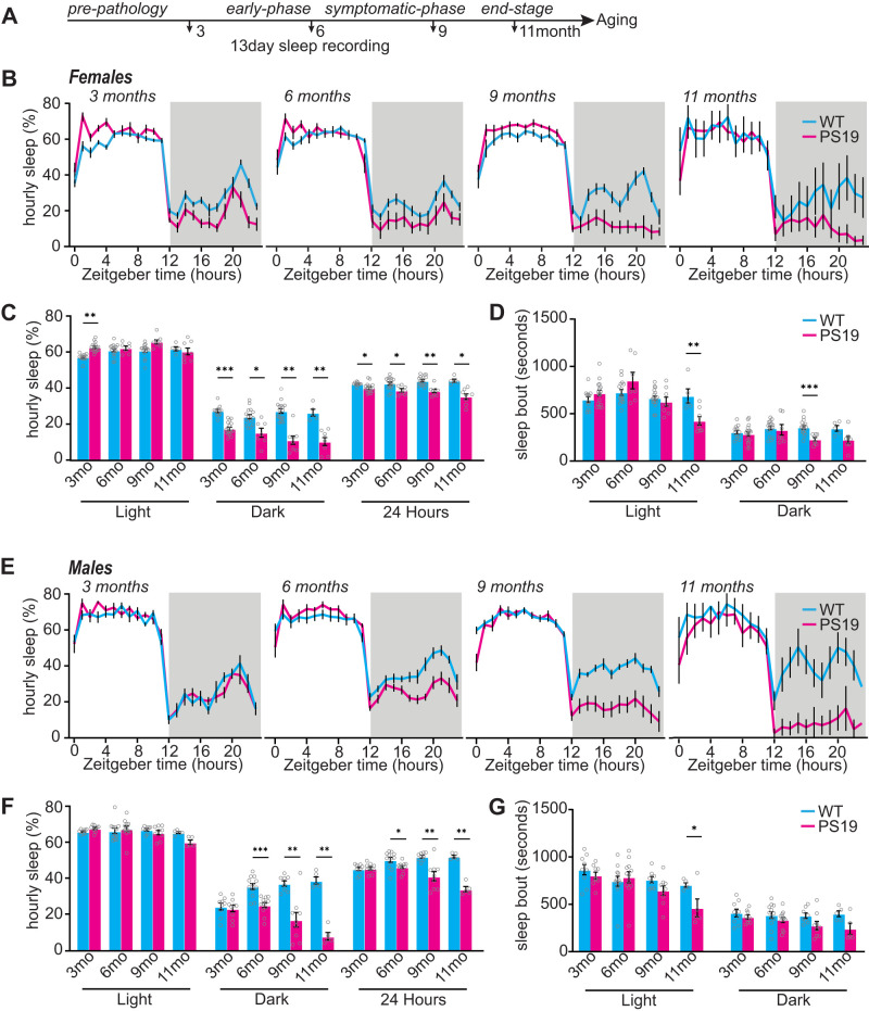
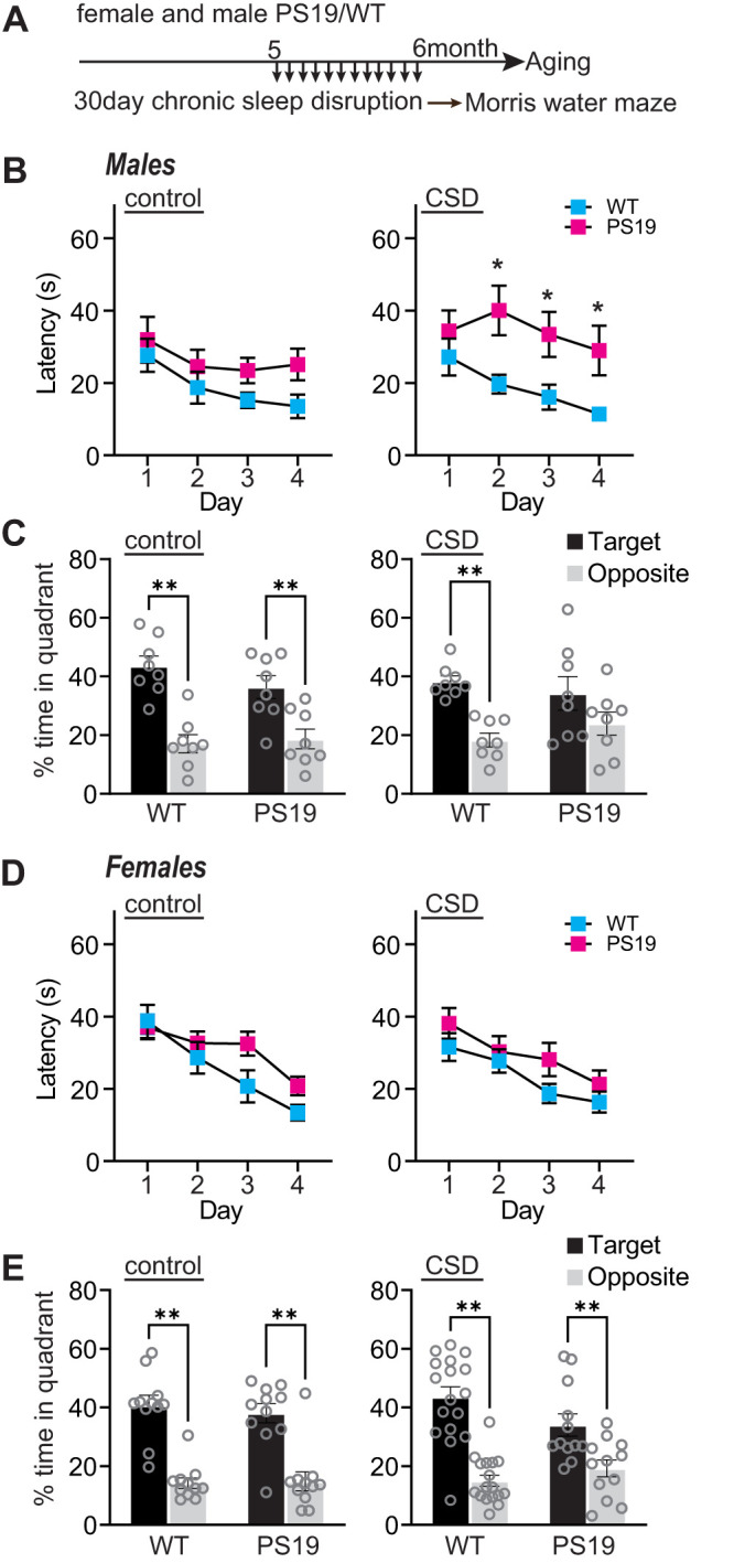
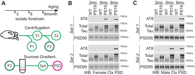
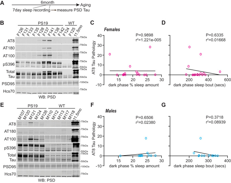
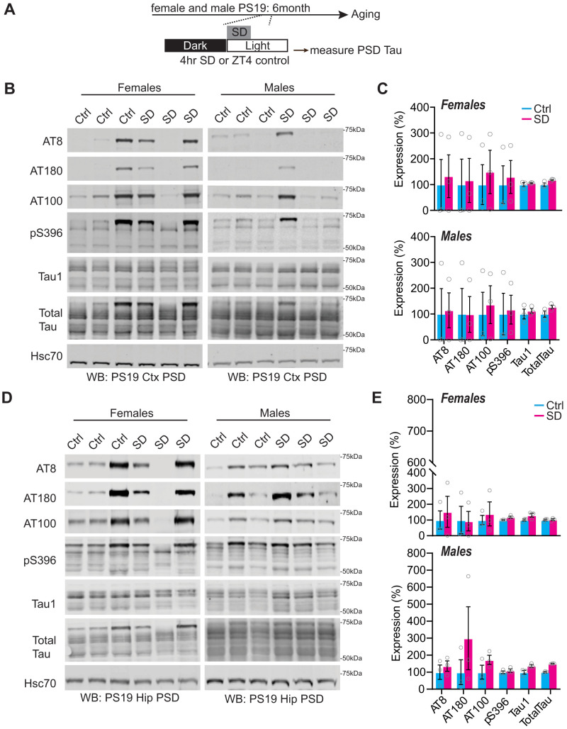
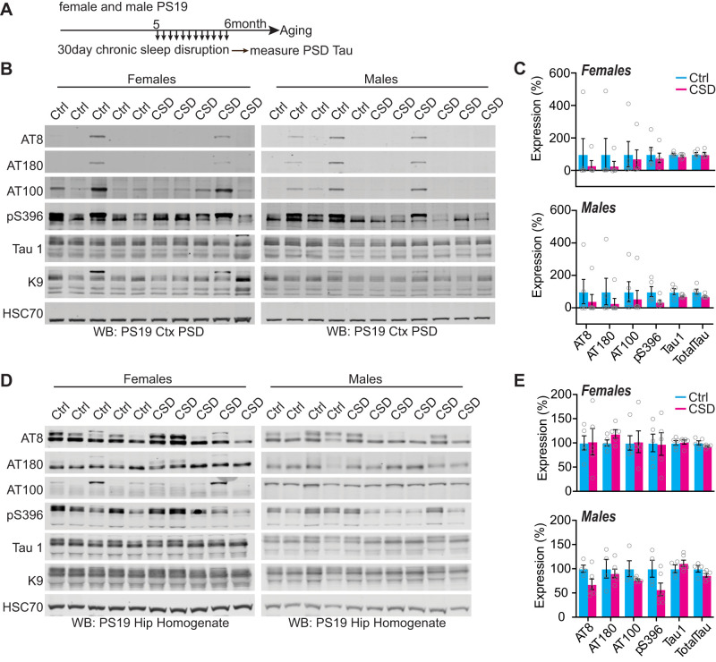
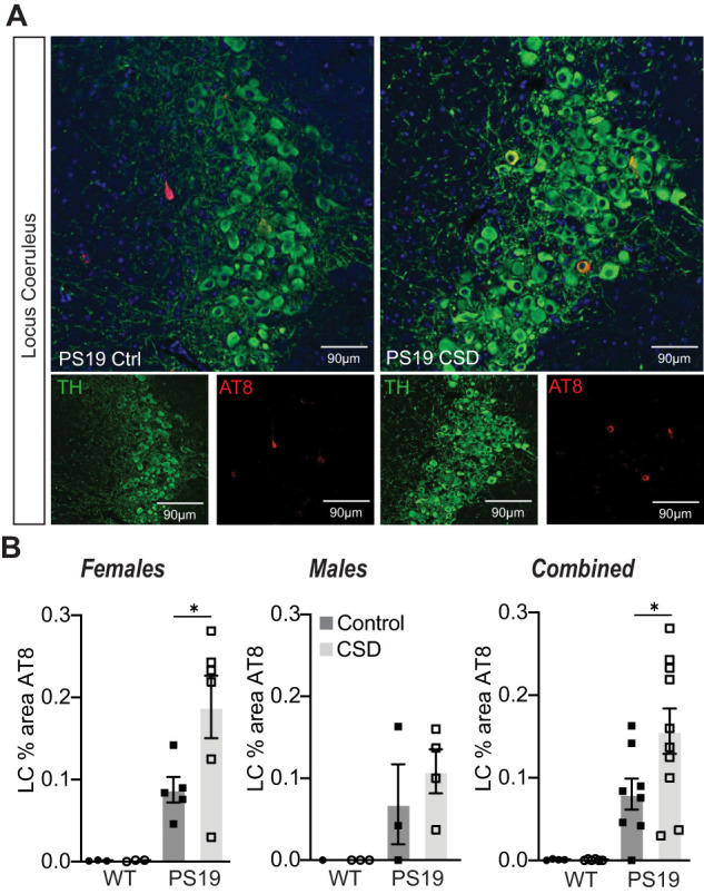
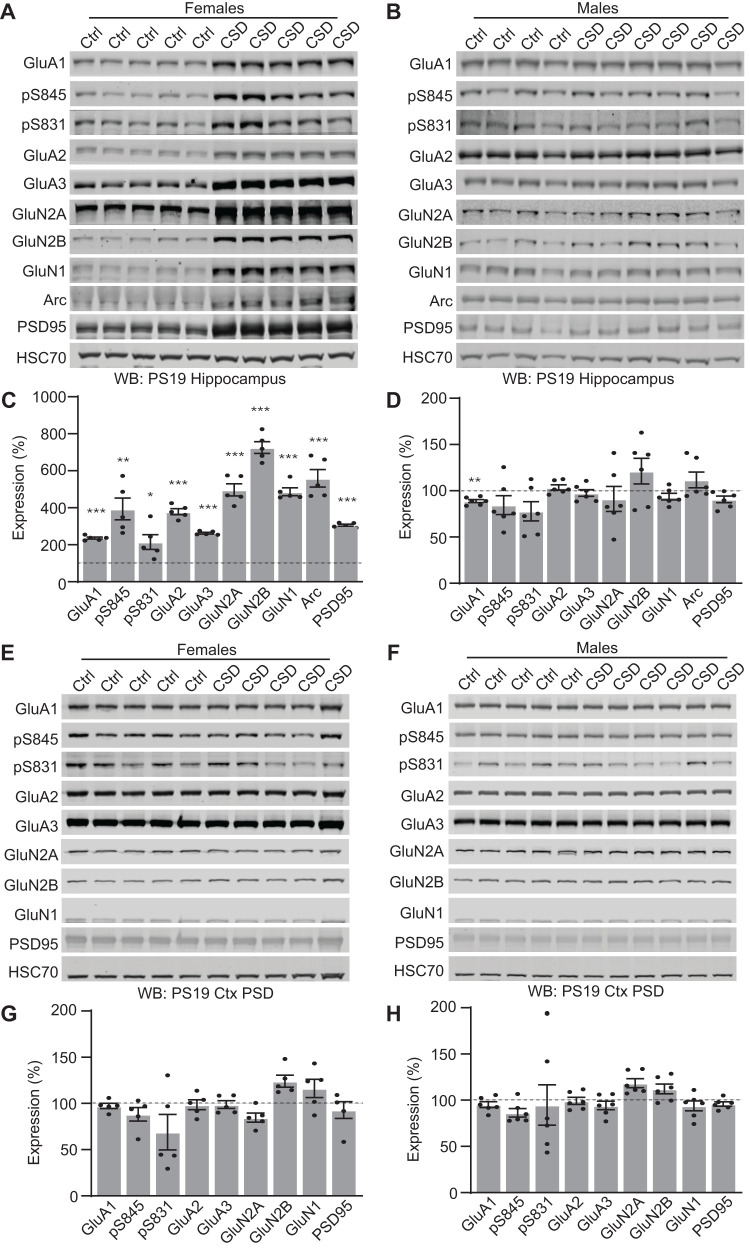
Sleep disruption and impaired synaptic processes are common features in neurodegenerative diseases, including Alzheimer's disease (AD). Hyperphosphorylated Tau is known to accumulate at neuronal synapses in AD, contributing to synapse dysfunction. However, it remains unclear how sleep disruption and synapse pathology interact to contribute to cognitive decline. Here, we examined sex-specific onset and consequences of sleep loss in AD/tauopathy model PS19 mice. Using a piezoelectric home-cage monitoring system, we showed PS19 mice exhibited early-onset and progressive hyperarousal, a selective dark-phase sleep disruption, apparent at 3 months in females and 6 months in males. Using the Morris water maze test, we report that chronic sleep disruption (CSD) accelerated the onset of decline of hippocampal spatial memory in PS19 males only. Hyperarousal occurs well in advance of robust forebrain synaptic Tau burden that becomes apparent at 6-9 months. To determine whether a causal link exists between sleep disruption and synaptic Tau hyperphosphorylation, we examined the correlation between sleep behavior and synaptic Tau, or exposed mice to acute or chronic sleep disruption at 6 months. While we confirm that sleep disruption is a driver of Tau hyperphosphorylation in neurons of the locus ceruleus, we were unable to show any causal link between sleep loss and Tau burden in forebrain synapses. Despite the finding that hyperarousal appears earlier in females, female cognition was resilient to the effects of sleep disruption. We conclude sleep disruption interacts with the synaptic Tau burden to accelerate the onset of cognitive decline with greater vulnerability in males.

Keywords: Alzheimer's disease; Tau; biological sex; sleep; sleep disruption; synapse.


Conflict of interest statement

The authors declare no competing financial interests.

Figures

Figure 1.
Figure 1.
PS19 mice have decreased sleep that worsens with age. A, Experimental design. B, Twenty-four hours of tracing the average hourly sleep of female WT (blue line) PS19 (pink line) mice at 3 months (prepathology), 6 months (early-phase), 9 months (symptomatic-phase), and 11 months (end-stage). The gray bars in sleep traces indicate a dark phase. C, D, Quantification of average hourly sleep (C) and sleep bout length in seconds (D). Data separated into 12 h of dark and light phases. E, Twenty-four hours of tracing the average hourly sleep of male WT (blue line) and PS19 (pink line) mice at 3, 6, 9, and 11 months. F, G, Quantification of average hourly sleep (F) and sleep bout length in seconds (G). Data separated into 12 h of dark and light phases. N = 5–17/age/sex/genotype. *p < 0.05, **p < 0.01, ***p < 0.001. Unpaired two-tailed Student's t test. Error bars indicate ± SEM. Estimated NREM and REM sleep % time in state and bout lengths are presented in Extended Data Figure 1-1 (females) and Extended Data Figure 1-2 (males).
Figure 2.
Figure 2.
Chronic sleep disruption accelerates cognitive decline in males. A, Experimental design. B, Males, Acquisition of spatial learning. Escape latencies during training in control and CSD treated mice. C, Males, Spatial memory retention during 1 min of probe trial, time in target or opposite quadrant in control and CSD treated mice. Target indicates the quadrant where the platform had been located, versus the opposite quadrant. D, Females, Acquisition of spatial learning. Escape latencies during training in control and CSD treated mice. E, Females, Spatial memory retention during 1 min of probe trial, time in target or opposite quadrant in control and CSD treated mice. N = 8–17 per group (sex, genotype, treatment). B, D, Data are means(±SEM) of four trials per day. C, E, Data are means (+SEM). *p < 0.05, **p < 0.01.
Figure 3.
Figure 3.
Tau in the cortex postsynaptic density (PSD). A, Subcellular fractionation technique to isolate PSD. The red line indicates the synaptosome fraction used to further isolate the PSD. B, C, Western blot analysis of cortex PSD samples showing soluble phosphorylated Tau (AT8) accumulation with age in PS19 and WT females (B) and males (C). Western blots also include total Tau and PSD95, a protein enriched in the PSD.
Figure 4.
Figure 4.
Decreased sleep amount in PS19 Tau tg mice is not predictive of AT8-Tau pathology in the cortex. A, Experimental design. B, Western blot analysis of AT8, AT180, AT100, pS396, total Tau, and PSD95 in 6-month-old (early-phase) PS19 females. C, D, Correlation of AT8-Tau hyperphosphorylation expression in the cortex of PS19 females with average dark-phase hourly sleep (C) or sleep bout length in seconds (D). Sleep data were separated into 12 h of dark and light phases. Dark-phase sleep is represented here. N = 16 PS19 females. E, Western blot analysis of AT8, AT180, AT100, pS396, total Tau, and PSD95 in 6-month-old PS19 males. F, G, Correlation of AT8-Tau hyperphosphorylation expression in the cortex of PS19 males with average dark-phase hourly sleep (F) or sleep bout length in seconds (G). Sleep data were separated into 12 h of dark and light phases. Dark-phase sleep is represented here. N = 11. All antibodies normalized to loading control. No significance (Pearson’s correlation). See Extended Data Figure 4-1 for further correlation analysis between sleep metrics and phosphorylated Tau.
Figure 5.
Figure 5.
Synaptic Tau is not increased in PS19 mice after acute sleep deprivation in the cortex and hippocampus. A, Experimental design. B, Western blot analysis of AT8, AT180, AT100, pS396, Tau1, and total Tau cortical expression in 6-month-old (early pathology) PS19 females and males. C, Quantification of cortical synaptic Tau proteins in PS19 females and males. N = 3 control; 3 CSD per sex. D, Western blot analysis of AT8, AT180, AT100, pS396, Tau1, and total Tau hippocampal expression in 6-month-old (early pathology) PS19 females and males. E, Western blot analysis of AT8, AT180, AT100, pS396, Tau1, and total Tau hippocampal expression in 6-month-old (early pathology) PS19 females and males. N = 3 control; 3 SD per sex. All antibodies normalized to loading control and then normalized to the control group. Unpaired two-tailed Student's t test between the control and treatment groups. Error bars indicate mean ± SEM.
Figure 6.
Figure 6.
Synaptic Tau is not increased in PS19 mice after chronic sleep disruption in the cortex and hippocampus. A, Experimental design. B, Western blot analysis of AT8, AT180, AT100, pS396, Tau1, and total Tau in cortical PSD fractions of 6-month-old (early pathology) PS19 females and males. C, Quantification of cortical synaptic Tau proteins in PS19 females and males. N = 5 control; 5 CSD females; N = 5 control; 6 CSD males. D, Western blot analysis of AT8, AT180, AT100, pS396, Tau1, and total Tau in the hippocampi of 6-month-old PS19 females and males. E, Quantification of hippocampal Tau proteins in PS19 females and males. N = 5 control; 5 CSD females; N = 4 control; 6 CSD males. All antibodies normalized to loading control and then normalized to the control group. Unpaired two-tailed Student's t test between the control and treatment groups. Error bars indicate mean ± SEM.
Figure 7.
Figure 7.
Chronic sleep disruption is a driver of Tau pathology in LC. A, Histology of locus ceruleus sections from PS19 mice under control or following 30 d of CSD treatment. Slices stained with TH to mark the noradrenergic neurons and AT8 to mark the Tau pathology. Example images from female PS19 mice. B, % area positive for AT8 in females, males, or combined. No AT8 signal was detected in WT littermates; therefore, statistical analysis was only conducted comparing control and CSD treatment in PS19 animals. CSD drove a significant increase in AT8 % area compared with control treatment in PS19 females or combined sexes. PS19 males showed a trend to increase. *p < 0.05 unpaired two-tailed Student's t test between the control and treatment groups for PS19 genotype. Error bars indicate mean ± SEM.
Figure 8.
Figure 8.
Chronic sleep disruption leads to changes in hippocampal synaptic protein expression in PS19 females but not males. A, B, Western blot analysis of hippocampal synaptic receptor expression in 6-month-old PS19 females (A) and males (B). C, Quantification of hippocampal protein expression in PS19 females. N = 5 control; 5 CSD. D, Quantification of hippocampal protein expression in PS19 males. N = 4 control; 6 CSD. All antibodies normalized to loading control and then normalized to the control group. Unpaired two-tailed Student's t test between the control and treatment groups for each genotype. *p < 0.05, **p < 0.01, ***p < 0.001. Error bars indicate mean ± SEM. E, F, Western blot analysis of cortical synaptic protein expression in 6-month-old PS19 females (E) and males (F). G, Quantification of cortical synaptic protein expression in PS19 females. N = 5 control; 5 CSD. H, Quantification of cortical synaptic protein expression in PS19 males. N = 4 control; 6 CSD. All antibodies normalized to loading control and then normalized to the control group. Unpaired two-tailed Student's t test between the control and treatment group for each genotype. Error bars indicate mean ± SEM.

Update of

Similar articles

Cited by

References

    1. Bombois S, Derambure P, Pasquier F, Monaca C (2010) Sleep disorders in aging and dementia. J Nutr Health Aging 14:212–217. 10.1007/s12603-010-0052-7 - DOI - PubMed
    1. Braak H, Thal DR, Ghebremedhin E, Del Tredici K (2011) Stages of the pathologic process in Alzheimer disease: age categories from 1 to 100 years. J Neuropathol Exp Neurol 70:960–969. 10.1097/NEN.0b013e318232a379 - DOI - PubMed
    1. Bruning F, Noya SB, Bange T, Koutsouli S, Rudolph JD, Tyagarajan SK, Cox J, Mann M, Brown SA, Robles MS (2019) Sleep-wake cycles drive daily dynamics of synaptic phosphorylation. Science 366. 10.1126/science.aav3617 - DOI - PubMed
    1. Chalermpalanupap T, Schroeder JP, Rorabaugh JM, Liles LC, Lah JJ, Levey AI, Weinshenker D (2018) Locus coeruleus ablation exacerbates cognitive deficits, neuropathology, and lethality in P301S Tau transgenic mice. J Neurosci 38:74–92. 10.1523/JNEUROSCI.1483-17.2017 - DOI - PMC - PubMed
    1. Crimins JL, Rocher AB, Luebke JI (2012) Electrophysiological changes precede morphological changes to frontal cortical pyramidal neurons in the rTg4510 mouse model of progressive tauopathy. Acta Neuropathol 124:777–795. 10.1007/s00401-012-1038-9 - DOI - PMC - PubMed

LinkOut - more resources