. 2024 Jul 10;44(28):e1215232024.
doi: 10.1523/JNEUROSCI.1215-23.2024.

G272V and P301L Mutations Induce Isoform Specific Tau Mislocalization to Dendritic Spines and Synaptic Dysfunctions in Cellular Models of 3R and 4R Tau Frontotemporal Dementia

Affiliations

G272V and P301L Mutations Induce Isoform Specific Tau Mislocalization to Dendritic Spines and Synaptic Dysfunctions in Cellular Models of 3R and 4R Tau Frontotemporal Dementia

Ke Yu et al. J Neurosci. .

Abstract

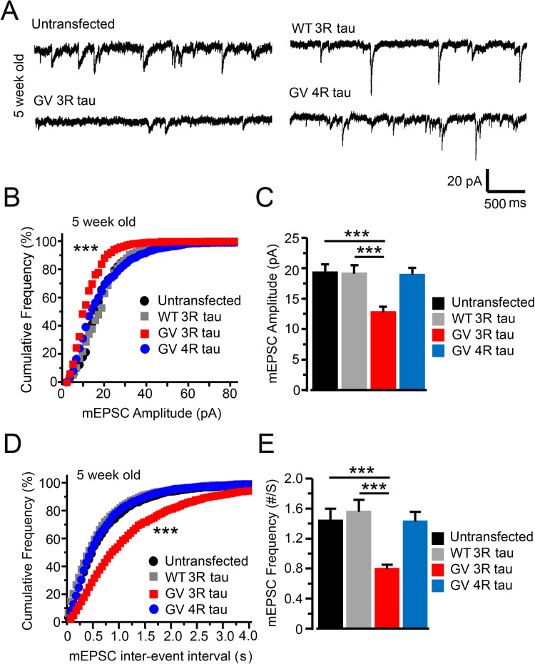
Tau pathologies are detected in the brains of some of the most common neurodegenerative diseases including Alzheimer's disease (AD), Lewy body dementia (LBD), chronic traumatic encephalopathy (CTE), and frontotemporal dementia (FTD). Tau proteins are expressed in six isoforms with either three or four microtubule-binding repeats (3R tau or 4R tau) due to alternative RNA splicing. AD, LBD, and CTE brains contain pathological deposits of both 3R and 4R tau. FTD patients can exhibit either 4R tau pathologies in most cases or 3R tau pathologies less commonly in Pick's disease, which is a subfamily of FTD. Here, we report the isoform-specific roles of tau in FTD. The P301L mutation, linked to familial 4R tau FTD, induces mislocalization of 4R tau to dendritic spines in primary hippocampal cultures that were prepared from neonatal rat pups of both sexes. Contrastingly, the G272V mutation, linked to familial Pick's disease, induces phosphorylation-dependent mislocalization of 3R tau but not 4R tau proteins to dendritic spines. The overexpression of G272V 3R tau but not 4R tau proteins leads to the reduction of dendritic spine density and suppression of mEPSCs in 5-week-old primary rat hippocampal cultures. The decrease in mEPSC amplitude caused by G272V 3R tau is dynamin-dependent whereas that caused by P301L 4R tau is dynamin-independent, indicating that the two tau isoforms activate different signaling pathways responsible for excitatory synaptic dysfunction. Our 3R and 4R tau studies here will shed new light on diverse mechanisms underlying FTD, AD, LBD, and CTE.

Keywords: AMPA receptors; Alzheimer's disease; dendritic spines; dynamin; frontotemporal dementia; tau mislocalization.


Conflict of interest statement

The authors declare no competing financial interests.

Figures

Figure 1.
Figure 1.
Isoform-specific tau mislocalization to dendritic spines induced by G272V and P301L mutations. A, A diagram showing the location of G272V and P301L mutations in tau proteins. The second microtubule-binding repeat (R2) was deleted in all our 3R tau constructs (denoted by the Δ symbol). Note that P301L mutation only exists in 4R tau as it is located in the R2 region (Wszolek et al., 2006; Ghetti et al., 2015). B, Representative images of 3-week-old cultured rat hippocampal neurons coexpressing DsRed and various GFP-tagged tau proteins (from top to bottom: wild-type 3R, wild-type 4R, P301L 4R, G272V 3R and G272V 4R tau proteins). The arrows denote dendritic spines that contain tau proteins, and the triangles denote dendritic spines devoid of tau. C, Comparisons between the proportions of dendritic spines that contained tau versus the total number of DsRed-labeled spines in dendrites from the above groups (n = 7 neurons in each group). ANOVA, NS (not significant), p > 0.05; ***, p < 0.001. D, Comparisons between the densities of dendritic spines (number of spines per 100 µm length of dendrites) of the above groups. ANOVA, NS (not significant), p > 0.05; mean ± standard error.
Figure 2.
Figure 2.
Expression of G272V 3R or 4R tau proteins does not impair synaptic function in 3-week-old neurons. A, Representative traces of mEPSCs recorded from untransfected neurons as well as neurons expressing GFP-tagged WT 3R tau, G272V 3R tau, and G272V 4R tau proteins. G272V is abbreviated as “GV” in the labeling. B, C, Cumulative frequency curves (B) and means (C) of mEPSC amplitudes in neurons of the above 4 groups (n = 10 neurons in each group). D, E, Cumulative frequency curves of interevent intervals (D) and means of mEPSC frequencies (E) in the above four groups. K–S tests were used in B and D. ANOVA tests were used in C and E, NS (no significant), p > 0.05; mean ± standard error.
Figure 3.
Figure 3.
Expression of G272V 3R tau but not 4R tau induces loss of dendritic spines in older primary rat hippocampal cultures (5-week-old). A, Representative images of 5-week-old cultured rat hippocampal neurons coexpressing DsRed and various GFP-tagged tau proteins (from top to bottom: wild-type 3R, wild-type 4R, P301L 4R, G272V 3R and G272V 4R tau proteins). The arrows denote dendritic spines that contain tau proteins, and the triangles denote dendritic spines devoid of tau. B, Comparisons between the proportions of dendritic spines that contained tau versus the total number of DsRed-labelled spines in dendrites from the above groups (n = 8 neurons in each group). ANOVA, ***, p < 0.001. C, Comparisons between the densities of dendritic spines (number of spines per 100 µm length of dendrites) of the above groups. n = 7 neurons in each group, ANOVA, ***, p < 0.001; mean ± standard error.
Figure 4.
Figure 4.
Expression of G272V 3R but not 4R tau proteins impaired synaptic function in older primary rat hippocampal cultures (5-week-old). A, Representative traces of mEPSCs recorded from untransfected neurons as well as neurons expressing GFP-tagged WT 3R tau, G272V 3R tau, and G272V 4R tau proteins. G272V is abbreviated as “GV” in the labeling. B, C, Cumulative frequency curves (B) and means (C) of mEPSC amplitudes in neurons of the above 4 groups (n = 10 neurons in each group). D, E, Cumulative frequency curves of interevent intervals (D) and means of mEPSC frequencies (E) in the above four groups. n = 10 neurons in each group. K–S test was used in B and D. ANOVA in C and E. ***, p < 0.001; mean ± standard error. The synaptic function is likely caused by the postsynaptic presence of tau due to the low transfection rate of the calcium phosphate precipitation method as presynaptic terminals are rarely transfected (see Materials and Methods and Teravskis et al., 2018, for previous characterizations).
Figure 5.
Figure 5.
Pharmacological blockade of both GSK3β and CDK5 tau kinases prevents mislocalization of G272V 3R tau proteins to dendritic spines and spine loss caused by this mutant protein. A, Five-week-old neurons that had been transfected with WT 3R tau with no drug treatment were used as the control (top row). Neurons coexpressing DsRed and GFP-tagged G272V 3R tau were untreated (middle row) or treated with a GSK3β inhibitor, CHIR99021 (500 nM), and a CDK5 inhibitor, Roscovitine (500 nM) for 24 h (bottom row). B, Comparisons between the proportions of dendritic spines that contained tau versus the total number of DsRed-labeled spines in dendrites from the above groups. C, Comparisons between the densities of dendritic spines (number of spines per 100 µm length of dendrites) of the above groups. ANOVA, n = 7 neurons in each group, ***, p < 0.001; mean ± standard error.
Figure 6.
Figure 6.
Pharmacological blockade of both GSK3β and CDK5 tau kinases rescues synaptic dysfunction caused by G272V 3R tau. A, Representative traces from 5-week-old neurons that had been transfected with no plasmid (top) or G272V 3R tau (middle, no drug treatment; lower, treated with GSK3β and CDK5 inhibitors). B, C, Cumulative curves and histograms of mEPSC amplitudes in three groups of neurons as described in A. In B, K–S tests were performed to compare neurons expressing G272V 3R tau with the untransfected control and the treated G272V 3R tau group. ***, p < 0.001 in both comparisons. In C, ANOVA analyses were performed to compare the means between the three groups (n = 8–12). *, p < 0.05; **, p < 0.01. D, E, Cumulative curves of interevent intervals and histograms of mEPSC frequencies in three groups of neurons as described in A. In D, K–S test; ***, p < 0.001 when the untreated G272V 3R was compared with the other two groups. In E, ANOVA test; *, p < 0.05; **, p < 0.01; mean ± standard error. F, Representative traces of untransfected neurons without (top) or with (bottom) cotreatment with GSK3β and CDK5 inhibitors. G, Cumulative frequency curves of the amplitudes (left) and interevent intervals (right) of mEPSCs from untreated and treated neurons (gray, no drug; black, with inhibitor treatment). H, I, Comparison of mEPSC amplitudes (H) and mEPSC frequency (I) between neurons without or with the treatment of inhibitors (gray and black, respectively). Two-group t test, n = 14 in each group, NS, not significant, p > 0.05; mean ± standard error.
Figure 7.
Figure 7.
The G272V mutation induces the phosphorylation-dependent formation of Pick body–like structures by causing asymmetric accumulation of 3R tau proteins next to the nucleus. A, B, Representative images of 5-week-old cultured neurons expressing various GFP-tagged 4 R (A) and 3 R (B) tau proteins, which had been fixed and stained with DAPI to label the nucleus (see Materials and Methods). The bottom row of B shows a neuron expressing 3R G272V tau that had been treated with CDK5 and GSK3β inhibitors. The arrows denote a Pick body–like structure on one side of the nucleus. C, The quantification of the asymmetric distributions of tau in neurons expressing various types of tau proteins without or with inhibitor treatment (see main text). ANOVA, n = 7 neurons in each group, **, p < 0.01; ***, p < 0.001; mean ± standard error.
Figure 8.
Figure 8.
The phosphorylation of the B-domain in 3R tau caused by the G272V mutation is slower than that in 4R tau caused by the P301L mutation. A, Representative images of 3-week-old cultured neurons expressing various GFP-tagged 3R (top 2 rows) and 4R (bottom 2 rows) tau proteins. The neurons were costained with the mouse AT8 antibody to detect the phosphorylation of Ser202/Ser205 in tau (part of the B-domain as described in Teravskis et al., 2021) and a rabbit polyclonal anti-GFP antibody to detect total tau, respectively. B, Images that are similar to those in A but at an older age of 5 weeks in vitro. C, Comparison of AT8 staining (normalized by GFP staining) between eight groups of neurons as described in A and B (see Materials and Methods for quantification equations). Two-way ANOVA, n = 8 neurons in each group; Factor 1 = 2 ages; Factor 2 = 4 tau variants. Overall results, Overall model, F = 8.5, p < 0.001; means in Factor 2, p < 0.001; interaction between two factors, p < 0.05. Individual group comparison in C, NS, not significant, p > 0.05; *, p < 0.05; **, p < 0.01; mean ± standard error.
Figure 9.
Figure 9.
G272V 3R tau and P301L 4R tau impair AMPAR-mediated synaptic responses through dynamin-dependent and dynamin-independent mechanisms, respectively. A, Representative traces from 4 to 5-week-old neurons that had been transfected with no plasmid (top), G272V 3R tau (middle), or P301L 4R tau (bottom) with (+) or without (−) dynasore treatment (15 μM; see Materials and Methods, Pharmacology). The arrows denote suppressed synaptic responses, and the triangle denotes a rescuing effect of dynasore. B, Cumulative curves of mEPSC amplitudes (left) and interevent time interval (right) from four groups of neurons (untransfected with or without dynasore; transfected with G272V 3R tau with or without dynasore; labeled with 4 colored symbols on the top). C, Similar cumulative curves as in B from four different groups of neurons (untransfected with or without dynasore; transfected with P301L 4R tau with or without dynasore). D, Comparison of mEPSC amplitudes between four groups of neurons in two sets of experiments [left four bars, untransfected (black), untransfected + dynasore treatment (gray), G272V 3R (red), and G272V 3R + dynasore treatment (blue); right four bars, untransfected (black), untransfected + dynasore treatment (gray), P301L 4R tau (red), and P301L 4R tau + dynasore treatment (blue)]. E, Comparison of mEPSC frequencies between 4 groups of neurons in two same sets of experiments as in D. In B and C, K–S tests were performed to compare the cumulative curves with the control curves (no transfection, no drug treatment). In D and E, one-way ANOVA tests were performed to compare the means of four groups in each set of experiments. ***, p < 0.001; NS, not significant, p > 0.05.
Figure 10.
Figure 10.
Hypothetical models for the roles of tau mislocalization in 4R tau FTD and 3R tau FTD (Pick's disease). A, In 4R tau FTDP-17 caused by P301L mutation, 4R tau proteins were phosphorylated and mislocalized to dendritic spines, which brings Fyn proteins to dendritic spines (Ittner et al., 2010; Xia et al., 2015; Padmanabhan et al., 2019). The postsynaptic nano-clustering of Fyn may lead to the loss of AMPARs via an unknown dynamin-independent mechanism. B, In familial Pick's disease (also called 3R tau FTDP-17), the phosphorylation of 3R tau leads to tau mislocalization to dendritic spines and subsequent loss of functional AMPARs in postsynaptic membranes via dynamin-dependent clathrin-mediated AMPAR endocytosis. The mislocalized 3R tau proteins may also directly or indirectly interact with actin, leading to the loss of dendritic spines. However, it remains to be determined whether 3R tau mislocalization is also associated with the postsynaptic nano-clustering of Fyn.

Similar articles

Cited by

References

    1. Andreadis A (2005) Tau gene alternative splicing: expression patterns, regulation and modulation of function in normal brain and neurodegenerative diseases. Biochim Biophys Acta 1739:91–103. 10.1016/j.bbadis.2004.08.010 - DOI - PubMed
    1. Andreadis A, Brown WM, Kosik KS (1992) Structure and novel exons of the human tau gene. Biochemistry 31:10626–10633. 10.1021/bi00158a027 - DOI - PubMed
    1. Anggono V, Huganir RL (2012) Regulation of AMPA receptor trafficking and synaptic plasticity. Curr Opin Neurobiol 22:461–469. 10.1016/j.conb.2011.12.006 - DOI - PMC - PubMed
    1. Arena JD, Smith DH, Lee EB, Gibbons GS, Irwin DJ, Robinson JL, Lee VM, Trojanowski JQ, Stewart W, Johnson VE (2020) Tau immunophenotypes in chronic traumatic encephalopathy recapitulate those of ageing and Alzheimer’s disease. Brain 143:1572–1587. 10.1093/brain/awaa071 - DOI - PMC - PubMed
    1. Bang J, Spina S, Miller BL (2015) Frontotemporal dementia. Lancet 386:1672–1682. 10.1016/S0140-6736(15)00461-4 - DOI - PMC - PubMed

MeSH terms

LinkOut - more resources