. 2024 Jun 10;7(8):e202402614.
doi: 10.26508/lsa.202402614. Print 2024 Aug.

The nucleolar phase of signal recognition particle assembly

Affiliations

The nucleolar phase of signal recognition particle assembly

Amani Issa et al. Life Sci Alliance. .

Abstract

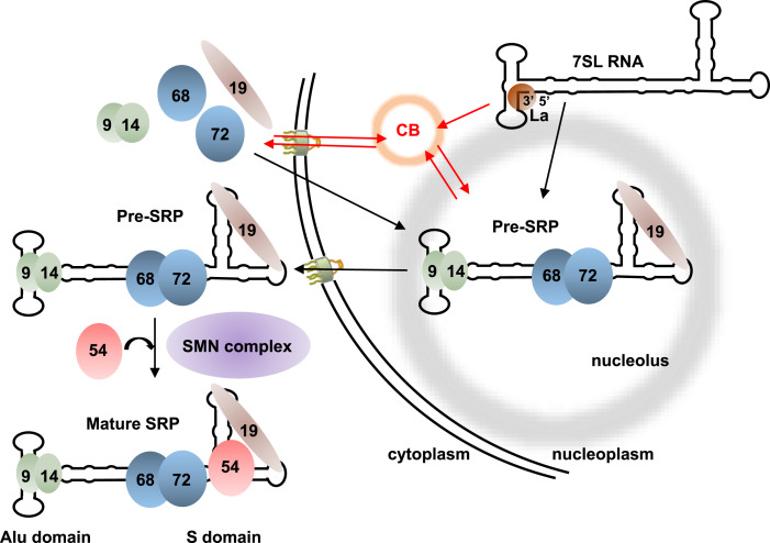
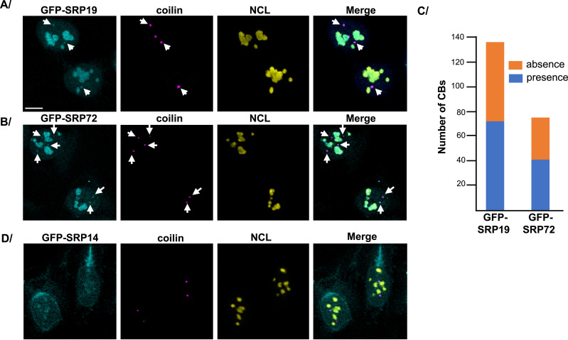
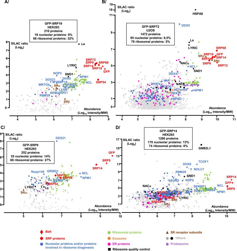
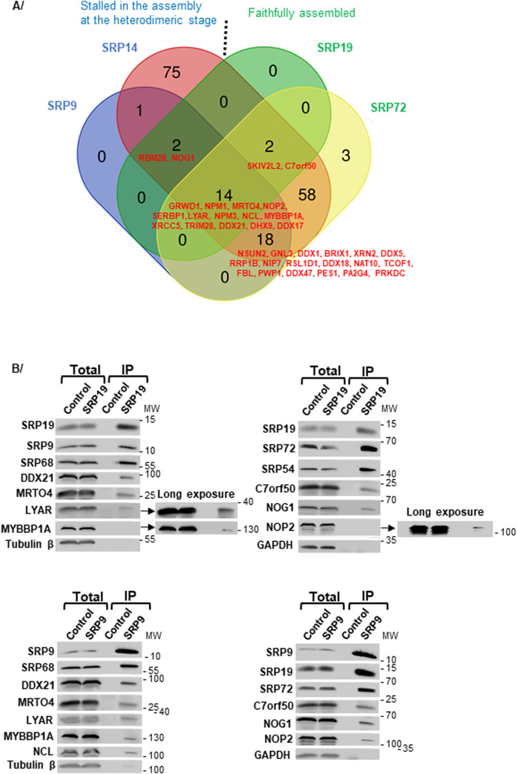
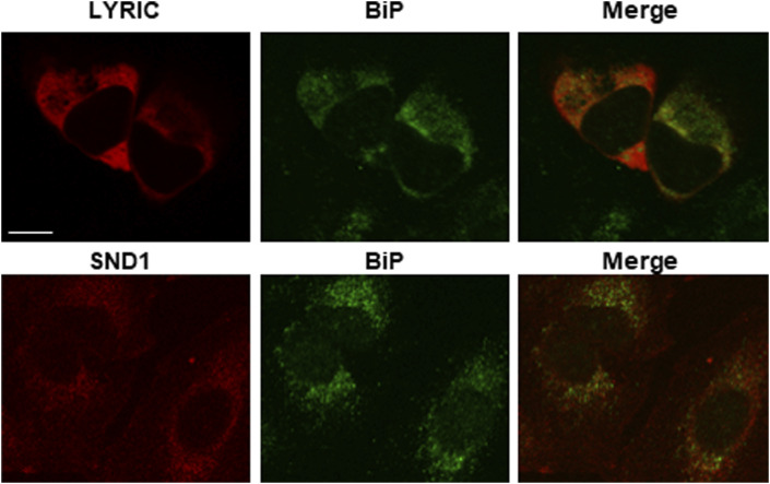
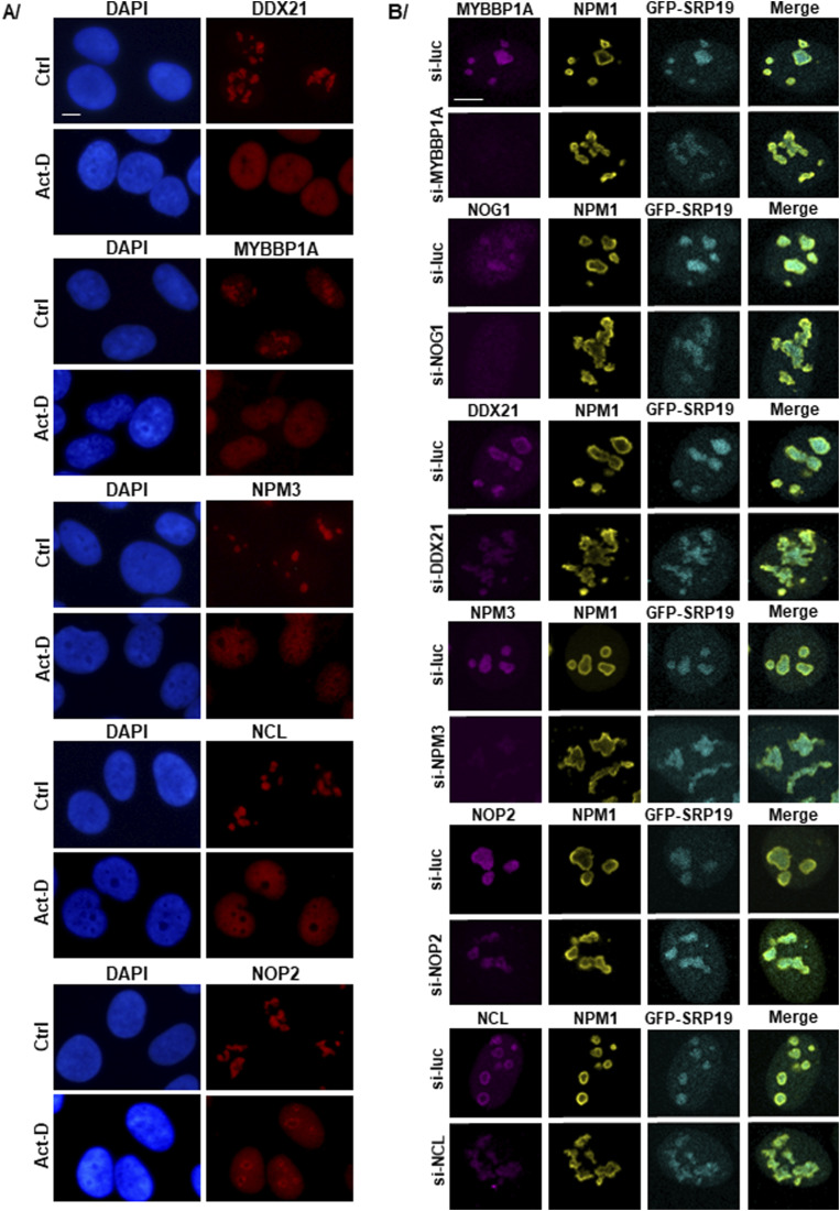
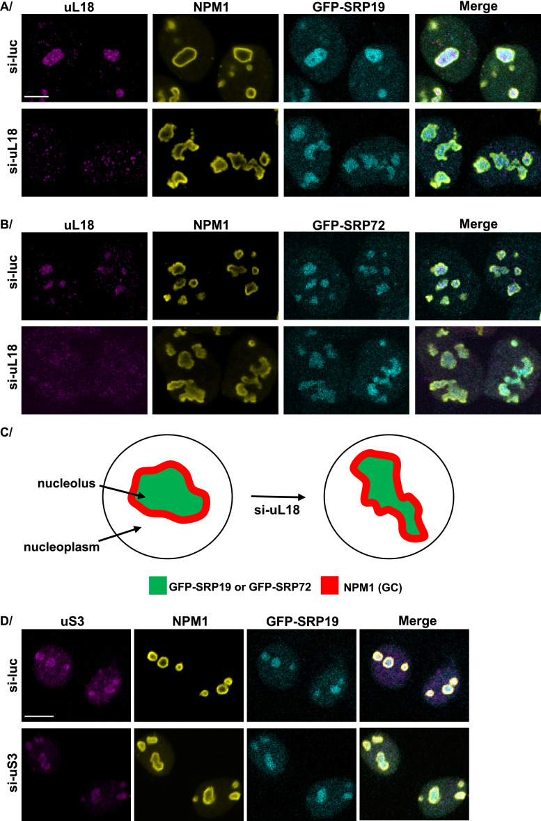
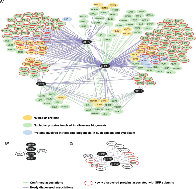
The signal recognition particle is essential for targeting transmembrane and secreted proteins to the endoplasmic reticulum. Remarkably, because they work together in the cytoplasm, the SRP and ribosomes are assembled in the same biomolecular condensate: the nucleolus. How important is the nucleolus for SRP assembly is not known. Using quantitative proteomics, we have investigated the interactomes of SRP components. We reveal that SRP proteins are associated with scores of nucleolar proteins important for ribosome biogenesis and nucleolar structure. Having monitored the subcellular distribution of SRP proteins upon controlled nucleolar disruption, we conclude that an intact organelle is required for their proper localization. Lastly, we have detected two SRP proteins in Cajal bodies, which indicates that previously undocumented steps of SRP assembly may occur in these bodies. This work highlights the importance of a structurally and functionally intact nucleolus for efficient SRP production and suggests that the biogenesis of SRP and ribosomes may be coordinated in the nucleolus by common assembly factors.


Conflict of interest statement

The authors declare that they have no conflict of interest.

Figures

Figure 1.
Figure 1.. Assembly pathway of mammalian SRP.
The RNA component of SRP, 7SL, is synthesized in the nucleoplasm by RNA polymerase III, where its 3′ end is bound by protein La. Other maturation steps occur in the nucleolus and/or putatively the Cajal bodies (CBs) as indicated. Five of the six SRP protein subunits (SRP9, SRP14, SRP19, SRP68, and SRP72) are assembled in the nucleolus (see text for details). After export to the cytoplasm, the sixth subunit (SRP54) is assembled, aided by the SMN complex, and mature SRP is produced. The Alu and S domains of 7SL are indicated.
Figure S1.
Figure S1.. Expression analysis of GFP-tagged SRP proteins in U2OS and HEK293 cell lines.
(A, B, C, D, E, F, G, H) For U2OS Flp-In T-REx cells (left column), the expression of the genes encoding GFP-SRP19 (A), GFP-SRP72 (B), GFP-SRP9 (C), and GFP-SRP14 (D) was induced by addition of Dox (1 μg/ml) to the medium and incubation for the indicated time. (E, F, G, H) Same concentration of Dox was used to induce the expression of the genes encoding GFP-SRP19 (E), GFP-SRP72 (F), GFP-SRP9 (G), and GFP-SRP14 (H) in HEK293 Flp-In T-REx cells (right column). Amounts of each GFP-SRP protein and the corresponding endogenous protein were analyzed by SDS–PAGE and WB with specific antibodies. GAPDH or tubulin β was used as a loading control. The expression of a given GFP-tagged SRP construct generally led to a reduced level of the corresponding endogenous protein. To us, this suggests that cells somehow regulate overall amounts of SRP proteins via a mechanism that remains to be determined. (I, J) Proteins SRP14 and SRP72 exist in different isoforms. U2OS Flp-In T-REx cells were transfected for 48 h with siRNAs specific to the mRNA encoding the SRP14 protein (si-SRP14) (I) or the SRP72 protein (si-SRP72) (J). Control siRNAs targeting the mRNA encoding luciferase (si-Luc) were used as negative controls. Total extracts were prepared and analyzed by SDS–PAGE and WB. Tubulin β was used as a loading control. The molecular weight ladder (MW) loaded in parallel with the samples is indicated.
Figure 2.
Figure 2.. Analysis of the SRP proteins associated with each expressed GFP-tagged SRP protein.
(A, B, C, D) Total extracts were produced from HEK293 Flp-In T-REx cells having expressed for 3 h one of the following proteins: GFP-SRP19 (A), GFP-SRP72 (B), GFP-SRP9 (C), or GFP-SRP14 (D). IPs were carried out, in the presence (+) and absence (−) of RNase A, with GFP-Trap beads and either one of these extracts or an extract of parental HEK293 Flp-In T-REx cells (Control). The immunoprecipitate (IP) and a fraction of the total cell extract (5%) (Total) were analyzed by SDS–PAGE and WB with antibodies against the indicated proteins. The molecular weight ladder (MW) loaded in parallel with the samples is indicated.
Figure S2.
Figure S2.. Analysis of the SRP proteins associated with each GFP-tagged SRP protein produced in U2OS cells.
(A, B, C, D) Synthesis of GFP-SRP19 (A), GFP-SRP72 (B), GFP-SRP9 (C), and GFP-SRP14 (D) was induced for 12 h in U2OS Flp-In T-REx cells. Total extracts were prepared, and IPs were carried out with GFP-Trap beads on extracts from parental U2OS Flp-In T-REx cells (Control) and U2OS Flp-In T-REx cells expressing one of the GFP-SRP proteins. The immunoprecipitate (IP) and a fraction (5%) of the total protein extract (Total) were analyzed by SDS–PAGE and WB with antibodies against the indicated proteins. The molecular weight ladder (MW) loaded in parallel with the samples is indicated.
Figure 3.
Figure 3.. Subcellular localization of the GFP-SRP proteins.
(A) Subcellular distribution of GFP-SRP19 and GFP-SRP72 in U2OS cells after 12 h of induction. Direct detection of GFP fluorescence by an Airyscan confocal microscopy. DNA was counterstained with 4′,6-diamidino-2-phenylindole (DAPI). Scale bar: 5 μm. (B) Schematics describing the main layers of the nucleolus. (C) Co-localization studies in U2OS cells expressing GFP-SRP19 (left) or GFP-SRP72 (right). Images acquired by a spinning disk confocal microscopy. Specific antibodies were used to detect RPA194 (labels the FC subcompartment), FBL (DFC), URB1 and NCL (PDFC), NST, and PES1 (GC) (see the Materials and Methods section for details). Scale bar: 5 μm.
Figure S3.
Figure S3.. Subcellular localization of GFP-SRP proteins in HEK293 and U2OS Flp-In T-REx cells.
(A, B, C, D, E, F) Expression of the gene encoding GFP-SRP19 (A), GFP-SRP72 (B), GFP-SRP9 (C), or GFP-SRP14 (E) in HEK293 Flp-In T-REx cells or GFP-SRP9 (D) or GFP-SRP14 (F) in U2OS Flp-In T-Rex cells was induced with Dox for 12 h. The subcellular localization of the indicated protein was then analyzed by direct fluorescence of the GFP tag. Nuclei were stained in blue with DAPI. Images were acquired with an epifluorescence microscope. GFP-SRP proteins are in green. Scale bar: 15 μm. (G, H) Expression of the genes encoding GFP-SRP19 and GFP-SRP72 was induced with Dox for 12 h in HEK293 Flp-In T-REx cells. Double IF experiments were performed with anti-coilin (a marker of CBs) and anti-NCL (a marker of nucleolus) antibodies. Images were acquired with a confocal microscope. GFP-SRP19 and GFP-SRP72 are in cyan, and their localization was determined by direct GFP fluorescence analysis. Coilin is in magenta, and NCL is in yellow. White arrows indicate the co-localization of the GFP-SRP19 or GFP-SRP72 protein with coilin. Scale bar: 7 μm.
Figure 4.
Figure 4.. Detection of SRP proteins in Cajal bodies.
(A, B, D) Expression of GFP-SRP19 (A), GFP-SRP72 (B), or GFP-SRP14 (D) was induced in U2OS Flp-In T-REx cells for 12 h. Double IF experiments were performed, using anti-coilin (a marker of CBs) and anti-NCL (a marker of nucleolus) antibodies. Images were acquired with a scanning confocal microscope. GFP-SRP19, GFP-SRP72, and GFP-SRP14 are in cyan and were located by direct GFP fluorescence detection. Coilin is in magenta, and NCL is in yellow. White arrows indicate co-localization of GFP-SRP19 or GFP-SRP72 with coilin. Scale bar: 8 μm. In (C), a graph is shown, representing the number of CBs containing (in blue) or not containing (in orange) GFP-SRP19 or GFP-SRP72, respectively, in 95 U2OS cells expressing GFP-SRP19 or 45 U2OS cells expressing GFP-SRP72. Counting was done manually by operators blinded to the samples observed.
Figure 5.
Figure 5.. Proteomic analysis of the partners of GFP-SRP proteins.
(A, B, C, D) IP-SILAC analyses performed on HEK293 Flp-In T-REx cells expressing GFP-SRP19 (A), GFP-SRP9 (C), or GFP-SRP14 (D) for 3 h or on U2OS Flp-In T-REx cells expressing GFP-SRP72 for 3 h (B). The graph displays the log2 of the SILAC ratio (y-axis, specific IP versus control IP performed with parental Flp-In T-REx cells) as a function of signal abundance (x-axis, log10(intensity)/MW). Each dot represents a protein. The labeled dots were arbitrarily selected to highlight proteins relevant to this study and families of proteins (see Key below the graphs) associated with GFP-SRP proteins. Analysis of the functions of the associated proteins was performed with the Gene Ontology Resource and UniProt. The full hit list with Significance B values is given in Table S1. The indicated percentage of nucleolar proteins and/or proteins involved in ribosome biogenesis, as well as the one of ribosomal proteins, represents the percentages in the number of these classes of proteins among all the associated proteins with the GFP-SRP protein analyzed and with a SILAC ratio above 1.
Figure 6.
Figure 6.. Nucleolar proteins associated with GFP-SRP proteins.
(A) Venn diagram showing the intersection between nucleolar proteins and/or proteins involved in ribosome biogenesis present in the GFP-SRP9, GFP-SRP14, GFP-SRP19, and GFP-SRP72 interactomes as determined by IP-SILAC analysis. The diagram includes all the proteins associated with a SILAC ratio up to 1. The ones associated with at least 3 GFP-SRP proteins are listed in red. (B) IPs were carried out on U2OS Flp-In T-REx cell total extracts, using anti-SRP19 (upper panels) and anti-SRP9 (lower panels) antibodies bound to magnetic beads with recombinant protein A (Dynabeads Protein A). Beads alone were used as a negative control (Control). The immunoprecipitate (IP) and a fraction of the total cell extract (5%) (Total) were analyzed by SDS–PAGE. The indicated proteins were revealed by WB. The molecular weight ladder (MW) loaded in parallel with the samples is indicated.
Figure S4.
Figure S4.. LYRIC and SND1 localize to the ER of U2OS cells.
The localization of SND1 was determined on untransfected U2OS cells. A double IF experiment was performed with anti-BiP (a marker of ER) and anti-SND1 antibodies. For LYRIC, U2OS cells were transfected for 48 h with a plasmid coding for GFP-tagged LYRIC. The subcellular localization of GFP-LYRIC was then determined by direct fluorescence of the GFP tag, and an IF experiment was performed with an anti-BiP antibody. Images were acquired with a confocal microscope. LYRIC and SND1 are in red, and BiP is in green. Scale bar: 7 μm.
Figure 7.
Figure 7.. Functionally intact nucleolus is required for proper localization of SRP proteins.
(A) Schematics illustrating the nucleolar redistribution of GFP-SRP19, GFP-SRP72, FBL, and NPM1 upon actinomycin D (Act-D) treatment. GFP-SRP19 and GFP-SRP72 were relocated from a DFC/GC distribution with rugged contours into a smooth, compact sphere; NPM1 was shifted from a discrete distribution in the GC (lining the periphery) to a distribution throughout the nucleoplasmic space; the distribution of FBL was shifted from bead-like in the DFC to caps. GFP-SRP19 and GFP-SRP72 are in green, FBL in red, and NPM1 in blue. (B, C, D, E, F) U2OS Flp-In T-REx cells expressing GFP-SRP19 (B, D, F) or GFP-SRP72 (C, E) for 12 h were treated with Act-D for 2 h. Cells not treated with Act-D were used as negative controls (Ctrl). IF experiments were performed with antibodies against FBL (B, C), NPM1 (D, E), or NOG1 (F). Images were acquired with a scanning confocal microscope. The localization of GFP-SRP19 and GFP-SRP72 (in green) was determined by direct GFP fluorescence analysis. NPM1, FBL, and NOG1 are shown in red. Scale bar: 8 μm (A, B, C, D, E) or 7 μm (F).
Figure S5.
Figure S5.. Nucleolar distributions of MYBBP1A, DDX21, NPM3, NOP2, and NCL are altered upon actinomycin D treatment, and their depletion induces a change in the nuclear localization of GFP-SRP19.
(A) U2OS cells were treated with Act-D for 2 h. Cells not treated with Act-D were used as a negative control (Ctrl). IF experiments were performed with antibodies against each protein (in red). Nuclei were stained in blue with DAPI. Images were acquired with an epifluorescence microscope. Scale bar: 6 μm. (B) U2OS Flp-In T-REx cells producing the GFP-SRP19 protein were transfected for 48 h with siRNAs targeting the mRNA encoding each indicated protein. siRNAs targeting luciferase mRNA (si-Luc) were used as a negative control. The expression of the gene encoding GFP-SRP19 was induced with Dox for 12 h. Double IF experiments were performed with antibodies against NPM1 (in yellow) and against each of the depleted nucleolar proteins (in magenta). The localization of GFP-SRP19 (in cyan) was determined by direct GFP fluorescence analysis. Images were acquired with a confocal microscope. Scale bar: 5 μm.
Figure 8.
Figure 8.. Nuclear localizations of GFP-SRP19 and GFP-SRP72 are disrupted upon uL18 depletion in U2OS Flp-In T-REx cells.
(A, B) U2OS Flp-In T-REx cells expressing the gene encoding GFP-SRP19 (A) or GFP-SRP72 (B) were transfected for 72 h with siRNAs targeting the mRNA encoding the ribosomal protein uL18 (si-uL18). Specific siRNAs against luciferase mRNA (si-Luc) were used as a negative control. The expression of the genes encoding GFP-SRP19 and GFP-SRP72 was induced with Dox for 12 h. Double IF experiments were performed with antibodies against the ribosomal protein uL18 and against NPM1, which marks the GC of the nucleolus. The localizations of GFP-SRP19 and GFP-SRP72 were determined by direct GFP fluorescence analysis. Images were acquired with a confocal microscope. uL18 is in magenta, NPM1 is in yellow, and GFP-SRP19 and GFP-SRP72 are in cyan. Scale bar: 7 μm. (C) Schematic representation of the disrupted nucleolus and altered nucleolar distribution of GFP-SRP19 and GFP-SRP72 after uL18 depletion. (D) U2OS Flp-In T-REx cells producing the GFP-SRP19 protein were transfected for 72 h with siRNAs targeting the mRNA encoding ribosomal protein uS3 of the small subunit (si-uS3). The legend is the same as in (A, B). Scale bar: 7 μm.
Figure 9.
Figure 9.. Network of associations with SRP9, SRP14, SRP19, and SRP72 proteins in humans.
(A) Associations between SRP proteins and nucleolar proteins involved in ribosome biogenesis (in green circles), nucleolar proteins with currently no known function in ribosome biogenesis (in yellow circles), and proteins involved in ribosome biogenesis in the nucleoplasm and cytoplasm (in blue circles). (B) Associations between SRP proteins and LYRIC and SND1. (C) Associations between SRP proteins and the RQC. The associations uncovered by our data are indicated by a purple line, and the confirmed ones by a green line. The schematics have been prepared with Cytoscape. The previously reported associations were extracted from BioGRID and were described in high-throughput screens (Hayano et al, 2003; Ewing et al, 2007; Todd & Picketts, 2012; Marcon et al, 2014; Hein et al, 2015; Huttlin et al, 2015, 2017, 2021; Kärblane et al, 2015; Boldt et al, 2016; Salvetti et al, 2016; Fasci et al, 2018; Horlbeck et al, 2018; Jang et al, 2018; Liu et al, 2018; Kim et al, 2021; Cho et al, 2022). The new SRP interactors discovered by our data are circled in red.

Similar articles

Cited by

References

    1. Abdel Ghafar MT, Soliman NA (2022) Metadherin (AEG-1/MTDH/LYRIC) expression: Significance in malignancy and crucial role in colorectal cancer. Adv Clin Chem 106: 235–280. 10.1016/bs.acc.2021.09.007 - DOI - PubMed
    1. Bassler J, Hurt E (2019) Eukaryotic ribosome assembly. Annu Rev Biochem 88: 281–306. 10.1146/annurev-biochem-013118-110817 - DOI - PubMed
    1. Bensaude O (2011) Inhibiting eukaryotic transcription: Which compound to choose? How to evaluate its activity? Transcription 2: 103–108. 10.4161/trns.2.3.16172 - DOI - PMC - PubMed
    1. Boldt K, van Reeuwijk J, Lu Q, Koutroumpas K, Nguyen T-MT, Texier Y, van Beersum SEC, Horn N, Willer JR, Mans DA, et al. (2016) An organelle-specific protein landscape identifies novel diseases and molecular mechanisms. Nat Commun 7: 11491. 10.1038/ncomms11491 - DOI - PMC - PubMed
    1. Boulon S, Pradet-Balade B, Verheggen C, Molle D, Boireau S, Georgieva M, Azzag K, Robert MC, Ahmad Y, Neel H, et al. (2010. a) HSP90 and its R2TP/Prefoldin-like cochaperone are involved in the cytoplasmic assembly of RNA polymerase II. Mol Cell 39: 912–924. 10.1016/j.molcel.2010.08.023 - DOI - PMC - PubMed

Substances

LinkOut - more resources