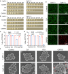
Thymoquinone as an electron transfer mediator to convert Type II photosensitizers to Type I photosensitizers
- PMID: 38858372
- PMCID: PMC11164902
- DOI: 10.1038/s41467-024-49311-z
Thymoquinone as an electron transfer mediator to convert Type II photosensitizers to Type I photosensitizers
Abstract
The development of Type I photosensitizers (PSs) is of great importance due to the inherent hypoxic intolerance of photodynamic therapy (PDT) in the hypoxic microenvironment. Compared to Type II PSs, Type I PSs are less reported due to the absence of a general molecular design strategy. Herein, we report that the combination of typical Type II PS and natural substrate carvacrol (CA) can significantly facilitate the Type I pathway to efficiently generate superoxide radical (O2-•). Detailed mechanism study suggests that CA is activated into thymoquinone (TQ) by local singlet oxygen generated from the PS upon light irradiation. With TQ as an efficient electron transfer mediator, it promotes the conversion of O2 to O2-• by PS via electron transfer-based Type I pathway. Notably, three classical Type II PSs are employed to demonstrate the universality of the proposed approach. The Type I PDT against S. aureus has been demonstrated under hypoxic conditions in vitro. Furthermore, this coupled photodynamic agent exhibits significant bactericidal activity with an antibacterial rate of 99.6% for the bacterial-infection female mice in the in vivo experiments. Here, we show a simple, effective, and universal method to endow traditional Type II PSs with hypoxic tolerance.
© 2024. The Author(s).
Conflict of interest statement
The authors declare no competing interests.
Figures


Similar articles
-
Electron Transfer Mediator Modulates Type II Porphyrin-Based Metal-Organic Framework Photosensitizers for Type I Photodynamic Therapy.Angew Chem Int Ed Engl. 2025 Feb 24;64(9):e202420643. doi: 10.1002/anie.202420643. Epub 2024 Nov 28. Angew Chem Int Ed Engl. 2025. PMID: 39560938
-
Constructing Heavy-Atom-Free Photosensitizers for Hypoxic Tumor Phototherapy Based on Donor-Excited Photoinduced Electron-Transfer-Driven Type-I and Type-II Mechanisms.ACS Appl Mater Interfaces. 2024 Aug 7;16(31):40428-40443. doi: 10.1021/acsami.4c02175. Epub 2024 Jul 23. ACS Appl Mater Interfaces. 2024. PMID: 39042585
-
BODIPY-Based Photodynamic Agents for Exclusively Generating Superoxide Radical over Singlet Oxygen.Angew Chem Int Ed Engl. 2021 Sep 1;60(36):19912-19920. doi: 10.1002/anie.202106748. Epub 2021 Aug 3. Angew Chem Int Ed Engl. 2021. PMID: 34227724
-
The Current Advances in Design Strategy (Indirect Strategy and Direct Strategy) for Type-I Photosensitizers.Adv Sci (Weinh). 2025 Feb;12(7):e2413365. doi: 10.1002/advs.202413365. Epub 2024 Dec 25. Adv Sci (Weinh). 2025. PMID: 39721012 Free PMC article. Review.
-
Recent Advances in Hypoxia-Overcoming Strategy of Aggregation-Induced Emission Photosensitizers for Efficient Photodynamic Therapy.Adv Healthc Mater. 2021 Dec;10(24):e2101607. doi: 10.1002/adhm.202101607. Epub 2021 Oct 27. Adv Healthc Mater. 2021. PMID: 34674386 Review.
Cited by
-
Nanocatalytic medicine enabled next-generation therapeutics for bacterial infections.Mater Today Bio. 2024 Sep 16;29:101255. doi: 10.1016/j.mtbio.2024.101255. eCollection 2024 Dec. Mater Today Bio. 2024. PMID: 39381264 Free PMC article. Review.
-
PhotoChem Interplays: Lighting the Way for Drug Delivery and Diagnosis.Adv Drug Deliv Rev. 2025 Apr;219:115549. doi: 10.1016/j.addr.2025.115549. Epub 2025 Feb 20. Adv Drug Deliv Rev. 2025. PMID: 39986440 Review.
-
pH-Responsive AIE Photosensitizers for Enhanced Antibacterial Therapy.Angew Chem Int Ed Engl. 2025 Jul;64(27):e202506505. doi: 10.1002/anie.202506505. Epub 2025 May 8. Angew Chem Int Ed Engl. 2025. PMID: 40299633 Free PMC article.
-
Nanoparticle-Mediated Cuproptosis and Photodynamic Synergistic Strategy: A Novel Horizon for Cancer Therapy.Cancer Med. 2025 Feb;14(3):e70599. doi: 10.1002/cam4.70599. Cancer Med. 2025. PMID: 39868904 Free PMC article. Review.
-
Nanoparticles for Photodynamic Therapy of Breast Cancer: A Review of Recent Studies.Molecules. 2025 Mar 31;30(7):1571. doi: 10.3390/molecules30071571. Molecules. 2025. PMID: 40286175 Free PMC article. Review.
References
-
- Kenry, Liu B. Enhancing the theranostic performance of organic photosensitizers with aggregation-induced emission. Acc. Mater. Res. 2022;3:721–734. doi: 10.1021/accountsmr.2c00039. - DOI
MeSH terms
Substances
Grants and funding
LinkOut - more resources
Full Text Sources

