. 2024 Jun 10;14(1):13303.
doi: 10.1038/s41598-024-61127-x.

Antiviral activity of pimecrolimus against dengue virus type 2 infection in vitro and in vivo

Affiliations

Antiviral activity of pimecrolimus against dengue virus type 2 infection in vitro and in vivo

Seong-Ryeol Kim et al. Sci Rep. .

Abstract

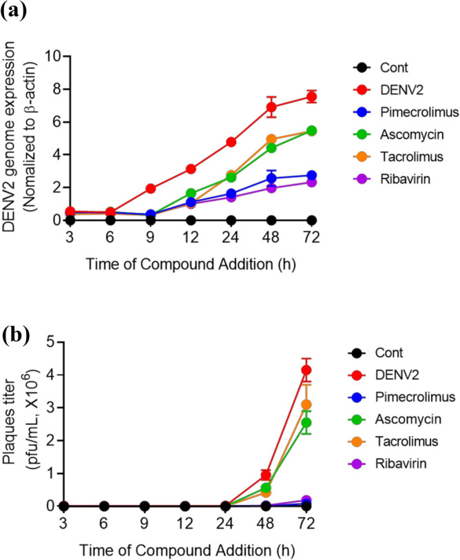
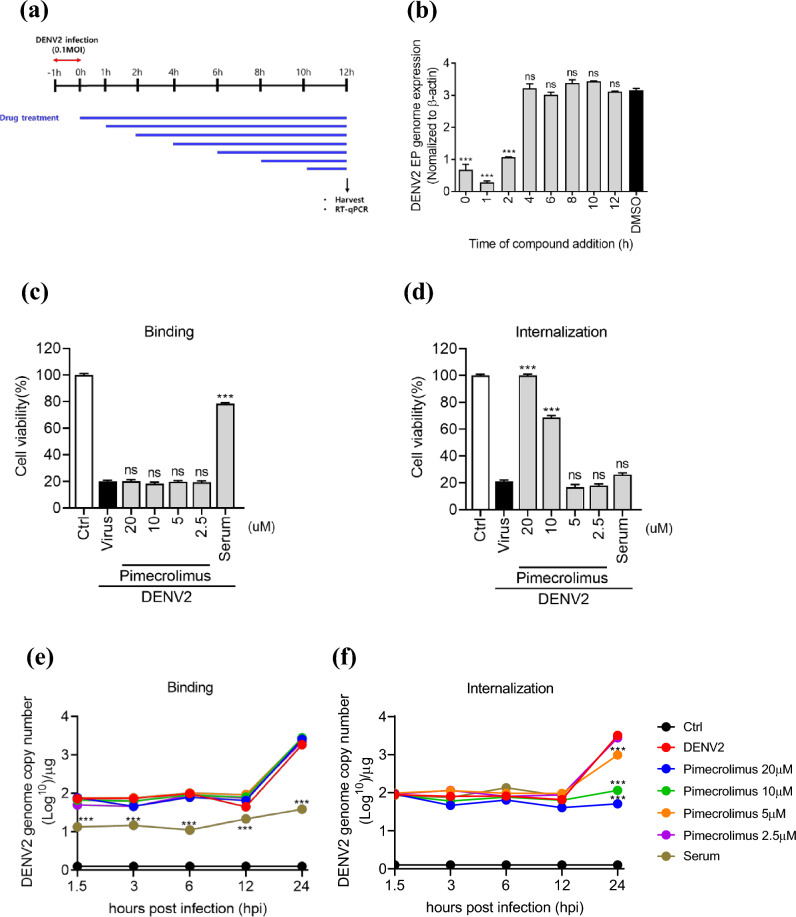
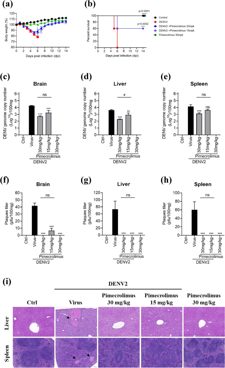
Dengue virus (DENV) infection is a public health concern in several countries and is associated with severe diseases, such as dengue hemorrhagic fever and dengue shock syndrome. DENVs are transmitted to humans via the bites of infected Aedes mosquitoes, and no antiviral therapeutics are currently available. In this work, we aimed to identify antiviral drugs against DENV type 2 (DENV2) infections and selected pimecrolimus as a potential antiviral drug candidate. Pimecrolimus significantly inhibited DENV2-mediated cell death and replication in vitro. We also confirmed a decrease in the number of plaques formed as well as in the envelope protein levels of DENV2. The time-of-addition and course experiments revealed that pimecrolimus inhibited DENV2 infection during the early stages of the virus replication cycle. In an experimental mouse model, orally administered pimecrolimus alleviated body weight loss and lethality caused by DENV2 infection, which we used as readouts of the drug's antiviral potency. Furthermore, pimecrolimus significantly inhibited the DENV2 load and ameliorated focal necrosis in the liver and spleen. Taken together, our in vitro and in vivo findings suggest that pimecrolimus is a promising antiviral drug candidate for the treatment of DENV2 infection.


Conflict of interest statement

The authors declare no competing interests.

Figures

Figure 1
Figure 1
Inhibition of dengue virus type 2 (DENV2) production by pimecrolimus, ascomycin, and tacrolimus in Vero cells,Vero cells were infected with DENV2 at 0.1 multiplicity of infection (MOI) and treated with 20 μΜ pimecrolimus, ascomycin, or tacrolimus and 500 μΜ ribavirin. (a) Relative DENV2 genome expression across the various treatments was determined via real-time PCR at different time points. (b) Culture supernatants at different time points were used to infect BHK-21 cells. DENV2 titers were determined using plaque assays. Data are expressed as mean ± SEM (n = 3 independent biological experiments, each conducted with triplicate technical repeats).
Figure 2
Figure 2
Pimecrolimus inhibits DENV2 replication at an early stage of infection. (a) Experimental scheme of the time-of-addition assay. (b) Vero cells infected with 0.1 MOI of DENV2. Pimecrolimus at 20 μΜ was added at the indicated time points, and genome expression was analyzed 12 h post-infection using real-time PCR. (c) Binding inhibition assay and (d) internalization inhibition assay of 0.1 MOI DENV2-infected Vero cells treated with the indicated concentrations of pimecrolimus at 96 hpi using cell viability assay. (e) Binding inhibition assay and (f) internalization inhibition assay of 0.1 MOI DENV2-infected Vero cells treated with the indicated concentrations of pimecrolimus at different time points via real-time PCR for DENV2 genome copy number. Data are expressed as mean ± SEM (n = 3 independent biological experiments, each conducted with triplicate technical repeats; ns, not significant; ***p < 0.001; comparisons were made against the DENV2-infected and non-infected control groups.
Figure 3
Figure 3
Pimecrolimus exhibits antiviral activity against DENV2 infection, (a) Viability of Vero cells treated with the indicated concentrations of pimecrolimus or ribavirin without virus for 4 dpi. (b) Vero cells infected with 0.1 MOI of DENV2 and treated with the indicated concentrations of pimecrolimus and ribavirin for 4 dpi. (c) Representative images of the crystal violet staining for visually comparing the viability of control and DENV2-infected Vero cells across the different compound concentrations used. (d) BHK-21 cells treated with the indicated concentrations of pimecrolimus and ribavirin without virus for 2 dpi. (e) BHK-21 cells infected with 0.1 MOI of DENV2 and treated with the indicated concentrations of pimecrolimus and ribavirin for 2 dpi. (f) Representative images of the crystal violet staining for visually comparing the viability of control and DENV2-infected BHK-21 cells across the different compound concentrations used. (g) Plaque reduction assay in DENV2-infected BHK-21 cells treated with the indicated concentrations of pimecrolimus and ribavirin. After 4 days, cells were stained with 0.2% crystal violet, and representative images of cell viability results are shown. (h) Comparison of the relative number of plaques formed in BHK-21 cells across the different pimecrolimus or ribavirin treatment concentrations used. (i) Plaque reduction assay in DENV2-infected Vero E6 cells treated with the indicated concentrations of pimecrolimus and ribavirin. After 7 days, cells were stained with 0.2% crystal violet, and representative images of cell viability results are shown. (j) Comparison of the relative number of plaques formed in Vero E6 cells across the different pimecrolimus or ribavirin treatment concentrations used. (k) Plaque reduction assay on DENV2-infected Huh-7 cells treated with the indicated concentrations of pimecrolimus and ribavirin. After 7 days, cells were stained with 0.2% crystal violet. Representative images of cell viability results are shown. (l) Comparison of the relative number of plaques formed in Huh-7 cells treated with different pimecrolimus and ribavirin concentrations. Data are expressed as mean ± SEM (n = 3 independent biological experiments, each conducted with triplicate technical repeats; ns, not significant; ***p < 0.001; comparisons were made against the DENV2-infected group).
Figure 4
Figure 4
Pimecrolimus limits DENV2 localization in Vero cells, (a) Immunofluorescence images of Vero cells infected with 0.1 MOI of DENV2 and treated with pimecrolimus or ribavirin at the indicated concentrations for 48 h. The expression of the DENV envelope (DENV E) protein was used to distinguish DENV2-positive cells. Nuclei are stained with DAPI. Scale bar: 50 μm. (b) Huh-7 cells infected with 0.1 MOI of DENV2 for 72 h at the indicated concentrations of pimecrolimus or ribavirin. The protein levels of DENV E were determined via western blotting. The expression of GAPDH was used as a loading control. (c) Quantification of the DENV E fold-change in protein levels relative to the DENV2 and vehicle-treated group. Data are expressed as mean ± SEM (n = 3 independent biological experiments, each conducted with triplicate technical repeats; ns, not significant; ***p < 0.001; comparisons made against the DENV2-infected and vehicle-treated group).
Figure 5
Figure 5
Antiviral effect of pimecrolimus against DENV2 in IFNAR1-transgenic mice. Six-week-old IFNAR1-transgenic mice were intraperitoneally inoculated with DENV2 (1.05 × 106 PFU/mouse). Pimecrolimus was orally administered in mice (15 or 30 mg/kg) twice daily for 14 days. (a) Graph depicting changes in the body weight of mice across the different treatment groups, monitored for 14 dpi. (b) Survival rate of mice infected with DENV2 and treated with the indicated pimecrolimus concentrations. The DENV2 genome copy number was estimated in the supernatants of (c) brain, (d) liver, and (e) spleen tissue homogenates at 6 dpi, between the various treatment groups. Accordingly, the DENV2 plaque titer was analyzed in the supernatants of (f) brain, (g) liver, and (h) spleen tissue homogenates at 6 dpi, between the various treatment groups. (i) Representative hematoxylin and eosin staining images of the murine liver and spleen tissues. Black arrows indicate focal necrosis. Scale bar: 100 μm. All data are expressed as mean ± SEM (n = 3 independent biological experiments, each conducted with triplicate technical repeats; ns, not significant; *p < 0.05; ***p < 0.001; ****p < 0.0001; all comparisons were made against the DENV2-infected group).

Similar articles

References

    1. Zhou L, et al. Identification of ascomycin against Zika virus infection through screening of natural product library. Antiviral Res. 2021;196:105210. doi: 10.1016/j.antiviral.2021.105210. - DOI - PubMed
    1. Henchal EA, Putnak JR. The dengue viruses. Clin. Microbiol. Rev. 1990;3:376–396. doi: 10.1128/CMR.3.4.376. - DOI - PMC - PubMed
    1. Bhatt S, et al. The global distribution and burden of dengue. Nature. 2013;496:504–507. doi: 10.1038/nature12060. - DOI - PMC - PubMed
    1. Stanaway JD, et al. The global burden of dengue: An analysis from the Global Burden of Disease Study 2013. Lancet Infect. Dis. 2016;16:712–723. doi: 10.1016/S1473-3099(16)00026-8. - DOI - PMC - PubMed
    1. Kok BH, et al. Dengue virus infection—a review of pathogenesis, vaccines, diagnosis and therapy. Virus Res. 2023;324:199018. doi: 10.1016/j.virusres.2022.199018. - DOI - PMC - PubMed