. 2024 Oct;16(10):1715-1722.
doi: 10.1038/s41557-024-01552-7. Epub 2024 Jun 10.

A folding motif formed with an expanded genetic alphabet

Affiliations

A folding motif formed with an expanded genetic alphabet

Bang Wang et al. Nat Chem. 2024 Oct.

Abstract

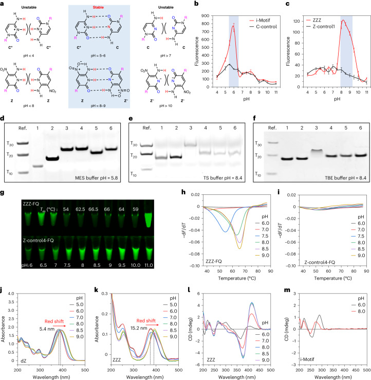
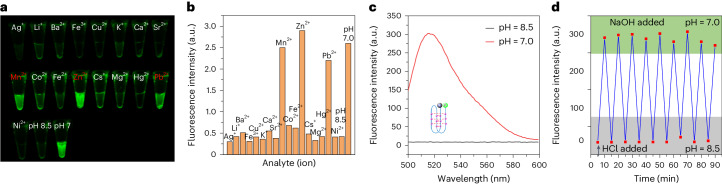
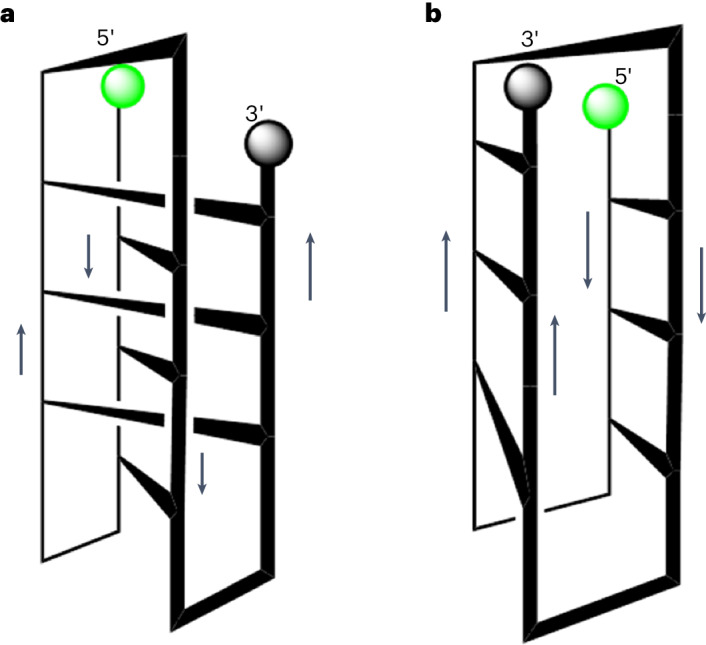
Adding synthetic nucleotides to DNA increases the linear information density of DNA molecules. Here we report that it also can increase the diversity of their three-dimensional folds. Specifically, an additional nucleotide (dZ, with a 5-nitro-6-aminopyridone nucleobase), placed at twelve sites in a 23-nucleotides-long DNA strand, creates a fairly stable unimolecular structure (that is, the folded Z-motif, or fZ-motif) that melts at 66.5 °C at pH 8.5. Spectroscopic, gel and two-dimensional NMR analyses show that the folded Z-motif is held together by six reverse skinny dZ-:dZ base pairs, analogous to the crystal structure of the free heterocycle. Fluorescence tagging shows that the dZ-:dZ pairs join parallel strands in a four-stranded compact down-up-down-up fold. These have two possible structures: one with intercalated dZ-:dZ base pairs, the second without intercalation. The intercalated structure would resemble the i-motif formed by dC:dC+-reversed pairing at pH ≤ 6.5. This fZ-motif may therefore help DNA form compact structures needed for binding and catalysis.


Conflict of interest statement

The authors declare no competing interests.

Figures

Fig. 1
Fig. 1. Definition of terms describing pairs between nucleobases in various orientations.
a, Canonical Watson–Crick pair joining antiparallel strands between dZ and dP, with deoxyribose or ribose (R, labelled pink) oriented down. b, Reverse Watson–Crick pair between dZ and dJ, with the 2′-deoxyribose (R) on the left (right) oriented down (up), holding together parallel strands. c, Skinny pair between dZ and dS, with both 2′-deoxyriboses (R) oriented down, holding together antiparallel strands. d, Reverse skinny pair between dZ and dC; the 2′-deoxyribose (R) on the left (right) is oriented down (up). e, Positively charged reverse skinny pair between charge-neutral and protonated C; the 2′-deoxyribose (R) on the left (right) is oriented down (up). f, Negatively charged reverse skinny pair between charge-neutral and deprotonated Z; the 2′-deoxyribose (R) on the left (right) is oriented down (up).
Fig. 2
Fig. 2. Characterization of fZ-motif.
a, Structural influence of pH on reverse skinny C:C and Z:Z pair formation in various charge conditions. b, The effect of pH on ThT fluorescence when intercalated into C-rich sequences able to form the i-motif via dC:dC+ pairing. c, The impact of pH on ThT fluorescence when intercalated into dZ-rich sequences able to form the fZ-motif via dZ:dZ pairing. There were n = 5 independent runs; error bars in b and c represent mean values ± s.d. The blue shaded areas represent the DNA forming the motif structure. df, DNA samples analysed by non-denaturing polyacrylamide gel electrophoresis (20%) in 2-(N-morpholino)ethanesulfonic acid (MES) (d), TS (e) and TBE (f) buffers. Left lane: T10, T20 and T30 oligonucleotides. Lane 1, i-motif; lane 2, C-control; lane 3, fZ-motif; lane 4, Z-control1; lane 5, Z-control2; lane 6, Z-control3. The gels were stained using the Stains-All dye. g, Photographs of fluorescence of DNA samples (1 μM) in 100 mM phosphate buffer with pHs ranging from 6 to 11 at 25 °C. h,i, Melting peaks of ZZZ-FQ (h) and Z-control4-FQ (i) reveal temperature responses across pHs 6 to 9 in phosphate buffer. j,k, Absorption spectra of dZ nucleoside (j) and ZZZ oligo (k) in phosphate buffer across pHs 5–9. l,m, Circular dichroism (CD) spectra compare ZZZ oligo (l) and i-motif (m) sequences, highlighting structural insights across pHs 6–9 in phosphate buffer. Source data
Fig. 3
Fig. 3. Characterization of fZ-motif structure by NMR.
a, 1H NMR spectra of ZZZ oligo at indicated pH in two indicated solvent systems. All samples contain 50 mM phosphate buffer. The asterisks represent DNA samples from which triethylamine was not fully removed by desalting. b, NOESY spectrum of ZZZ at pH = 8.5 in H2O/D2O (9:1). Box a (red) holds the cross-peak assigned to neighbouring [Z]N1H and [Z]NH2. Box b (blue) holds the cross-peak assigned to neighbouring [Z]N1H and [Z-]NH2. c, 1H NMR spectrum assignment of ZZZ at pH = 8.5 in D2O solution or in H2O/D2O (9:1), ZZZ at pH = 7 in D2O solution and free dZ over a range of pHs (6, 7, 8.5 and 9 in D2O). The primes indicate protons in sugar. Signal multiplicity is abbreviated as follows: s, singlet; d, doublet; t, triplet; m, multiplet; and br, broad.
Fig. 4
Fig. 4. Using fZ-Motif as a sensor.
a, Photographs of ZZZ-FQ samples (1 µM) with various metal ion species added (5 mM) at pH 8.5 (50 mM phosphate buffer) at 25 °C (incubated overnight). The red-highlighted ions represent the ions that disrupt the DNA fold. b, Quantitation of the fluorescence using a quantitative PCR instrument. In several cases, precipitates of the metal phosphates were seen, lowering the effective metal ion concentration. c, Fluorescence emission spectra at 25 °C of the ZZZ oligonucleotide as it is reversibly converted from its closed folded state (at pH 8.5) to its open unfolded state (pH 7). Excitation was performed at 488 nm (1 µM DNA sample in 50 mM phosphate buffer at pH 8.5 or 7). d, Cycle system of fZ-motif was monitored by fluorescence spectroscopy with excitation at 488 nm and emission at 517 nm. Source data
Fig. 5
Fig. 5. Two down–up–down–up topologies possible for the fZ-motif.
a, Topology 1: a motif structure with intercalated pairs, as in the i-motif with skinny reverse C+:C pairs. b, Topology 2: a motif structure with non-intercalated edge-on pairs.

References

    1. Crawford, J. L. et al. The tetramer d(CpGpCpG) crystallizes as a left-handed double helix. Proc. Natl Acad. Sci. USA77, 4016–4020 (1980). - PMC - PubMed
    1. Conner, B. N., Takano, T., Tanaka, S., Itakura, K. & Dickerson, R. E. The molecular structure of d(ICpCpGpG), a fragment of right-handed double helical A-DNA. Nature295, 294–299 (1982). - PubMed
    1. Panayotatos, N. & Wells, R. D. Cruciform structures in supercoiled DNA. Nature289, 466–470 (1981). - PubMed
    1. Bikard, D., Loot, C., Baharoglu, Z. & Mazel, D. Folded DNA in action: hairpin formation and biological functions in prokaryotes. Microbiol. Mol. Biol. Rev.74, 570–588 (2010). - PMC - PubMed
    1. Chakraborty, S., Sharma, S., Maiti, P. K. & Krishnan, Y. The poly dA helix: a new structural motif for high performance DNA-based molecular switches. Nucleic Acids Res.37, 2810–2817 (2009). - PMC - PubMed

LinkOut - more resources