Review
. 2024 Jun 10;22(1):327.
doi: 10.1186/s12951-024-02616-z.

Magnetogenetics as a promising tool for controlling cellular signaling pathways

Affiliations
Review

Magnetogenetics as a promising tool for controlling cellular signaling pathways

Anastasiia A Latypova et al. J Nanobiotechnology. .

Abstract

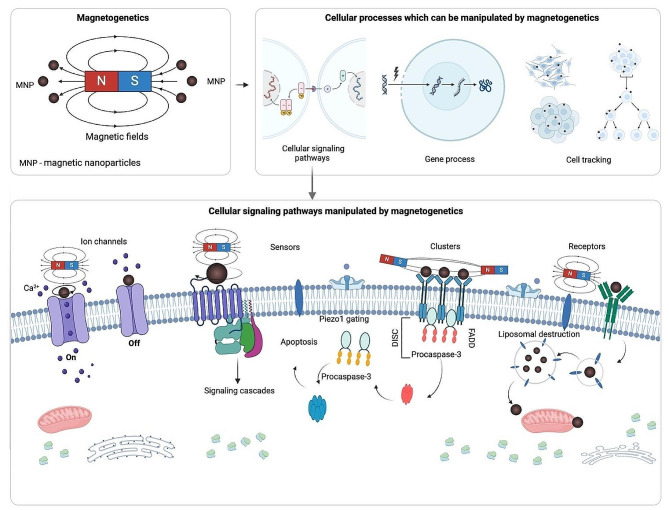
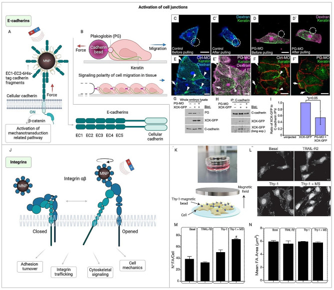
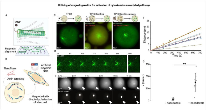
Magnetogenetics emerges as a transformative approach for modulating cellular signaling pathways through the strategic application of magnetic fields and nanoparticles. This technique leverages the unique properties of magnetic nanoparticles (MNPs) to induce mechanical or thermal stimuli within cells, facilitating the activation of mechano- and thermosensitive proteins without the need for traditional ligand-receptor interactions. Unlike traditional modalities that often require invasive interventions and lack precision in targeting specific cellular functions, magnetogenetics offers a non-invasive alternative with the capacity for deep tissue penetration and the potential for targeting a broad spectrum of cellular processes. This review underscores magnetogenetics' broad applicability, from steering stem cell differentiation to manipulating neuronal activity and immune responses, highlighting its potential in regenerative medicine, neuroscience, and cancer therapy. Furthermore, the review explores the challenges and future directions of magnetogenetics, including the development of genetically programmed magnetic nanoparticles and the integration of magnetic field-sensitive cells for in vivo applications. Magnetogenetics stands at the forefront of cellular manipulation technologies, offering novel insights into cellular signaling and opening new avenues for therapeutic interventions.

Keywords: Cell signaling; Magnetic nanoparticles; Magnetogenetics; Mechanosensitivity; Mechanotransduction.


Conflict of interest statement

The authors declare no competing interests.

Figures

Fig. 1
Fig. 1
Exploring the potential of magnetogenetics in cellular modulation. This review outlines the current state of magnetogenetics as a versatile tool for the precise control of cellular signaling pathways. It details the application of magnetic fields and nanoparticles for the direct manipulation of ion channels, the activation of mechanosensitive sensors, the clustering of membrane proteins, and the targeting of cellular receptors. The review also addresses how magnetogenetics influences gene expression, orchestrates signaling cascades, and facilitates cell tracking. It presents the strategies for nanoparticle encapsulation and discusses their effects on cellular processes such as apoptosis and liposomal destruction. The potential of magnetogenetics is posited to be transformative for precision medicine and bioengineering, setting the stage for future innovations [–67]
Fig. 2
Fig. 2
Overview of magnetogenetic principles and applications. (A) Schematic of the basic principles of magnetogenetics showing the interaction of a magnetic field with both exogenous and endogenous magnetic nanoparticles (MNPs). (B) Properties of magnetic actuators classified according to size, composition, and interaction types with magnetic fields, illustrated with examples of superparamagnetism (SPM), ferrimagnetism (FM), and varying coercivity and saturation magnetization (Ms) based on MNP diameter [65, 81]. (C) Methods to control magnetic actuators with no field, DC magnetic field, and AC magnetic field, showing the mechanical effects (e.g., pulling movement, torque, relaxation process, and ROS generation) [65]. (D) Different configurations of DC magnetic field applicators and their corresponding magnetic orientations and interactions (gradient, uniform, rotating uniform) [65]. (E) A magnetic field map highlighting non-heating and heating magnetic field intensity limits across a wide frequency range [82]
Fig. 3
Fig. 3
Utilizing magnetogenetics to activate mechano- and thermosensitive pathways. (A) Magnetic forces applied to the membrane region near mechanosensitive channels PIEZO1/PIEZO2 can activate them. This activation can occur through torque exerted by uniform magnetic fields or by pulling membrane sections tethered to nanoparticles in magnetic field gradients [164]. (B The role of PIEZO channels in human physiology and medical applications [165, 166]. (C) Using the Piezo1 channel in magnetogenetics for CRISPR gene editing [93]. (D-E) Stimulation of mechano-thermosensitive channels: K2P and TRP. (D) (Left) Information about that TRPV1 regulates processes such as inflammatory, pain from different etiology, migraine. (Right) Thermally gated ion channels, like TRPV1, activate in response to the hysteretic heating of nearby magnetic nanoparticles when exposed to magnetic fields alternating at frequencies exceeding 100 kHz [164]. (E) Activation of TRPV4 channels through ferritin magnetocalorics [142]. (Left) The diagram illustrates the mechanism by which the magnetocaloric effect in ferritin can trigger nearby temperature-sensitive ion channels like TRPV4. When a magnetic field is applied, it aligns the magnetic moments within paramagnetic ferritin nanoparticles, thereby lowering the magnetic entropy. This reduction in magnetic entropy produces heat (Q) through the magnetocaloric effect, which in turn can activate a nearby temperature-sensitive ion channel. Although ferritin had been represented as a paramagnet here, the computations remain the same for superparamagnetic particles. (Right) TRPV4 channels indirectly participate in various processes [–160]
Fig. 4
Fig. 4
Application of magnetogenetics for activation of cell junction and cytoskeletal associated pathways. (A) Magneto-mechanical actuation via cadherin-nanoparticle bioconjugates. MNP – magnetic nanoparticles [185]. (B) Polarized cell behavior and migration directed by mechanoresponsive cadherin-keratin complex [186]. (C-I) PG necessity for Cadherin/Keratin Link. (C, D) Isolated cells marked with Alexa-dextran, showcasing GFP-XCK1(8) (green) expression and cultured on fibronectin. (C) and (C′) depict a standard cell (blue dextran), while (D) and (D′) display a PG-deficient cell (magenta dextran). C-cadFc bead (circle) attaches (C and D), then is pulled away (C′ and D′). (E, E’) Control (blue dextran) (E) and PG-deficient (magenta dextran) (E’) mesendoderm samples expressing GFP-XCK1(8) (green). (F, F’) Control (F) and PG-deficient (F’) mesendoderm in full embryos, stained for XCK1(8) (green) and β-catenin (red). (E–F’) Arrows indicate cabling at the leading-edge cells’ front, and arrowheads point to KIF clusters near intercellular junctions. All measurement bars represent 25 μm. (G-I) Embryos received injections of XCK1(8)-GFP, with or sans PG morpholino. (G) Embryo extract immunoblots reveal XCK1(8)-GFP and innate PG levels, with or without PG morpholino (PG-MO). (H) Immunoblots of C-cadherin co-precipitates for XCK1(8)-GFP and C-cadherin, with or without PG-MO. (I) Three separate co-immunoprecipitation experiments quantified, presented as average ± SEM [186]. (B-I) reprinted from [186]. © 2011 Cell Press (Open Access). (J) Activation and regulation of Integrin by MNP [–189]. (K-N) Indirect immunofluorescence analysis of focal adhesions in DITNC1 cells. (K) The top image displays the setup with magnets below the tissue culture plate, generating a magnetic field. This diagram details the experimental setup: Cells were seeded on coverslips (light blue) and treated with serum-free medium for 30 min. DITNC1 cells (beige) were then exposed to either TRAIL-R2- (as a control) or Thy-1-coated Protein A magnetic beads (blue balls with yellow projections), with or without mechanical stress (MS) induced by a magnet (gray) for 5 min. (L) Focal Adhesions (FA) were identified using an antibody for phospho-tyrosine and a secondary antibody. Scale bar = 10 μm. (M, N) The data in the graph represent average + s.e.m. for at least 30 cells under each condition in three separate experiments (n = 3), showing the count (N°) of FAs per cell (M) and the mean area (µm²) of the FAs (N). Statistical significance was assessed using the Kruskal–Wallis non-parametric test followed by Dunn’s multiple comparisons test. #p < 0.05, indicating significance against the Thy-1 condition [190]. (K-N) reprinted from [190]. © 2021 Frontiers (Open Access)
Fig. 5
Fig. 5
Utilizing of magnetogenetics for activation of cytoskeleton associated pathways. (A) Magnetic alignment of microtubules with CoPt nanoparticle-encapsulating Tau peptides [209]. (B) Magnetic-field-guided polarization of stem cells via supramolecular nanofibers [72]. (C-G): Initiation and precise control over the formation and movement of microtubule structures using magnetic fields. (C) Using confocal microscopy, the formation of microtubules was observed in Xenopus egg extract droplets, initiated by FKBP-TPX2. These microtubules and ferritins were marked with fluorescein-tagged tubulin and mCherry, respectively. Sequentially, the formation of microtubule networks was induced by FKBP-TPX2, followed by mCherry/TPX2-ferritins, and then ferritin-TPX2 aggregates. (D) A sequence of images showing the expansion of microtubules from a central point, known as an aster, prompted by clusters of ferritin. EB1-GFP served as a marker for the growing ends of the microtubules. (E) Selected moments capturing the movements of an aster’s center driven by a magnetic field. (F) Illustration of how aster centers move along a magnetic field gradient. This is shown by a graph depicting the path length of an aster over time under the influence of magnetic forces. (G) The average speed of asters moving towards a magnet, measured with and without the application of a microtubule-disassembling agent (nocodazole) [210]. (C-G) reprinted from [210]. © 2017 Springer Nature (Open Access). FKBP – FK506 binding protein; TPX2 – targeting protein for Xklp2; EB1 – end binding protein 1; GFP - green fluorescent protein
Fig. 6
Fig. 6
Ligand-free induction of ligand-mediated signaling. (A-B) Ligand-free receptor dimerization and clustering [51]. (A) The diagram depicts the process of labeling human adipose-derived stem cells (hASCs) with magnetic nanoparticles (MNPs) targeting the activin A receptor (ActRIIA). Upon stimulation with a magnetic field, the labeled cells activate the intracellular signaling pathway, specifically TGF-β/Smad2/3, leading to the promotion of tenogenesis. (B) Smad2/Smad3 Phosphorylation: The first chart on the right top illustrates the phosphorylation level of Smad2/Smad3 over time, following stimulation with activin A and two different types of MNP complexes targeted to the activin A type IIA receptor (ActRIIA). Collagen Production: The second chart on the right bottom shows collagen production in cell cultures measured at day 0–14. The quantification is based on Sirius Red staining, which binds to collagen fibers. The graph compares collagen levels across different culture conditions, including control groups, activin A-treated groups, and MNP-ActRIIA complex-treated groups, demonstrating how collagen production changes over the culture period and under different stimulatory conditions. Reprinted with permission from [51]. © 2018 Elsevier Inc. (C-D) SPION magnetic switches for EGFR activation (C) and Shc activation as a result of magnetic activation of EGFR (D). Confocal immunofluorescence displays cells post 15-minute incubation at 37 °C subsequent to 0–180 s (0, 30, 180 s) of magnetic field application. From left to right, the columns show: MS Alexa-488 biocytin labeling (green); indirect immunofluorescence with monoclonal antibody specific to pY-317 Shc protein paired with GARIG-CY5 (red); a composite of the first two columns; two-dimensional colocalization scatter plots of magnetic switches (MS) and pY317-Shc fluorescence signals after refining 50 optical slices with SVI Huygens software [56]. (C-D) reprinted from [56]. © 2013 PLOS One (Open Access). (E-J) Spatial and temporal control of Notch signaling transduction with MPNs. (E–J) Dynamics of H2B-mCherry Expression in UAS-Gal4 Reporter Cells Following MPN-Triggered Notch Signaling. (E) Showcases a typical fluorescence image alongside a temporal data graph. (F) Compilation of time series data from multiple cells. (G) Evaluates the initiation timing (ton) and production speed (RmC) of mCherry. (H) Deliberate spatial activation of Notch signaling. (I and J) Regulation over time of Notch pathway activity. (I) Sequential images over time and (J) tracks of mCherry fluorescence intensity for three randomly selected cells (labeled a, b, and c) within a group, following staggered stimulations at 2-hour intervals [57]. (E-J) reprinted from [57]. © 2016 Cell Press (Open Access)
Fig. 7
Fig. 7
G protein-coupled receptors activation (GPCR family). (A-C) A schematic representation of the use of MNP and the sequential steps of cell labeling and stimulation or cell injection into femurs targeting Wnt signaling. Initially, MNPs are tailored with specific binding agents like peptides (A). Subsequently, cells are marked with these bespoke MNPs. Post-labeling, the cells can either be exposed to fluctuating magnetic fields in a laboratory setting to trigger receptor aggregation and the commencement of signaling pathways (B), or they can be introduced as a cell-MNP mixture into tissue engineering constructs, such as a fetal chick femur, and then magnetically manipulated to modulate bone development in an ex vivo environment (C) [251]. (D-E) Activation of Notch signaling by mechanical force. (D) A graphical depiction of the live-cell assay designed to observe the activation of the Notch receptor triggered by mechanical stress. The engineered receptor is linked to streptavidin-coated paramagnetic beads via a SNAP-biotin connector. (E) Application of mechanical force across the receptor results in the severance of its C-terminal domain. In this particular setup, the C-terminal domain has been substituted with Gal4 to serve as a transcriptional indicator, which in turn promotes the synthesis of mCherry-H2B [244]. (F) Magnetic sorting of cells with/without A3AR overexpression. Ligands for the A3 adenosine receptor attached to iron-filled carbon nanotubes were created to specifically target certain cancer cell types for magnetic cell separation and thermal treatment in cancer therapy. Although these nanostructures could effectively attach to the A3 adenosine receptors, they failed to demonstrate selective binding to cells that overexpressed the receptor upon cellular interaction [245]

Similar articles

Cited by

References

    1. Aplin AE, Howe A, Alahari SK, Juliano RL. Signal Transduction and Signal Modulation by Cell Adhesion receptors: the role of integrins, cadherins, immunoglobulin-cell adhesion molecules, and Selectins. Pharmacol Rev. 1998;50:197–264. - PubMed
    1. Kefauver JM, Ward AB, Patapoutian A. Discoveries in structure and physiology of mechanically activated ion channels. Nature. 2020;587:567–76. doi: 10.1038/s41586-020-2933-1. - DOI - PMC - PubMed
    1. Xiao R, Xu XZS. Temperature sensation: from molecular thermosensors to neural circuits and Coding principles. Annu Rev Physiol. 2021;83:205–30. doi: 10.1146/annurev-physiol-031220-095215. - DOI - PMC - PubMed
    1. Lingueglia E. Acid-sensing Ion channels in sensory perception *. J Biol Chem. 2007;282:17325–9. doi: 10.1074/jbc.R700011200. - DOI - PubMed
    1. Sun Y, Liu W-Z, Liu T, Feng X, Yang N, Zhou H-F. Signaling pathway of MAPK/ERK in cell proliferation, differentiation, migration, senescence and apoptosis. J Recept Signal Transduction. 2015;35:600–4. doi: 10.3109/10799893.2015.1030412. - DOI - PubMed

Substances

LinkOut - more resources