. 2024 Jun 10;14(1):78.
doi: 10.1186/s13578-024-01260-2.

CDK7/CDK9 mediates transcriptional activation to prime paraptosis in cancer cells

Affiliations

CDK7/CDK9 mediates transcriptional activation to prime paraptosis in cancer cells

Shih-Kai Chiang et al. Cell Biosci. .

Abstract

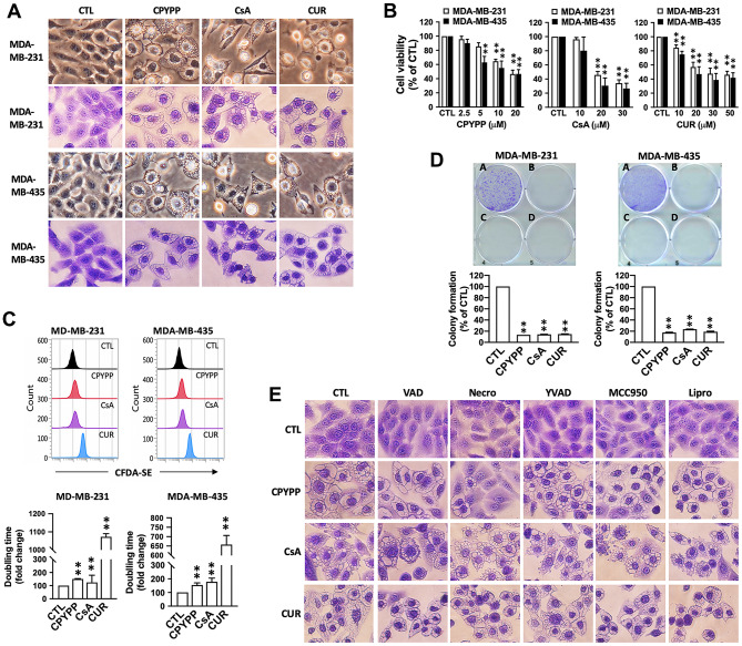
Background: Paraptosis is a programmed cell death characterized by cytoplasmic vacuolation, which has been explored as an alternative method for cancer treatment and is associated with cancer resistance. However, the mechanisms underlying the progression of paraptosis in cancer cells remain largely unknown.

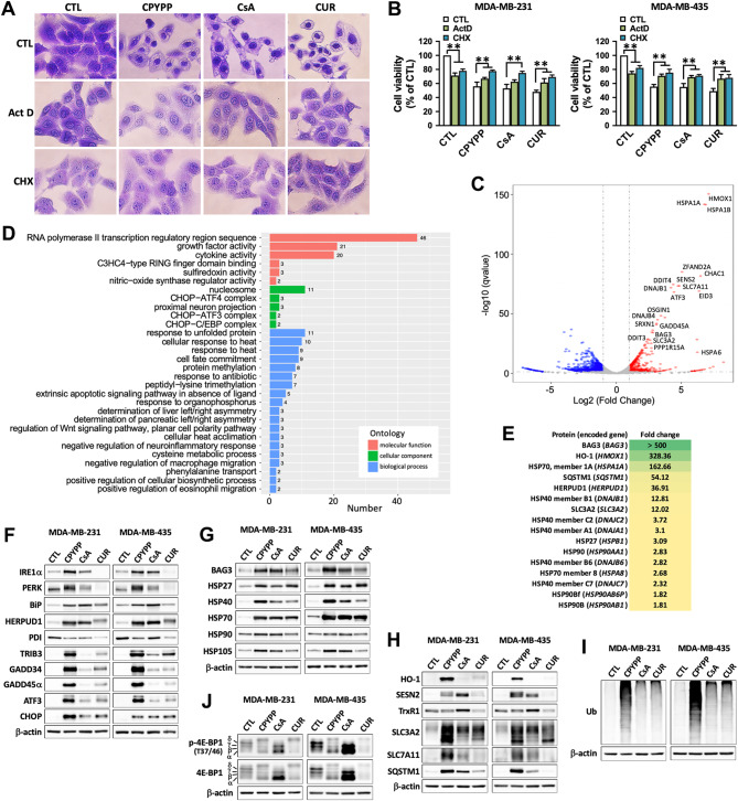
Methods: Paraptosis-inducing agents, CPYPP, cyclosporin A, and curcumin, were utilized to investigate the underlying mechanism of paraptosis. Next-generation sequencing and liquid chromatography-mass spectrometry analysis revealed significant changes in gene and protein expressions. Pharmacological and genetic approaches were employed to elucidate the transcriptional events related to paraptosis. Xenograft mouse models were employed to evaluate the potential of paraptosis as an anti-cancer strategy.

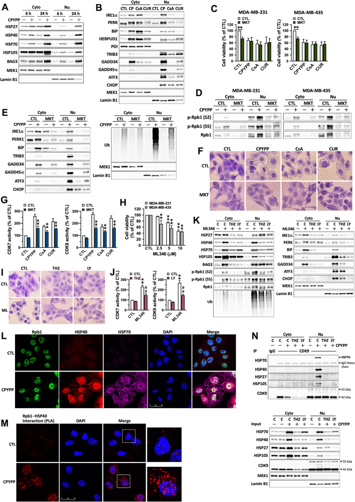
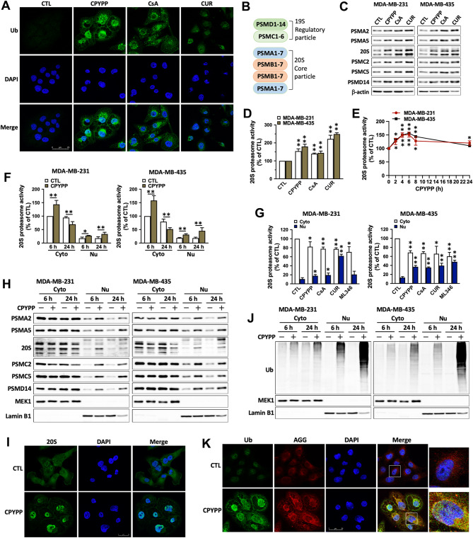
Results: CPYPP, cyclosporin A, and curcumin induced cytoplasmic vacuolization and triggered paraptosis in cancer cells. The paraptotic program involved reactive oxygen species (ROS) provocation and the activation of proteostatic dynamics, leading to transcriptional activation associated with redox homeostasis and proteostasis. Both pharmacological and genetic approaches suggested that cyclin-dependent kinase (CDK) 7/9 drive paraptotic progression in a mutually-dependent manner with heat shock proteins (HSPs). Proteostatic stress, such as accumulated cysteine-thiols, HSPs, ubiquitin-proteasome system, endoplasmic reticulum stress, and unfolded protein response, as well as ROS provocation primarily within the nucleus, enforced CDK7/CDK9-Rpb1 (RNAPII subunit B1) activation by potentiating its interaction with HSPs and protein kinase R in a forward loop, amplifying transcriptional regulation and thereby exacerbating proteotoxicity leading to initiate paraptosis. The xenograft mouse models of MDA-MB-231 breast cancer and docetaxel-resistant OECM-1 head and neck cancer cells further confirmed the induction of paraptosis against tumor growth.

Conclusions: We propose a novel regulatory paradigm in which the activation of CDK7/CDK9-Rpb1 by nuclear proteostatic stress mediates transcriptional regulation to prime cancer cell paraptosis.

Keywords: Cancer; Cyclin-dependent kinase 7/9; Heat shock proteins; Nuclear stress; Paraptosis; Protein kinase R; Reactive oxygen species.


Conflict of interest statement

The authors declare that they have no competing interests.

Figures

Fig. 1
Fig. 1
CPYPP, cyclosporin A, and curcumin induce paraptotic death in cancer cells. (AD) Human cancer cells MDA-MB-231 and MDA-MB-435 were treated with CPYPP (10 µM), cyclosporin A (CsA, 20 µM), or curcumin (CUR, 30 µM) at the indicated concentrations for 24 h. Cells were then assessed for morphological alterations in cytoplasmic vacuolization (upper panel, phase-contrast; lower panel, crystal violet staining, A), cell viability (MTT assay, B), proliferation (CFDA-SE staining, C), and clonogenic activity (A, CTL; B, CPYPP; C, CsA; D, CUR) (D). (E) MDA-MB-435 cells pretreated with various inhibitors of programmed cell death, including z-VAD-FMK (VAD, 10 µM, apoptosis), necrostatin-1 (Necro, 100 µM, necroptosis), Ac-YVAD-CHO (YVAD, 10 µM, pyroptosis), MCC950 (10 µM, pyroptosis), or liproxstatin-1 (Lipro, 2 µM, ferroptosis), for 30 min were treated with CPYPP (10 µM), cyclosporin A (CsA, 20 µM), or curcumin (CUR, 30 µM) for 24 h to trigger cell paraptosis. Cells were collected for morphological vacuolization analysis. The results are expressed as the mean ± SD from three independent experiments. **P < 0.01 compared to the control group (CTL)
Fig. 2
Fig. 2
Paraptosis proceeds with activation of proteostatic dynamics and transcriptional regulation involved in redox homeostasis and proteostasis. (A, B) Cancer cells were pretreated with the transcription inhibitor actinomycin A (ActD, 1 µM) or the translation inhibitor cycloheximide (CHX, 20 µM) for 30 min, followed by treatment with CPYPP (10 µM), cyclosporin A (CsA, 20 µM), or curcumin (CUR, 30 µM) for 24 h. The cells were then assessed for morphological vacuolization alterations using crystal violet staining (A, MDA-MB-435 cells) or for cell viability via MTT assay (B). The results are expressed as the mean ± SD from three independent experiments. **P < 0.01, compared to the control group (CTL). (C, D) CPYPP-treated MDA-MB-435 cells underwent next-generation sequencing (NGS) to analyze differential gene expressions (DGEs) The volcano plot represents the distribution of DGEs based on the FPKM analysis with at least 2-fold changes (C). Results from the Gene Ontology (GO) analysis classify molecular function, cellular component, and biological process (D), along with the number of related significant changed genes. (E) CPYPP-treated MDA-MB-435 cells were subjected to mass spectrometric analysis for proteomic profiling with at least 1.5-fold changes. (FJ) Western blot analysis confirmed the involvement of differentially expressed genes in ER stress (F), HSP chaperones (G), redox homeostasis (H), protein ubiquitination (I), and translational activity (J) in cancer cells treated with CPYPP, CsA, or CUR for 24 h
Fig. 3
Fig. 3
CDK7/CDK9 mediates the transcriptional regulation for paraptotic development. (AC) MDA-MB-435 cells pretreated with the CDK7 inhibitor THZ1 (THZ, 0.2 µM) or the CDK9 inhibitor LY2857785 (LY, 1 µM) for 30 min were treated with CPYPP (10 µM), cyclosporin A (CsA, 20 µM), or curcumin (CUR, 30 µM) for 24 h. Cells were then collected for vacuolization (A), cell viability (B), and protein expression (C) analyses. (DF) MDA-MB-435 cells were treated with control shRNA (Void), shCDK7, or shCDK9 for 4 days and then with CPYPP (10 µM) for 24 h. Cells were harvested for Western blot analysis (D) and vacuolization alterations (E, F). (G, H) MDA-MB-435 cells were treated with CPYPP (10 µM) for the indicated time and then collected for Western blot and CDK7/CDK9 enzyme activity analysis. (I, J) MDA-MB-435 cancer cells were treated with CPYPP for 6–24 h, then harvested to isolate cytosolic and nuclear fractions for Rpb1 phosphorylation (I) and CDK7–Rpb1 complex activation by co-immunoprecipitation method (J). (K) The co-localization of Rpb1 and CDK9 was detected through co-immunofluorescence staining using anti-Rpb1 antibody (shown in green) and anti-CDK9 antibody (shown in red), followed by secondary antibody staining, along with DAPI (shown in blue) for nuclei counter staining. The images were captured using confocal microscopy at 630× magnification. The scale bar represents 25 μm. (L) The interaction and distribution between Rpb1 and CDK9 were assessed using the proximity ligation assay. (MO) MDA-MB-435 cells pretreated with the CDK7 inhibitor THZ1 (THZ, 0.2 µM) or the CDK9 inhibitor LY2857785 (LY, 1 µM) for 30 min were treated with CPYPP (10 µM), cyclosporin A (CsA, 20 µM), or curcumin (CUR, 30 µM) for 24 h. Cells were collected for CDK7/CDK9 activity analysis (M) or to isolate cytosolic and nuclear fractions for Rpb1 phosphorylation (N) and CDK9–Rpb1 complex activation by co-immunoprecipitation method (O). **P < 0.01, compared to the corresponding control group (CTL)
Fig. 4
Fig. 4
HSPs interact with the CDK7/CDK9–Rpb1 complex to prime stress-specific transcription in a forward loop and reciprocally regulated manner. (AB) MDA-MB-435 cells were treated with CPYPP (10 µM) for 6–24 h, or with CPYPP, cyclosporin A (CsA, 20 μM), or curcumin (CUR, 30 μM) for 24 h, and then collected for cytosolic (Cyto) and nuclear (Nu) fraction isolation for Western blot analysis. (CG) MDA-MB-435 and/or MDA-MB-231 cells were pretreated with MKT-077 (MKT) for 30 min followed by CPYPP (10 µM), cyclosporin A (CsA, 20 µM), or curcumin (CUR, 30 µM) for 24 h. Cells were then collected for cell viability (C), RNA polymerase II activation (D), ER stress, UPR, and protein ubiquitination (E), paraptotic vacuolization (F), and CDK7 and CDK9 activity (G) analysis. The results are expressed as the mean ± SD from three independent experiments. **P < 0.01, compared to the corresponding control group. (HI) Cancer cells treated with indicated concentrations of ML346 for 24 h promoted cell death (H) and paraptotic vacuolization (I, 10 µM). (JKN) MDA-MB-435 cells were pretreated with THZ1 (THZ, 0.2 µM) or LY2857785 (LY, 1 µM) for 30 min followed by ML346 treatment for 24 h. Total cell lysates, cytosolic and nuclear fractions were used for CDK7 and CDK9 activity (J), protein expression (K), and the interaction of HSPs with CDKs by co-immunoprecipitation (N) analysis. **P < 0.01, compared to the corresponding control group (CTL). (L) The co-localization of Rpb1, HSP40, and HSP70 was examined in CPYPP-treated MDA-MB-435 cells co-stained with anti-Rpb1 (shown in green), anti-HSP40 (shown in red), and anti-HSP70 (shown in magenta) antibodies, followed by individual secondary antibodies. DAPI staining (shown in blue) was used for nuclei counterstaining. Confocal microscopy was utilized to capture the images at 630× magnification. The scale bar represents 25 μm. (M) The interaction and distribution between Rpb1 and HSP40 were evaluated using the proximity ligation assay
Fig. 5
Fig. 5
Overload of HSPs and the ubiquitin-proteasome system elicits unclear stress and promotes CDK7/CDK9–Rpb1 activation for transcriptional regulation. (A) MDA-MB-435 cells were treated with CPYPP (10 µM), cyclosporin A (CsA, 20 µM), or curcumin (CUR, 30 µM) for 24 h. Protein ubiquitination was visualized by immunofluorescence using an anti-ubiquitin antibody (shown in green), with DAPI (shown in blue) used for nuclei counterstaining. Results were imaged under a confocal microscopy at 630× magnification. Scale bar = 25 μm. (B) The subunits of the 19S and 20S proteasome complex. (C, D, F, G, H, J) Cancer cells were treated with CPYPP, CsA, CUR, or ML346 (10 µM) for 6 h (D, G) or 24 h and then harvested for whole lysate collection or cytosolic and nuclear fractionation for proteasome subunit abundance determination by Western blot (C, H), 20 S proteasome activity (D, F, G), and protein ubiquitination analysis (J). (E) Cancer cells were treated with CPYPP for the indicated time period (h). The results are expressed as the mean ± SD from three independent experiments. *P < 0.05, **P < 0.01, compared to the corresponding control group (CTL). (I,K) CPYPP-treated MDA-MB-435 cells were imaged by immunofluorescence staining for nuclear accumulation of proteasomes using an anti-20S core subunit antibody (green) and DAPI (blue) for nuclei counterstaining (I), or for colocalization of ubiquitins and aggresomes using an anti-ubiquitin antibody (green) with aggresome staining by ProteoStat® dye (red), and DAPI (blue) for nuclei counterstaining (K)
Fig. 6
Fig. 6
ROS ignites the paraptotic process by CPYPP. (AF, HL) MDA-MB-435 and/or MDA-MB-231 cells were pretreated with NAC (3 mM) for 30 min followed by CPYPP (10 µM), cyclosporin A (CsA, 20 µM), or curcumin (CUR, 30 µM) treatment for 24 h. Cells were collected for analyses, including morphological vacuolization (A), ROS generation (B), abundance of ER stress and UPR proteins, HSPs (C), CDK7 and CDK9 activity (D), RNA polymerase II activation (E), and cell viability (F), proteasome subunit abundances (H), 20S proteasome activity (I), nuclear hydrogen peroxide generation (J), free cysteine-thiol levels (K), and CDK7 interaction with HSPs by co-immunoprecipitation (L). (G) Cancer cells pretreated with THZ1 (THZ, 0.2 µM) or LY2857785 (LY, 1 µM) for 30 min followed by CPYPP treatment for 24 h were assessed for cellular ROS generation. The results are expressed as the mean ± SD from three independent experiments. *P < 0.05 or **P < 0.01, compared to the corresponding control group (CTL)
Fig. 7
Fig. 7
Protein kinase R (PKR) interacts with the CDK7/CDK9–Rpb1 complex to enhance transcription activation and exacerbate paraptotic progression. (AF, I, J) MDA-MB-435 cells were pretreated with PKR inhibitor (PKRi, 2 µM), trans-ISRIB (Trans, 3 µM), GCN2-IN-1 (GCN2i, 5 µM), or GSK2656157 (GSK, 5 µM) for 30 min followed by CPYPP (10 µM), CsA (20 µM), or curcumin (CUR, 30 µM) for 24 h. Cells were harvested and assessed for cell viability (A), morphological vacuolization (B), protein abundance of ER stress, UPR, and HSPs (C), CDK7 and CDK9 activity (D), RNA polymerase II activation (E), PKR interaction with CDK7/CDK9 by co-immunoprecipitation (F), cellular ROS (I), and nuclear hydrogen peroxide generation (J). The results are expressed as the mean ± SD from three independent experiments. *P < 0.05 or **P < 0.01, compared to the corresponding control group (CTL). (G, H) MDA-MB-435 cells pretreated with ActD (1 µM), CHX (20 µM), THZ1 (0.2 µM), or LY2857785 (1 µM) for 30 min followed by CPYPP treatment for 24 h. Cytosolic and nuclear fractions were isolated for PKR abundance and activation analysis by Western blot. (K, L) MDA-MB-435 cells pretreated with NAC (3 mM) for 30 min followed by CPYPP treatment for 24 h were used for PKR abundance and activation (K) and interaction with CDK7 by co-immunoprecipitation (L)
Fig. 8
Fig. 8
CPYPP and curcumin-induced paraptosis suppress tumor growth in xenograft mice. (AH) MDA-MB-231 breast cancer xenograft mouse models treated with vehicle (CTL) or CPYPP. n = 5. (A) Representative image of the tumor masses in mice treated with different regimens. (B, C) Tumor size and tumor weight. (D) Hematoxylin and eosin (H&E) staining, Ki67, CDK7, and CDK9 expression in tumor samples. (E–G) CDK7, CDK9, and 20S proteasome activities in tumor samples. (H) Changes in mouse body weight throughout the 14-day experimental period. (I, J) Docetaxel-resistant head and neck cancer cell lines, OECM1-DTX and SAS-DTX, were treated with indicated concentrations of CPYPP or curcumin (CUR) for 24 h, followed by morphological vacuolization assessment (I) and cell viability analysis (J). Results are expressed as the mean ± SD from three independent experiments. ** P < 0.01, compared to the control group (CTL). (KQ) OECM1-DTX tumor xenograft mouse models treated with vehicle (CTL), CPYPP, or curcumin (CUR). (K) Representative images of the tumor masses in mice receiving different treatments. (L, M) Tumor volume and tumor weight at experiment endpoint. n = 5. (NP) CDK7, CDK9, and 20S proteasome activities in tumor samples. (Q) Changes in mouse body weight throughout the 12-day experimental period
Fig. 9
Fig. 9
The proposed mechanism of paraptotic progression. Parts of the figure were created using images from Servier Medical Art, which is licensed under a Creative Commons Attribution 4.0 Unported License (https://creativecommons.org/licenses/by/4.0/)

Similar articles

Cited by

References

    1. Sperandio S, de Belle I, Bredesen DE. An alternative, nonapoptotic form of programmed cell death. Proc Natl Acad Sci USA. 2000;97:14376–81. - PMC - PubMed
    1. Shubin AV, Demidyuk IV, Komissarov AA, Rafieva LM, Kostrov SV. Cytoplasmic vacuolization in cell death and survival. Oncotarget. 2016;7(34):55863–89. - PMC - PubMed
    1. Fricker M, Tolkovsky AM, Borutaite V, Coleman M, Brown GC. Neuronal cell death. Physiol Rev. 2018;98(2):813–80. - PMC - PubMed
    1. Monel B, Compton AA, Bruel T, Amraoui S, Burlaud-Gaillard J, Roy N, et al. Zika virus induces massive cytoplasmic vacuolization and paraptosis-like death in infected cells. EMBO J. 2017;36(12):1653–68. - PMC - PubMed
    1. Wang Y, Li X, Wang L, Ding P, Zhang Y, Han W, et al. An alternative form of paraptosis-like cell death, triggered by TAJ/TROY and enhanced by PDCD5 overexpression. J Cell Sci. 2004;117(8):1525–32. - PubMed