. 2024 Jun 11;15(1):86.
doi: 10.1186/s40104-024-01045-0.

The walnut-derived peptide TW-7 improves mouse parthenogenetic embryo development of vitrified MII oocytes potentially by promoting histone lactylation

Affiliations

The walnut-derived peptide TW-7 improves mouse parthenogenetic embryo development of vitrified MII oocytes potentially by promoting histone lactylation

Yaozong Wei et al. J Anim Sci Biotechnol. .

Abstract

Background: Previous studies have shown that the vitrification of metaphase II (MII) oocytes significantly represses their developmental potential. Abnormally increased oxidative stress is the probable factor; however, the underlying mechanism remains unclear. The walnut-derived peptide TW-7 was initially isolated and purified from walnut protein hydrolysate. Accumulating evidences implied that TW-7 was a powerful antioxidant, while its prospective application in oocyte cryopreservation has not been reported.

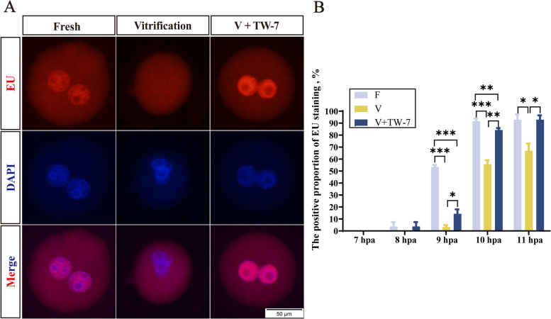
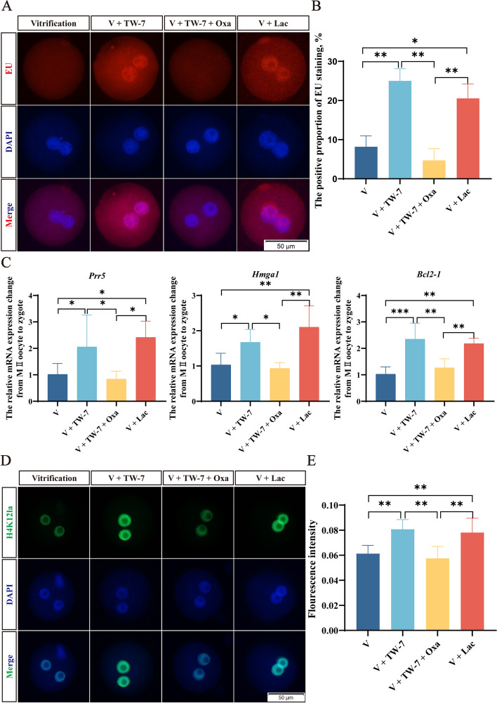
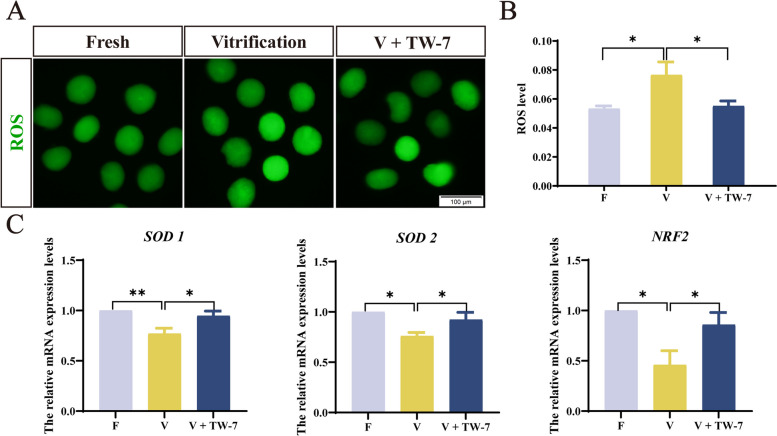
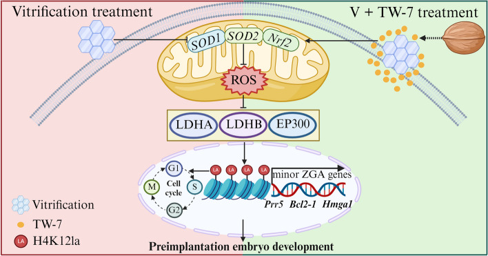
Result: Here, we found that parthenogenetic activation (PA) zygotes derived from vitrified MII oocytes showed elevated ROS level and delayed progression of pronucleus formation. Addition of 25 μmol/L TW-7 in warming, recovery, PA, and embryo culture medium could alleviate oxidative stress in PA zygotes from vitrified mouse MII oocytes, furtherly increase proteins related to histone lactylation such as LDHA, LDHB, and EP300 and finally improve histone lactylation in PA zygotes. The elevated histone lactylation facilitated the expression of minor zygotic genome activation (ZGA) genes and preimplantation embryo development.

Conclusions: Our findings revealed the mechanism of oxidative stress inducing repressed development of PA embryos from vitrified mouse MII oocytes and found a potent and easy-obtained short peptide that could significantly rescue the decreased developmental potential of vitrified oocytes, which would potentially contribute to reproductive medicine, animal protection, and breeding.

Keywords: Histone lactylation; Oocyte; TW-7; Vitrification; Zygotic genome activation.


Conflict of interest statement

The authors declare that they have no competing interests.

Figures

Fig. 1
Fig. 1
TW-7 improves the developmental potential of the PA embryos from vitrified mouse MII oocytes. A Representative images of 2-cell embryos, 4-cell embryos, and blastocysts in the Fresh, Vitrification, V + 25 μmol/L TW-7, V + 50 μmol/L TW-7 and V + 100 μmol/L TW-7 groups. Scale bar, 100 μm. B The percentage of embryo development rate of oocytes in Fresh, Vitrification, V + 25 μmol/L TW-7, V + 50 μmol/L TW-7, and V + 100 μmol/L TW-7 groups. Fresh (n = 116), Vitrification (n = 95), V + 25 μmol/L TW-7 (n = 80), V + 50 μmol/L TW-7 (n = 84), V + 100 μmol/L TW-7 (n = 85). C The rate of pronuclear formations in the Fresh, Vitrification, and V + TW-7 groups from 2 to 8 hpa. The dotted lines indicate the time after activation corresponding to the 10% and 50% rates of pronuclear formation in each group. D Time required for 50% of pronuclear formation. Fresh (n = 146), Vitrification (n = 143), V + TW-7 (n = 127). Data in (B), (C), and (D) were presented as mean percentage (mean ± SEM) of at least three independent experiments. *P < 0.05, **P < 0.01, ***P < 0.001
Fig. 2
Fig. 2
TW-7 promotes the new transcripts generation of the PA zygotes derived from vitrified mouse MII oocytes. A Representative images of EU staining in PA zygotes of Fresh, Vitrification, and V + TW-7 groups. Scale bar, 50 μm. B The positive proportion of EU staining in PA zygotes of Fresh, Vitrification, and V + TW-7 groups. Fresh (n = 225), Vitrification (n = 220), V + TW-7 (n = 192). Data in (B) were presented as mean percentage (mean ± SEM) of at least three independent experiments. *P < 0.05, **P < 0.01, ***P < 0.001
Fig. 3
Fig. 3
TW-7 promotes minor ZGA initiation in PA zygotes from vitrified mouse MII oocytes. A Heatmap of genes in the MII oocytes and PA zygotes (9 hpa). OF: MII oocytes (0 hpa), ZF: PA zygotes (9 hpa) of fresh group, ZV: PA zygotes (9 hpa) of vitrification group, ZT: PA zygotes (9 hpa) of V + TW-7 group. B–D Volcano map of differentially expressed genes in different groups during the development of MII oocytes to the zygote (9 hpa). E Venn diagram of activated ZGA genes in zygotes of Fresh, Vitrification, and V + TW-7 groups. F GO BP enrichment analysis of 129 ZGA genes suppressed by vitrification at the zygotes. G GO BP enrichment analysis of 108 ZGA genes activated by TW-7 at the zygotes. GO, Gene Ontology. BP, Biological Process. H Histogram of Prr5, Bcl2-1, and Hmga1 expression in MII oocytes and at the zygotic stage
Fig. 4
Fig. 4
TW-7 regulates the protein expression levels of histone lactylation in PA zygotes derived from vitrified mouse MII oocytes. A Representative images of Pan Kla and H4K12la protein expression in PA zygotes of Fresh, Vitrification, and V + TW-7 groups. Scale bar, 50 μm. B The fluorescence intensity of Pan Kla and H4K12la in PA zygotes of Fresh, Vitrification, and V + TW-7 groups. Number of PA zygotes involved in Pan Kla immunofluorescence staining: Fresh (n = 22), Vitrification (n = 24), V + TW-7 (n = 27). Number of PA zygotes involved in H4K12la immunofluorescence staining: Fresh (n = 50), Vitrification (n = 38), V + TW-7 (n = 42). C Representative images of EP300 and HDAC3 protein expression in PA zygotes of Fresh, Vitrification, and V + TW-7 groups. Scale bar, 50 μm. D The fluorescence intensity of EP300 and HDAC3 in PA zygotes of Fresh, Vitrification, and V + TW-7 groups. Number of PA zygotes involved in EP300 immunofluorescence staining: Fresh (n = 46), Vitrification (n = 49), V + TW-7 (n = 44). Number of PA zygotes involved in HDAC3 immunofluorescence staining: Fresh (n = 25), Vitrification (n = 21), V + TW-7 (n = 24). E Representative images of LDHA and LDHB protein expression in PA zygotes of Fresh, Vitrification, and V + TW-7 groups. Scale bar, 100 μm. F The fluorescence intensity of LDHA and LDHB in PA zygotes of Fresh, Vitrification, and V + TW-7 groups. Number of PA zygotes involved in LDHA immunofluorescence staining: Fresh (n = 37), Vitrification (n = 38), V + TW-7 (n = 38). Number of PA zygotes involved in LDHB immunofluorescence staining: Fresh (n = 36), Vitrification (n = 31), V + TW-7 (n = 37). G Representative images of LDHB Western blotting in parthenogenetic zygotes of Fresh, Vitrification, and V + TW-7 groups. H The relative grayscale values of LDHB in PA zygotes of the Fresh, Vitrification, V + TW-7 groups. Fresh (n = 400), Vitrification (n = 400), V + TW-7 (n = 400). Data in (B), (D), (F), and (H) were presented as mean percentage (mean ± SEM) of at least three independent experiments. *P < 0.05, **P < 0.01
Fig. 5
Fig. 5
TW-7 may promote minor ZGA initiation in PA zygotes from vitrified mouse MII oocytes through histone lactylation. A Representative images of EU staining in PA zygotes of Vitrification, V + TW-7, V + TW-7 + Oxamate, and V + Lactate groups. Scale bar, 50 μm. B The positive proportion of EU staining in PA zygotes of Vitrification, V + TW-7, V + TW-7 + Oxamate, and V + Lactate groups. Vitrification (n = 36), V + TW-7 (n = 45), V + TW-7 + Oxamate (n = 48), V + Lactate (n = 40). C The relative mRNA expression changes from MII oocytes (Fresh group) to zygotes (Vitrification, V + TW-7, V + TW-7 + Oxamate, and V + Lactate groups). D Representative images of H4K12la protein expression in PA zygotes of Vitrification, V + TW-7, V + TW-7 + Oxamate, and V + Lactate groups. Scale bar, 50 μm. E The fluorescence intensity of H4K12la in PA zygotes of Vitrification, V + TW-7, V + TW-7 + Oxamate, and V + Lactate groups. Number of PA zygotes involved in Pan Kla immunofluorescence staining: Vitrification (n = 38), V + TW-7 (n = 34), V + TW-7 + Oxamate (n = 43), V + Lactate (n = 29). Data in (B), (C), and (E) were presented as mean percentage (mean ± SEM) of at least three independent experiments. *P < 0.05, **P < 0.01, ***P < 0.001
Fig. 6
Fig. 6
TW-7 promoted PA embryo development derived from vitrified MII oocytes potentially by regulating histone lactylation. A Representative images of 2-cell embryos, 4-cell embryos, and blastocysts in the Vitrification, V + TW-7, V + TW-7 + Oxamate, and V + Lactate groups. Scale bar, 100 μm. B The percentage of embryo development rate of oocytes in Vitrification, V + TW-7, V + TW-7 + Oxamate, and V + Lactate groups. Vitrification (n = 42), V + TW-7 (n = 44), V + TW-7 + Oxamate (n = 44), V + Lactate (n = 44). Data in (B) were presented as mean percentage (mean ± SEM) of at least three independent experiments. *P < 0.05, **P < 0.01, ***P < 0.001
Fig. 7
Fig. 7
TW-7 modulates antioxidant levels in PA embryos from vitrified mouse MII oocytes. A Representative images of zygotes with DCHFDA staining. Scale bar, 100 μm. B The fluorescence intensity of ROS levels in zygotes. Fresh (n = 33), Vitrification (n = 43), V + TW-7 (n = 43). C The relative mRNA expression levels of antioxidant genes SOD1, SOD2, and NRF2 of zygotes and 2-cell embryos in the Fresh, vitrification, and V + TW-7 groups. Data in (B) and (C) were presented as mean percentage (mean ± SEM) of at least three independent experiments. *P < 0.05, **P < 0.01
Fig. 8
Fig. 8
TW-7 may increase the level of histone lactylation of the PA zygotes derived from vitrified mouse MII oocytes via antioxidant pathways. A Representative images of DCHFDA staining in PA zygotes of Fresh, F + H2O2 and F + H2O2 + TW-7 groups. Scale bar, 50 μm. B The fluorescence intensity of ROS levels in PA zygotes of Fresh, F + H2O2, F + H2O2 + TW-7 groups. Fresh (n = 48), F + H2O2 (n = 47), F + H2O2 + TW-7 (n = 44). C Representative images of Pan Kla and H4K12la protein expression in PA zygotes of Fresh, F + H2O2, F + H2O2 + TW-7 groups. Scale bar, 50 μm. D The fluorescence intensity of LDHA and LDHB in PA zygotes of Fresh, F + H2O2, F + H2O2 + TW-7 groups. Number of PA zygotes involved in Pan Kla immunofluorescence staining: Fresh (n = 57), F + H2O2 (n = 54), F + H2O2 + TW-7 (n = 45). Number of PA zygotes involved in H4K12la immunofluorescence staining: Fresh (n = 39), F + H2O2 (n = 45), F + H2O2 + TW-7 (n = 40). E Representative images of EP300 and HDAC3 protein expression in PA zygotes of Fresh, F + H2O2, F + H2O2 + TW-7 groups. Scale bar, 50 μm. F The fluorescence intensity of EP300 and HDAC3 in PA zygotes of Fresh, F + H2O2, F + H2O2 + TW-7 groups. Number of PA zygotes involved in EP300 immunofluorescence staining: Fresh (n = 37), F + H2O2 (n = 31), F + H2O2 + TW-7 (n = 36). Number of PA zygotes involved in HDAC3 immunofluorescence staining: Fresh (n = 28), F + H2O2 (n = 33), F + H2O2 + TW-7 (n = 30). G Representative images of LDHA and LDHB protein expression in PA zygotes of Fresh, F + H2O2, F + H2O2 + TW-7 groups. Scale bar, 100 μm. H The fluorescence intensity of LDHA and LDHB in PA zygotes of Fresh, F + H2O2, F + H2O2 + TW-7 groups. Number of PA zygotes involved in LDHA immunofluorescence staining: Fresh (n = 36), F + H2O2 (n = 38), F + H2O2 + TW-7 (n = 38). Number of PA zygotes involved in LDHB immunofluorescence staining: Fresh (n = 51), F + H2O2 (n = 50), F + H2O2 + TW-7 (n = 52). Data in (B), (D), (F), and (H) were presented as mean percentage (mean ± SEM) of at least three independent experiments. *P < 0.05, **P < 0.01
Fig. 9
Fig. 9
The walnut-derived peptide TW-7 improves mouse parthenogenetic embryo development of vitrified MII oocytes potentially by promoting histone lactylation. Created with BioRender.com

Similar articles

Cited by

References

    1. Borrás A, Manau D, Fabregues F, Peralta S, Calafell JM, Casals G, et al. Comparison between slow freezing and vitrification of ovarian tissue cryopreservation in assigned female at birth transgender people receiving testosterone therapy: data on histological and viability parameters. J Assist Reprod Genet. 2022;39(2):527–541. doi: 10.1007/s10815-021-02386-9. - DOI - PMC - PubMed
    1. Hafezi M, Zameni N, Nemati Aghamaleki SZ, Omani-Samani R, Vesali S. Awareness and attitude toward oocyte cryopreservation for non-medical reasons: a study on women candidates for social egg freezing. J Psychosom Obstet Gynaecol. 2022;43(4):532–540. doi: 10.1080/0167482x.2022.2090332. - DOI - PubMed
    1. Barrozo LG, Paulino L, Silva BR, Barbalho EC, Nascimento DR, Neto MFL, et al. N-acetyl-cysteine and the control of oxidative stress during in vitro ovarian follicle growth, oocyte maturation, embryo development and cryopreservation. Anim Reprod Sci. 2021;231:106801. doi: 10.1016/j.anireprosci.2021.106801. - DOI - PubMed
    1. de Fried EP, Ross P, Zang G, Divita A, Cunniff K, Denaday F, et al. Human parthenogenetic blastocysts derived from noninseminated cryopreserved human oocytes. Fertil Steril. 2008;89(4):943–947. doi: 10.1016/j.fertnstert.2007.04.045. - DOI - PubMed
    1. Imesch P, Scheiner D, Xie M, Fink D, Macas E, Dubey R, et al. Developmental potential of human oocytes matured in vitro followed by vitrification and activation. J Ovarian Res. 2013;6:30. doi: 10.1186/1757-2215-6-30. - DOI - PMC - PubMed

LinkOut - more resources