. 2024 Jun 10;22(1):318.
doi: 10.1186/s12964-024-01683-x.

The IL-33-ST2 axis plays a vital role in endometriosis via promoting epithelial-mesenchymal transition by phosphorylating β-catenin

Affiliations

The IL-33-ST2 axis plays a vital role in endometriosis via promoting epithelial-mesenchymal transition by phosphorylating β-catenin

Jingyao Ruan et al. Cell Commun Signal. .

Abstract

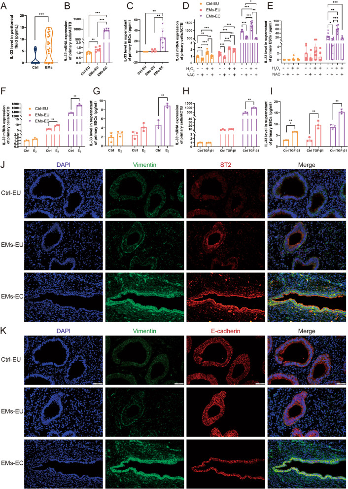
Objectives: Interleukin 33 (IL-33) is a crucial inflammatory factor that functions as an alarm signal in endometriosis (EMs). Epithelial-mesenchymal transition (EMT), a process related to inflammatory signals, intracellular reactive oxygen species (ROS) production, and lipid peroxidation, have been proposed as potential mechanisms that contribute to the development and progression of EMs. IL-33 is highly upregulated in the ectopic milieu. Moreover, ectopic endometrial cells constitutively express interleukin-33 receptor ST2 (IL-33R). However, the role of IL-33/ST2 in the EMT of EMs remains largely unknown. In this study, we aimed to mechanistically determine the role of IL-33/ST2 in EMs-associated fibrosis.

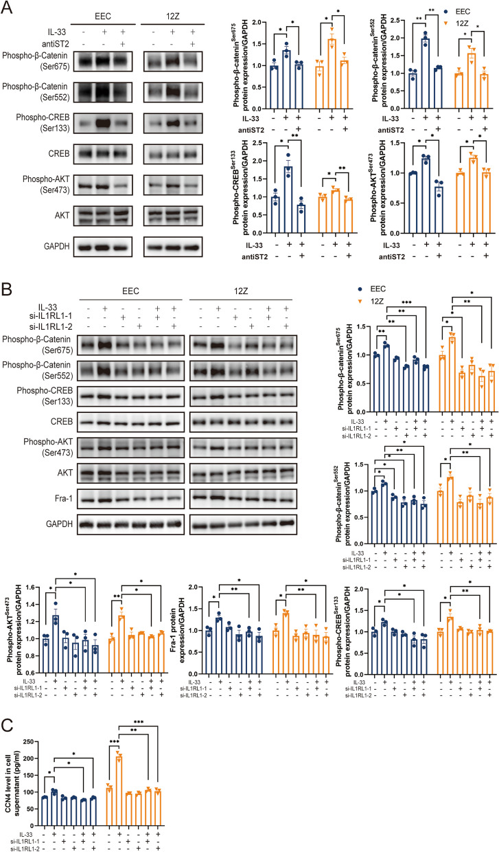
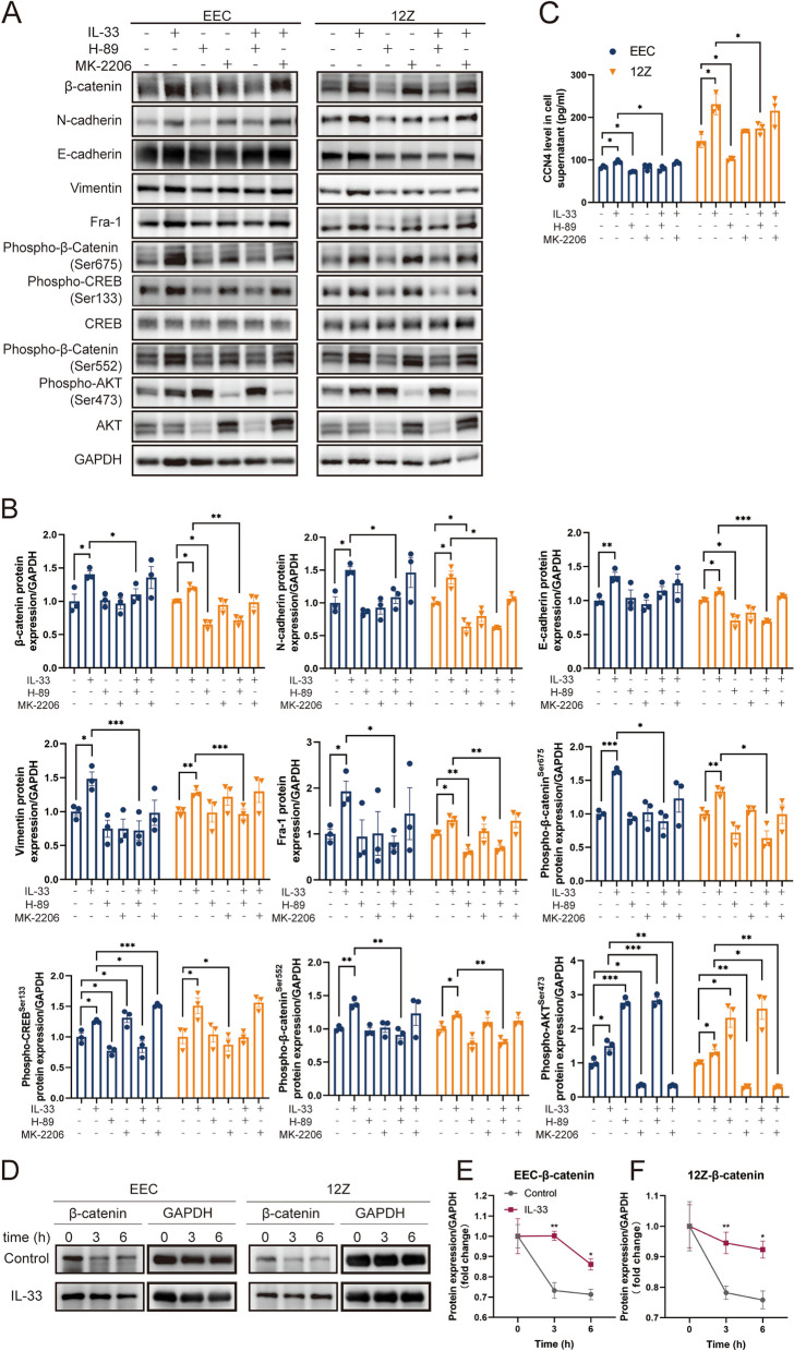
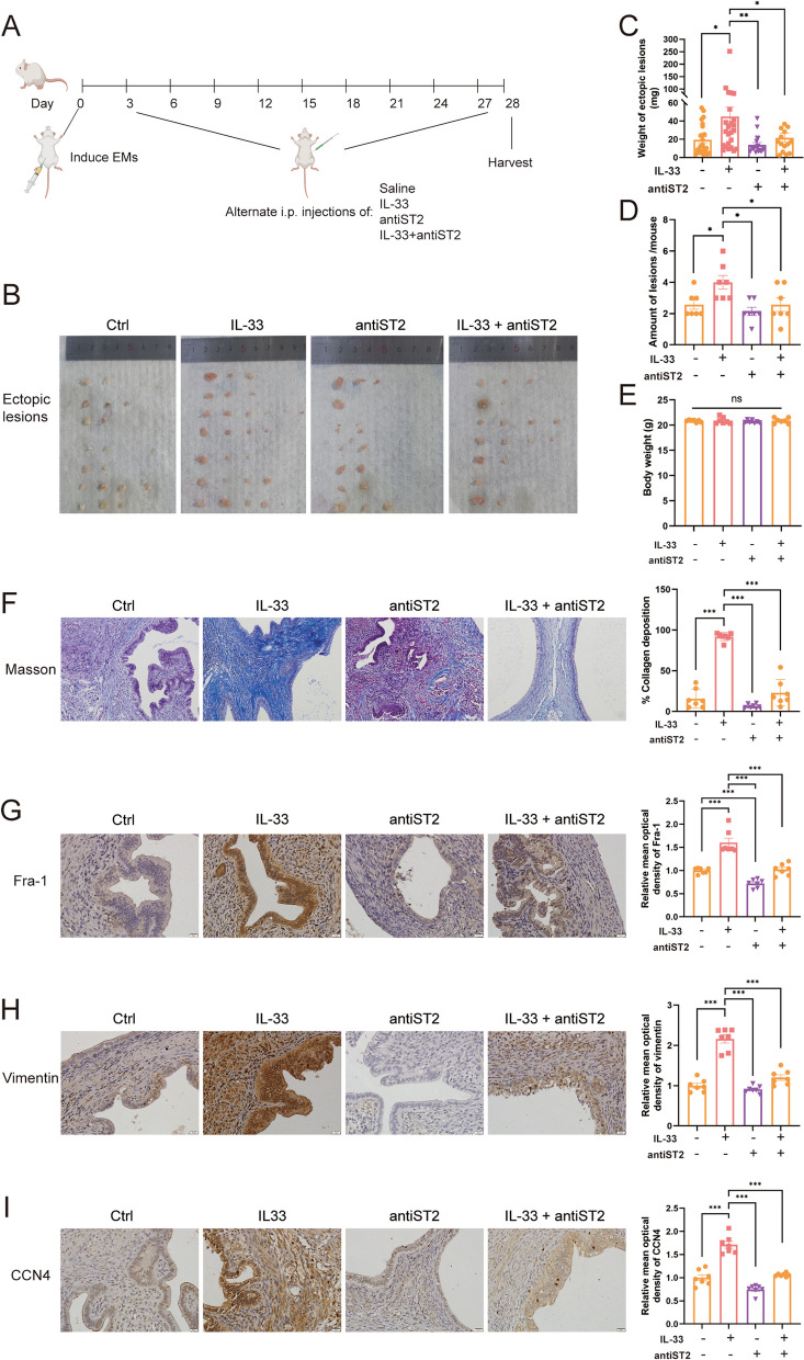
Materials and methods: We established a non-lethal oxidative stress model to explore the conditions that trigger IL-33 induction. We performed α-smooth muscle actin (α-SMA) protein detection, cell counting kit-8 (CCK-8) assays, and scratch assays to analyze the impact of IL-33 on primary endometrial stromal cells (ESCs) proliferation and invasion. Clinical samples from patients with or without EMs were subjected to immunohistochemical (IHC) and and immunofluorescence(IF) staining to assess the clinical relevance of IL-33 receptor ST2 and EMT-related proteins. Furthermore, we used the ectopic human endometrial epithelial cell line 12Z and normal human epithelial cell line EEC to evaluate the effects of IL-33 on Wnt/β-catenin signaling. The effect of IL-33 on EMT-associated fibrosis was validated in vivo by intraperitoneal injections of IL-33 and antiST2.

Results: We observed that ectopic milieu, characterized by ROS, TGF-β1, and high level of estrogen, triggers the secretion of IL-33 from ectopic ESCs. Ectopic endometrial lesions exhibited higher level of fibrotic characteristics and ST2 expression than that in the normal endometrium. Exogenous recombinant human (rhIL-33) enhanced ESC migration and survival. Similarly, 12Z cells displayed a higher degree of EMT characteristics with elevated expression of CCN4 and Fra-1, downstream target genes of the WNT/β-catenin pathway, than that observed in EECs. Conversely, blocking IL-33 with neutralizing antibodies, knocking down ST2 or β-catenin with siRNA, and β-catenin dephosphorylation abolished its effects on EMT promotion. In vivo validation demonstrated that IL-33 significantly promotes EMs-related fibrosis through the activation of Wnt/β-catenin signaling.

Conclusion: Our data strongly support the vital role of the IL-33/ST2 pathway in EMs-associated fibrosis and emphasize the importance of the EMT in the pathophysiology of fibrosis. Targeting the IL-33/ST2/Wnt/β-catenin axis may hold promise as a feasible therapeutic approach for controlling fibrosis in EMs.


Conflict of interest statement

The authors declare no competing interests.

Figures

Fig. 1
Fig. 1
Expression Patterns and Inducing Factors of Interleukin-33 (IL-33) in Endometriotic milieu. A Levels of IL-33 were detected by ELISA in the peritoneal fluid of patients with EMs and controls. B Real-time PCR for analyzing the mRNA expression of IL-33 in primary endometrial stromal cells (ESCs). C Levels of IL-33 secretion in the cell supernatant of different primary ESCs. D-I: Changes of IL-33 expression in ESC after different treatments: mRNA (D) and protein (E) level of IL-33 after H2O2 and/or NAC treatment; mRNA (F) and protein (G) levels of IL-33 after estradiol (E2) treatment; mRNA (H) and protein (I) levels of IL-33 after TGF-β1 treatment. J Immunofluorescence co-localization of vimentin and ST2 (IL-33 receptor) in endometrial tissues. (blue:DAPI, green:vimentin, red: ST2) (Scale bars, 20 μm). K Immunofluorescence co-localization of vimentin and E-cadherin in endometrial tissues. (blue:DAPI, green:vimentin, red: E-cadherin) (Scale bars, 20 μm). Ctrl-EU, eutopic endometrium of controls; EMs-EU, eutopic endometrium of patients with endometriosis; EMs-EC, ectopic lesions. All data were analyzed using one-way ANOVA followed by Dunnett’s post-hoc test. Spearman’s correlation analysis was used to analyze the correlation between ST2 and vimentin expression in humans.* P < 0.05, ** P < 0.01, *** P < 0.001, data are presented as mean ± SEM
Fig. 2
Fig. 2
IL-33 is Crucial for the Epithelial–Mesenchymal Transition (EMT) Process in EECs. A RNA expression of EMT-related genes (VIM, CDH1, CDH2, and CTNNB1) in EECs and 12Z cells. B EMT-related protein (vimentin, E-cadherin, N-cadherin, and β-catenin) were detected using western blot in EECs and 12Z cells. C mRNA expression of EMT-related genes (VIM, CDH1, CDH2, and CTNNB1) in EECs and 12Z cells after IL-33 or/and ST2 neutralizing antibody treatment. D Protein expression of EMT-related genes (vimentin, E-cadherin, N-cadherin, and β-catenin) in EEC and 12Z treated by IL-33 or/and ST2 neutralizing antibody. E Protein expression of EMT-related genes (vimentin, E-cadherin, N-cadherin, and β-catenin) in EECs and 12Z cells treated by IL-33 or/and knocking down ST2 by siRNA. Data are presented as mean ± SEM. All data were analyzed using one-way ANOVA followed by Dunnett’s post hoc test and Student’s t-test; * P < 0.05, ** P < 0.01, *** P < 0.001
Fig. 3
Fig. 3
IL-33 Enhances the Expression of Downstream Target Genes of WNT/β-catenin Pathway. A RNA expression of downstream target genes of WNT/β-catenin (AXIN2, CCN4, CCND1, CD44, Fra-1, JUN, LGR5, MYC, and PPARD) were detected via real-time PCR in EECs and 12Z cells. B Fra-1 expression in EECs and 12Z cells was detected by western blotting. C CCN4 secretion in the cell supernatant of EECs and 12Z cells was detected via ELISA. D-F After IL-33 or/and ST2 neutralizing antibody treatment, the mRNA expression and protein levels of Fra-1 and CCN4 in EECs and 12Z cells. G Immunohistochemical staining of Fra-1 and CCN4 in eutopic endometrium and ectopic lesions. Data are presented as mean ± SEM. All data were analyzed using one-way ANOVA followed by Dunnett’s post hoc test and Student’s t-test; *P < 0.05, **P < 0.01, ***P < 0.001
Fig. 4
Fig. 4
IL-33 obviously Induced β-catenin Phosphorylation. A Protein expression of phospho-β-catenin (Ser675 and Ser552), phospho-CREB (Ser133), CREB, phospho-AKT(Ser473) and AKT in EECs and 12Z cells treated by IL-33 or/and ST2 neutralizing antibody. B Protein expression of phospho-β-catenin (Ser675 and Ser552), phospho-CREB (Ser133), CREB, phospho-AKT (Ser473), AKT, and Fra-1 in EECs and 12Z cells treated by IL-33 or/and knocking down ST2 by siRNA. C Expression of CCN4 in supernatant of EEC and 12Z treated by IL-33 or/and knocking down ST2 by siRNA. Data are presented as mean ± SEM. All data were analyzed using one-way ANOVA followed by Dunnett’s post hoc test and Student’s t-test; * p < 0.05, ** p < 0.01, *** p < 0.001
Fig. 5
Fig. 5
IL-33 Mediates β-catenin Phosphorylation at Ser675 and Ser552 by PKA. A, B Protein expression of EMT-related proteins (vimentin, E-cadherin, N-cadherin, and β-catenin), Fra-1,phospho-β-catenin (Ser675 and Ser552), phospho-CREB (Ser133), CREB, phospho-AKT (Ser473) and AKT in EEC and 12Z treated by IL-33, PKA inhibitor-H89 or/and AKT inhibitor- MK2206. C Expression of CCN4 in supernatant of EEC and 12Z treated by IL-33, PKA inhibitor- H89 or/and AKT inhibitor- MK2206. D, F Cyclohexane chase assay of β-catenin in EECs and 12Z cells treated by IL-33. Data are presented as mean ± SEM. All data were analyzed using one-way ANOVA followed by Dunnett’s post hoc test and Student’s t-test; * P < 0.05, ** P < 0.01, *** P < 0.001
Fig. 6
Fig. 6
IL-33-ST2 Signal Blocking or β-catenin Knockout Weakens EMT in EEC and 12Z. A. Immunofluorescence staining of phospho-β-catenin at Ser675 and Ser552 (Scale bars, 20 μm). B, C Protein expression of EMT-related proteins (Vimentin, E-cadherin, N-cadherin, and β-catenin) and Fra-1 in EECs and 12Z cells treated by IL-33 or/and knocking β-catenin by siRNA. D Expression of CCN4 in supernatant of EEC and 12Z treated by IL-33 or/and knocking β-catenin by siRNA. Data are presented as mean ± SEM. All data were analyzed using one-way ANOVA followed by Dunnett’s post hoc test and Student’s t-test; * P < 0.05, ** P < 0.01, *** P < 0.001
Fig. 7
Fig. 7
IL-33 Promotes Fibrosis via EMT in Allograft Mouse Model of Endometriosis. A Schematic representation of the experimental outline shows the induction of endometriosis (day 0), i.p. injections of saline, IL-33, antiST2 or IL33 + anti ST2 every two days (beginning on day 3), and euthanasia (day 28). B Macroscopic view of ectopic endometriotic lesions in each group of mouse model. C, D Weight and number of lesions in mice treated with IL-33 or/and antiST2. E Body weights of mice in different groups. F-I. Masson, Fra1, vimentin, and CCN4 staining of lesions in different groups of mice. Data are presented as mean ± SEM. All data were analyzed using one-way ANOVA followed by Dunnett’s post hoc test and Student’s t-test; * p < 0.05, ** p < 0.01, *** p < 0.001
Fig. 8
Fig. 8
Schematic illustration of IL-33 Promoting EMT Process in Endometriotic Milieu. Interleukin 33 (IL-33) is highly expressed in ectopic ESCs, acting via the receptor ST2. Ectopic milieu, characterized by ROS, TGF-β1, and high level of estrogen, triggers secretion of IL-33 in ESCs, which in turn, enhanced the aggressive implantation and survival of ESCs. Meanwhile, elevated IL-33 in ectopic milieu also activated WNT/β-catenin pathway in EECs by phosphorylating β-catenin (Ser675 and Ser552), which primes exresssion of ECM related genes (CCN4 and Fra-1) and mesenchymal markers, enhanceing the EMT process and extracellular matrix produnction. Thus, IL-33/ST2 axis plays a pivotal role in endometriosis progress by promoting EMT. This figure was created with biorender.com

Similar articles

Cited by

References

    1. Saunders PTK, Horne AW. Endometriosis: etiology, pathobiology, and therapeutic prospects. Cell. 2021;184(11):2807–2824. doi: 10.1016/j.cell.2021.04.041. - DOI - PubMed
    1. Zondervan KT, Becker CM, Missmer SA. Endometriosis. N Engl J Med. 2020;382(13):1244–1256. doi: 10.1056/NEJMra1810764. - DOI - PubMed
    1. Doroftei B, Ilie OD, Balmus IM, Ciobica A, Maftei R, Scripcariu I, Simionescu G, Grab D, Stoian I, Ilea C. Molecular and clinical insights on the complex interaction between oxidative stress, apoptosis, and Endobiota in the pathogenesis of endometriosis. Diagnostics (Basel) 2021;11(8):1434. doi: 10.3390/diagnostics11081434. - DOI - PMC - PubMed
    1. Song Y, Liu J, Qiu Z, Chen D, Luo C, Liu X, Hua R, Zhu X, Lin Y, Li L, et al. Advanced oxidation protein products from the follicular microenvironment and their role in infertile women with endometriosis. Exp Ther Med. 2018;15(1):479–486. - PMC - PubMed
    1. Winarto H, Tan MI, Sadikin M, Wanandi SI. ARID1A expression is down-regulated by oxidative stress in endometriosis and endometriosis-associated ovarian cancer. Transl Oncogenomics. 2017;9:1177272716689818. doi: 10.1177/1177272716689818. - DOI - PMC - PubMed

Publication types

MeSH terms