

Echocardiogram by apical-subcostal protocol in prone position during invasive mechanical ventilation in cardiovascular intensive care unit
- PMID: 38858752
- PMCID: PMC11163713
- DOI: 10.1186/s12947-024-00326-y
Echocardiogram by apical-subcostal protocol in prone position during invasive mechanical ventilation in cardiovascular intensive care unit
Abstract
Aims: To evaluate the feasibility of a transthoracic echocardiogram using an apical-subcostal protocol in invasive mechanical ventilation (IMV) and prone position.
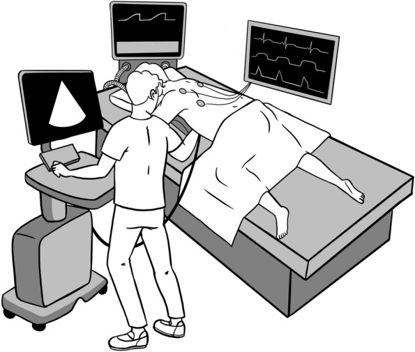
Methods: Prospective study of adults who required a prone position during IMV. A pillow was placed only under the left hemithorax in the prone position to elevate and ease the apical and subcostal windows. A critical care cardiologist (prone group) acquired and evaluated the images using the apical-subcostal protocol. Besides, we used ambulatory echocardiograms performed as a comparative group (supine group).
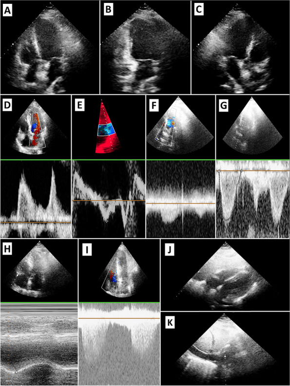
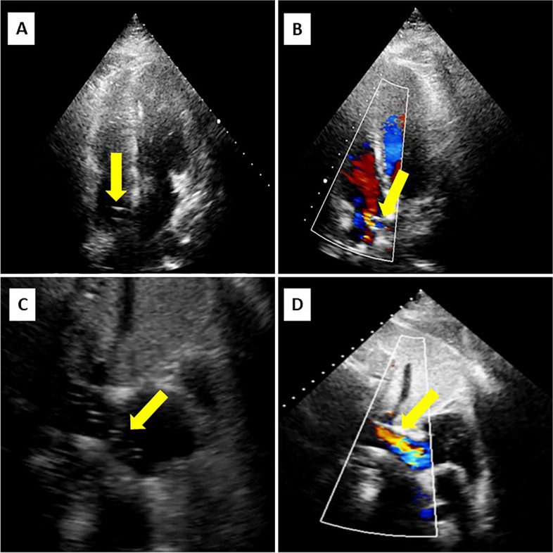
Results: 86 patients were included, 43 in the prone and 43 in the supine. In the prone group, the indication to perform an echocardiogram was hemodynamic monitoring. All patients were ventilated with protective parameters, and the mean end-expiratory pressure was 10.6 cmH2O. The protocol was performed entirely in 42 of 43 patients in the prone group because one patient did not have any acoustic window. In the 43 patients in the prone group analyzed and compared to the supine group, global biventricular function was assessed in 97.7% (p = 1.0), severe heart valve disease in 88.4% (p = 0.055), ruled out of the presence of pulmonary hypertension in 76.7% (p = 0.80), pericardial effusion in 93% (p = 0.12), and volume status by inferior vena cava in 93% (p = 0.48). Comparing prone versus supine position, a statistical difference was found when evaluating the left ventricle apical 2-chamber view (65.1 versus 100%, p < 0.01) and its segmental function (53.4 versus 100%, p < 0.01).
Conclusion: The echocardiogram using an apical-subcostal protocol is feasible in patients in the IMV and prone position.
© 2024. The Author(s).
Conflict of interest statement
Nothing to declare.
Figures
References
-
- Lancellotti P, Price S, Edvardsen T, et al. The use of echocardiography in acute cardiovascular care: recommendations of the European Association of Cardiovascular Imaging and the Acute Cardiovascular Care Association. Eur Heart J Cardiovasc Imaging. 2015;16(2):119–146. doi: 10.1093/ehjci/jeu210. - DOI - PubMed
-
- Fortuni F, Zilio F, Iannopollo G, et al. Management of temporary mechanical circulatory support devices in cath-lab and cardiac intensive care unit. European Heart Journal - Imaging Methods and Practice. 2024; 1(1).
MeSH terms
LinkOut - more resources
Full Text Sources
