. 2024 Jun 10;29(1):319.
doi: 10.1186/s40001-024-01921-5.

Exploration of tissue fixation methods suitable for digital pathological studies of the testis

Affiliations

Exploration of tissue fixation methods suitable for digital pathological studies of the testis

Pengxiang Tian et al. Eur J Med Res. .

Abstract

Background: The way of testicular tissue fixation directly affects the correlation and structural integrity between connective tissue and seminiferous tubules, which is essential for the study of male reproductive development. This study aimed to find the optimal fixative and fixation time to produce high-quality testicular histopathological sections, and provided a suitable foundation for in-depth study of male reproductive development with digital pathology technology.

Methods: Testes were removed from both sides of 25 male C57BL/6 mice. Samples were fixed in three different fixatives, 10% neutral buffered formalin (10% NBF), modified Davidson's fluid (mDF), and Bouin's Fluid (BF), for 8, 12, and 24 h, respectively. Hematoxylin and eosin (H&E) staining, periodic acid Schiff-hematoxylin (PAS-h) staining, and immunohistochemistry (IHC) were used to evaluate the testicle morphology, staging of mouse seminiferous tubules, and protein preservation. Aperio ScanScope CS2 panoramic scanning was used to perform quantitative analyses.

Results: H&E staining showed 10% NBF resulted in an approximately 15-17% reduction in the thickness of seminiferous epithelium. BF and mDF provided excellent results when staining acrosomes with PAS-h. IHC staining of synaptonemal complexes 3 (Sycp3) was superior in mDF compared to BF-fixed samples. Fixation in mDF and BF improved testis tissue morphology compared to 10% NBF.

Conclusions: Quantitative analysis showed that BF exhibited a very low IHC staining efficiency and revealed that mouse testes fixed for 12 h with mDF, exhibited morphological details, excellent efficiency of PAS-h staining for seminiferous tubule staging, and IHC results. In addition, the morphological damage of testis was prolonged with the duration of fixation time.

Keywords: Digital pathology; Fixative; HE staining; IHC staining; Mouse testis; PAS staining.


Conflict of interest statement

All authors have been notified of the article and declared no conflicts of interest.

Figures

Fig. 1
Fig. 1
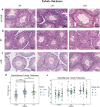
Representative images of hematoxylin and eosin (HE) stained mouse testis sections prepared with different fixatives. Before hematoxylin and eosin (H&E) staining mouse testis samples were fixed in 10%NBF (10% neutral buffered formalin) (a), BF (Bouin’s fixative) (b) and mDF (modified Davidson’s fluid) (c) solution for 8 h, 12 h, and 24 h. Arrows and asterisks highlight area of artefact including: disrupted seminiferous epithelium integrity (blank space; red asterisks), cell nuclei condensation (black arrow), and disrupted cell plasma integrity (blank space; red arrow). Bars = 50 μm
Fig. 2
Fig. 2
Measurements of seminiferous tubule thickness. ac Representative images of measured stage VI–VIII tubules. The average thickness of each tubule was generated from three lengths (from the periphery of the tubule to elongated spermatids), which are annotated by colored lines and numbers. d Dot plots show the thickness of the three fixatives, all fixation times were included. e Dot plots show thickness between fixatives at different durations. Each dot represents an independent stage VI–VIII tubule, data represent the mean ± standard deviation (SD) of 298 tubules from 30 sections, which were added as a pointrange. One-way ANOVA was used to detect the effect of fixative and duration on seminiferous epithelial thickness, significance was defined as ( ∗)P < 0.05, (∗ ∗)P < 0.01, (∗ ∗ ∗)P < 0.001, (∗ ∗  ∗ ∗)P < 0.0001
Fig. 3
Fig. 3
Periodic acid Schiff–hematoxylin (PAS-h) stain of mouse testis tissue prepared with different fixatives. Periodic acid Schiff–hematoxylin (PAS-h) staining was performed on the mouse testis, samples were fixed in 10%NBF (aa″), BF (bb″), and mDF (cc″) for 12 h, the red line annotate each stage VII tubules. a, b, c (left panel): representative testis sections, tubules that require further demonstrated are point out by black arrows, bars = 500 μm; aʹ, bʹ, cʹ (middle panel): a representative stage VII seminiferous tubule, the original (left) and markup (right) images, bars = 50 μm; a″, b″, c″ (right panel): further magnification of the tubule, red arrows point out the arcuate acrosomal caps, bars = 25 μm. The markup image was generated by Leica Aperio Positive Pixel Count v9 algorithm; positivity pixels are represented by: strong positive (Sp) (red), positive (p) (orange), and weak positive (Wp), (yellow)
Fig. 4
Fig. 4
Acrosomic granules assessments of fixative conditions using PAS-h stained mouse testis sections. Representative testis sections from different samples were assessed after performing PAS-h. Samples were fixed in 10%NBF (a), BF (b), and mDF (c) for 12 h, the red line annotate each stage VII tubule that needs to be analyzed, positive data were collected from each markup tubule. D, E Box plot show the Sycp3 positivity (%) of acrosomic granules of different fixatives, arranged by fixatives (d) and samples (e) (Each individual box represents data collection from one testis of the corresponding sample). Data represent the mean ± SD of 100 seminiferous tubules from 5 mouse. (∗ ∗)P < 0.01, (∗ ∗ ∗)P < 0.001, (∗ ∗  ∗ ∗)P < 0.0001, generated by One-Way ANOVA. Scale bar = 50 μm
Fig. 5
Fig. 5
Detection of Sycp3 using immunohistochemistry (IHC) in mouse testis fixed with different fixatives. Following the fixation of testis in 10%NBF (aa″), BF (bb″) and mDF (cc″) for 12 h, immunohistochemistry (IHC) was implemented on mouse testis sections. a, b, c (left panel): Representative testis sections, bars = 500 μm; aʹ–cʹ (middle panel) and a″–c″ (right panel; markup): Representative seminiferous tubules, black arrows highlighted the Sycp3 (a meiosis marker), which was detected in spermatocytes. Red arrows in B2 and B3 indicate poor DAB coloration in BF-fixed samples. The markup image was generated by Leica Aperio Positive Pixel Count v9 algorithm; positivity pixels are represented by: strong positive (Sp) (red), positive (p) (orange), and weak positive (Wp), (yellow)
Fig. 6
Fig. 6
IHC positivity assessments of mouse testis sections were preserved with different fixatives. Representative testis sections from different samples were analyzed after performing IHC. Samples were fixed in 10%NBF (a), BF (b), and mDF (c) for 12 h, the red line annotate random tubule that needs to be analyzed, positive data were collected from each markup tubule. D, E Box plot show the Sycp3 positivity (%) of different fixatives, arranged by fixatives (d) and samples (e) (Each individual box represents data collection from one testis of the corresponding sample). Data represent the mean ± SD of 160 seminiferous tubules from 5 mouse. (∗ ∗)P < 0.01, (∗ ∗  ∗ ∗)P < 0.0001, generated by One-Way ANOVA. Scale bar = 50 μm

Similar articles

Cited by

References

    1. Green CD, Ma Q, Manske GL, Shami AN, Zheng X, Marini S, Moritz L, Sultan C, Gurczynski SJ, Moore BB, et al. A Comprehensive roadmap of murine spermatogenesis defined by single-cell RNA-seq. Dev Cell. 2018;46:651–667.e610. doi: 10.1016/j.devcel.2018.07.025. - DOI - PMC - PubMed
    1. Ernst C, Eling N, Martinez-Jimenez CP, Marioni JC, Odom DT. Staged developmental mapping and X chromosome transcriptional dynamics during mouse spermatogenesis. Nat Commun. 2019;10:1251. doi: 10.1038/s41467-019-09182-1. - DOI - PMC - PubMed
    1. Fraenkel-Conrat H, Olcott HS. The reaction of formaldehyde with proteins; cross-linking between amino and primary amide or guanidyl groups. J Am Chem Soc. 1948;70:2673–2684. doi: 10.1021/ja01188a018. - DOI - PubMed
    1. Howat WJ, Wilson BA. Tissue fixation and the effect of molecular fixatives on downstream staining procedures. Methods. 2014;70:12–19. doi: 10.1016/j.ymeth.2014.01.022. - DOI - PMC - PubMed
    1. Liang JH, Sankai T, Yoshida T, Yoshikawa Y. Comparison of the effects of two fixatives for immunolocalization of testosterone in the testes of the cynomolgus monkey, mouse and rat. Exp Anim. 2000;49(4):301–304. doi: 10.1538/expanim.49.301. - DOI - PubMed

LinkOut - more resources