

Remarkable Recovery After Severe Gunshot Brain Injury: A Comprehensive Case Study of Functional Rehabilitation
- PMID: 38859569
- PMCID: PMC11180484
- DOI: 10.12659/AJCR.941601
Remarkable Recovery After Severe Gunshot Brain Injury: A Comprehensive Case Study of Functional Rehabilitation
Abstract
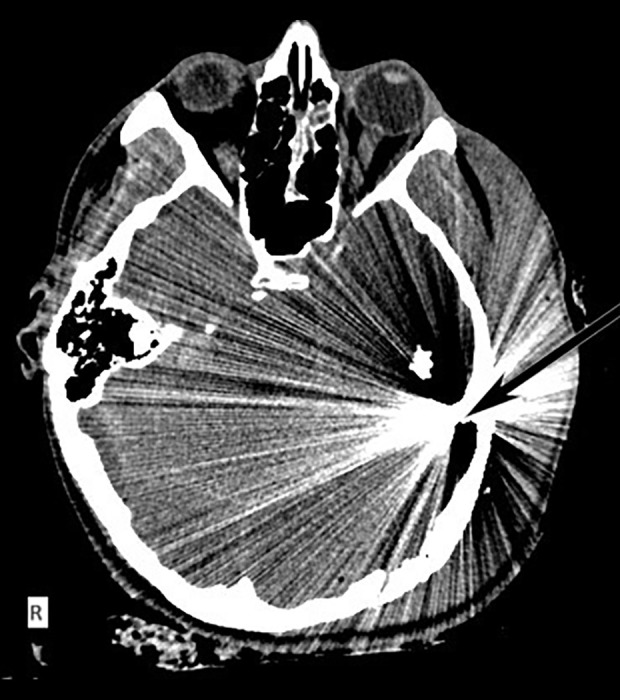
BACKGROUND Penetrating traumatic brain injury (TBI) caused by gunshots is a rare type of TBI that leads to poor outcomes and high mortality rates. Conducting a formal neuropsychological evaluation concerning a patient's neurologic status during the chronic recovery phase can be challenging. Furthermore, the clinical assessment of survivors of penetrating TBI has not been adequately documented in the available literature. Severe TBI in patients can provide valuable information about the functional significance of the damaged brain regions. This information can help inform our understanding of the brain's intricate neural network. CASE REPORT We present a case of a 29-year-old right-handed man who sustained a left-hemisphere TBI after a gunshot, causing extensive diffuse damage to the left cerebral and cerebellar hemispheres, mainly sparing the right hemisphere. The patient survived. The patient experienced spastic right-sided hemiplegia, facial hemiparesis, left hemiparesis, and right hemianopsia. Additionally, he had severe global aphasia, which caused difficulty comprehending verbal commands and recognizing printed letters or words within his visual field. However, his spontaneous facial expressions indicating emotions were preserved. The patient received a thorough neuropsychological assessment to evaluate his functional progress following a severe TBI and is deemed to have had a favorable outcome. CONCLUSIONS Research on cognitive function recovery following loss of the right cerebral hemisphere typically focuses on pediatric populations undergoing elective surgery to treat severe neurological disorders. In this rare instance of a favorable outcome, we assessed the capacity of the fully developed right hemisphere to sustain cognitive and emotional abilities, such as language.
Conflict of interest statement
Figures


Similar articles
-
[Cerebral lateralization in two cases of crossed dextral aphasia with right-hemisphere arteriovenous malformation].No To Shinkei. 1988 Nov;40(11):1027-33. No To Shinkei. 1988. PMID: 3219238 Japanese.
-
Recovery of injured Broca's portion of arcuate fasciculus in the dominant hemisphere in a patient with traumatic brain injury.Medicine (Baltimore). 2017 Dec;96(51):e9183. doi: 10.1097/MD.0000000000009183. Medicine (Baltimore). 2017. PMID: 29390458 Free PMC article.
-
["Left unilateral agraphia with right hemiparesis" after occlusion of the left middle cerebral artery].No To Shinkei. 1992 Jul;44(7):661-6. No To Shinkei. 1992. PMID: 1419344 Japanese.
-
SURGICAL MANAGEMENT OF A PENETRATING BRAIN WOUND AND ASSOCIATED PERFORATING OCULAR INJURY CAUSED BY A LOW-VELOCITY SHARP METALLIC OBJECT: A CASE REPORT AND LITERATURE REVIEW.Acta Clin Croat. 2022 Nov;61(3):537-546. doi: 10.20471/acc.2022.61.03.21. Acta Clin Croat. 2022. PMID: 37492370 Free PMC article. Review.
-
[Crossed aphasia in neurosurgical practice: case report and literature review].Zh Vopr Neirokhir Im N N Burdenko. 2022;86(1):103-111. doi: 10.17116/neiro202286011103. Zh Vopr Neirokhir Im N N Burdenko. 2022. PMID: 35170283 Review. Russian.
References
-
- Deng H, Yue JK, Winkler EA, et al. Adult firearm-related traumatic brain injury in United States trauma centers. J Neurotrauma. 2019;36(2):322–37. - PubMed
-
- Kim TW, Lee JK, Moon KS, et al. Penetrating gunshot injuries to the brain. J Trauma. 2007;62(6):1446–51. - PubMed
-
- Hofbauer M, Kdolsky R, Figl M, et al. Predictive factors influencing the outcome after gunshot injuries to the head-a retrospective cohort study. J Trauma. 2010;69(4):770–75. - PubMed
-
- Dikmen SS, Corrigan JD, Levin HS, et al. Cognitive outcome following traumatic brain injury. J Head Trauma Rehabil. 2009;24(6):430–38. - PubMed
-
- DuBose JJ, Barmparas G, Inaba K, et al. Isolated severe traumatic brain injuries sustained during combat operations: Demographics, mortality outcomes, and lessons to be learned from contrasts to civilian counterparts. J Trauma. 2011;70(1):11–16. ; discussion 16–18. - PubMed
Publication types
MeSH terms
LinkOut - more resources
Full Text Sources
