. 2024 Aug 1;128(30):7291-7303.
doi: 10.1021/acs.jpcb.4c01454. Epub 2024 Jun 10.

Robust Quantification of Live-Cell Single-Molecule Tracking Data for Fluorophores with Different Photophysical Properties

Affiliations

Robust Quantification of Live-Cell Single-Molecule Tracking Data for Fluorophores with Different Photophysical Properties

Amy N Moores et al. J Phys Chem B. .

Abstract

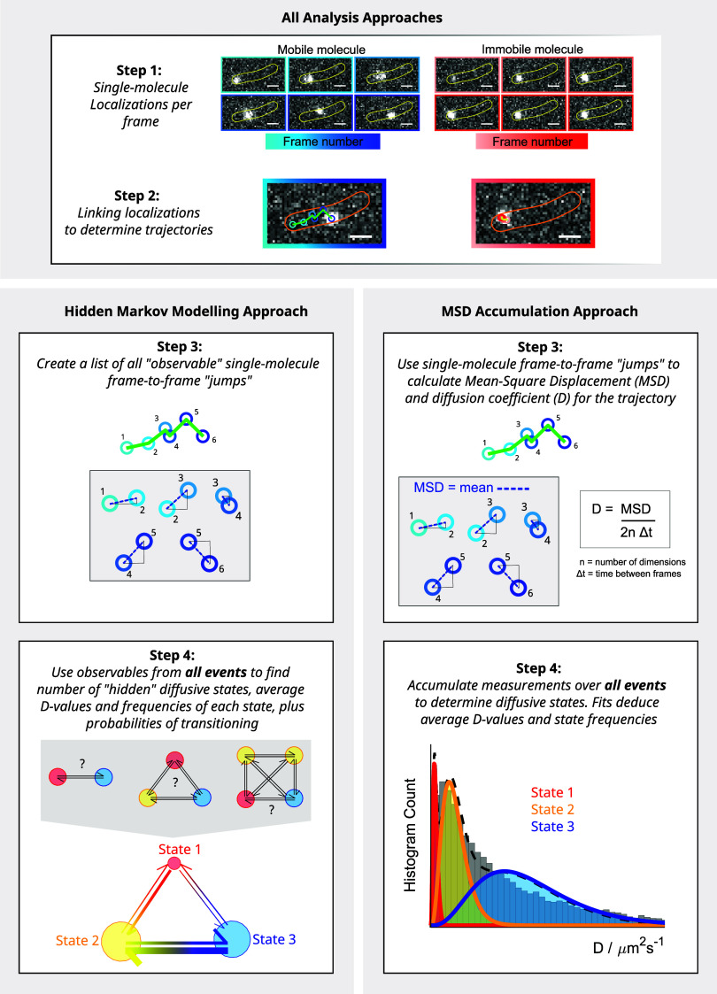
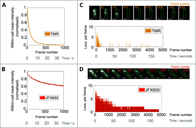
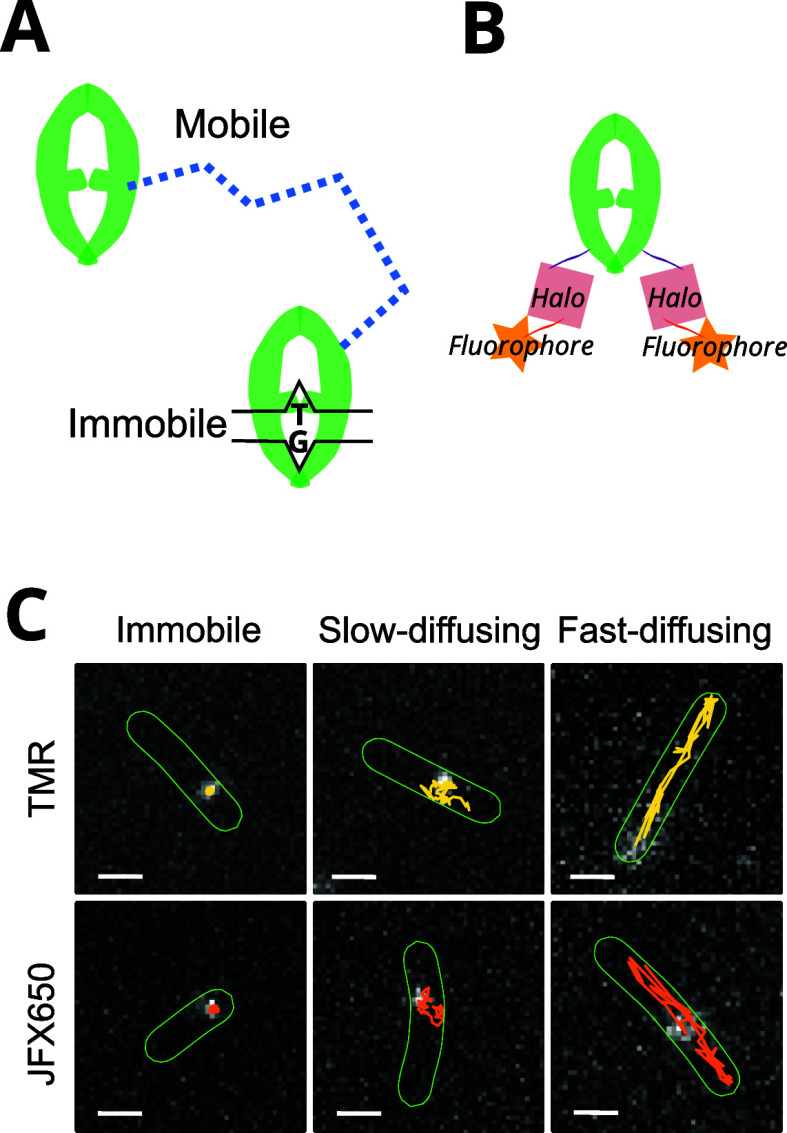
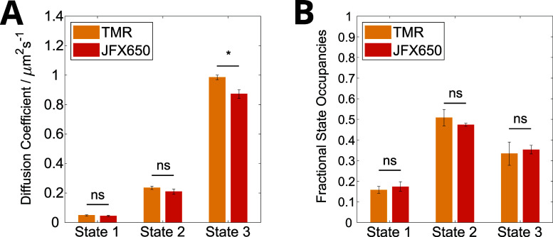
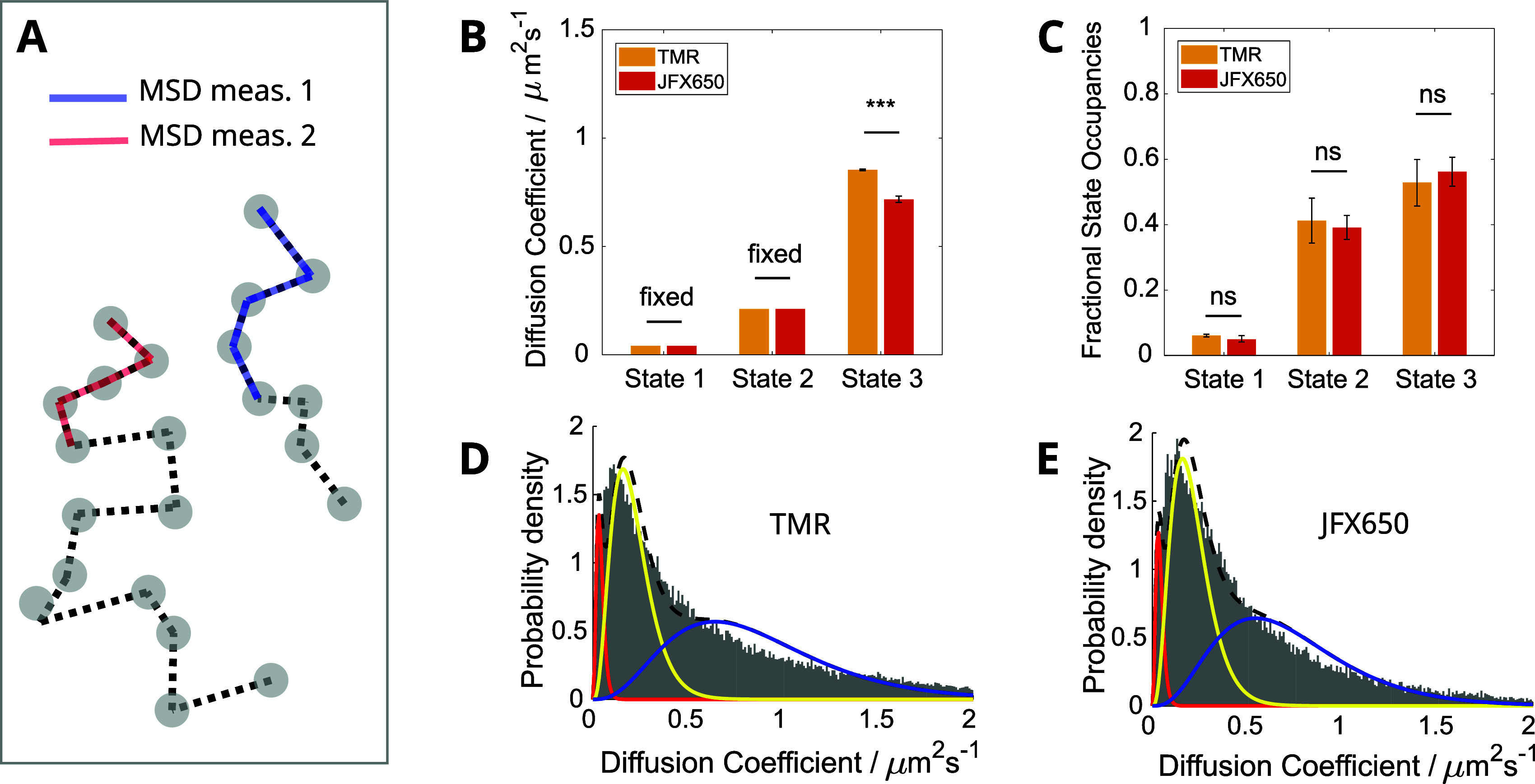
High-speed single-molecule tracking in live cells is becoming an increasingly popular method for quantifying the spatiotemporal behavior of proteins in vivo. The method provides a wealth of quantitative information, but users need to be aware of biases that can skew estimates of molecular mobilities. The range of suitable fluorophores for live-cell single-molecule imaging has grown substantially over the past few years, but it remains unclear to what extent differences in photophysical properties introduce biases. Here, we tested two fluorophores with entirely different photophysical properties, one that photoswitches frequently between bright and dark states (TMR) and one that shows exceptional photostability without photoswitching (JFX650). We used a fusion of the Escherichia coli DNA repair enzyme MutS to the HaloTag and optimized sample preparation and imaging conditions for both types of fluorophore. We then assessed the reliability of two common data analysis algorithms, mean-square displacement (MSD) analysis and Hidden Markov Modeling (HMM), to estimate the diffusion coefficients and fractions of MutS molecules in different states of motion. We introduce a simple approach that removes discrepancies in the data analyses and show that both algorithms yield consistent results, regardless of the fluorophore used. Nevertheless, each dye has its own strengths and weaknesses, with TMR being more suitable for sampling the diffusive behavior of many molecules, while JFX650 enables prolonged observation of only a few molecules per cell. These characterizations and recommendations should help to standardize measurements for increased reproducibility and comparability across studies.


Conflict of interest statement

The authors declare no competing financial interest.

Figures

Figure 1
Figure 1
Schematic describing the analysis of single-molecule tracking data to quantify subpopulations of molecules with different diffusion coefficients. Two examples of MutS-Halo-TMR single-molecule fluorescence events are shown (associated with mobile and immobile molecules), which appeared in the same cell at different time points in a movie. We show how localizations are linked across frames to determine the MSD and subsequent diffusion coefficient estimates from each trajectory. Repeat observations are made to accumulate diffusion coefficient measurements. In this example, we see three diffusive states in the diffusion coefficient histogram: fast-diffusing (blue), immobile (red), and a slowly diffusing state (yellow). Relative abundances of the three subpopulations could be quantified by fitting the histogram. In HMM analysis, diffusive states are inferred from the molecule displacements between successive frames without averaging over trajectories, and the state occupancies are estimated from the transition probabilities between the states.
Figure 2
Figure 2
Comparison of TMR- and JFX650-labeled MutS-HaloTag photobleaching. (A, B) Mean intensity of cells with (A) TMR-labeled or (B) JFX650-labeled MutS-Halo for 1000 frames (≈35 s) from laser turn-on. The labeling concentrations used for both TMR and JFX650 were 2.5 μM. The mean background intensity outside cells was subtracted from the intensity values of all pixels within the segmented cell masks and averaged across 506 and 1477 cells. Curves were normalized by the maximum value (from 3 and 7 movies) for TMR and JFX650, respectively. (C, D) The number of localizations within one representative cell expressing (C) TMR-labeled or (D) JFX650-labeled MutS-HaloTag for a duration of 5000 frames (≈174 s) from laser turn-on. Snapshots of the movie with segmented cell masks at the indicated frames are shown. We note that the high emitter density prior to photobleaching will prevent accurate localization and likely leads to an underestimation of the true number of localizations per frame at the start of the acquisitions. Scale bars 1 μm.
Figure 3
Figure 3
Single-molecule tracking of MutS-Halo. (A) Schematic of MutS undergoing target search and binding a DNA mismatch, corresponding to mobile and immobile diffusive states, respectively. (B) Schematic of a fluorescently labeled MutS-Halo dimer. (C) Example MutS-Halo trajectories for TMR (yellow) or JFX650 (orange) display three distinct diffusive states: immobile, slow-diffusing, and fast-diffusing. All example trajectories have the same duration of 40 frames. Trajectories are overlaid onto a fluorescence image corresponding to the final position of the molecule, on which the cell segmentation masks are also displayed (green). Scale bars 1 μm.
Figure 4
Figure 4
Comparison of TMR- and JFX650-labeled MutS-Halo diffusive state analysis using HMM. Shown are the diffusion coefficient values (A) and the state occupancies (B) for the 3-state model. Error bars show the standard error of the mean across 3x (TMR) and 6x (JFX650) independent experimental repeats, and bar heights are weighted means according to the total number of trajectories per repeat.
Figure 5
Figure 5
Comparison of TMR- and JFX650-labeled MutS-Halo diffusive state analysis using the MSD calculation approach. (A) Schematic showing the frames over which the MSD is measured for each example trajectory. (B, C) Estimated diffusion coefficients and state occupancies for the 3-state model. Error bars show the standard error of the mean across 3x (TMR) and 6x (JFX650) independent experimental repeats, and bar heights are weighted means according to the total number of trajectories per repeat. (D, E) Averaged diffusion coefficient histograms from each experiment repeat for TMR (D) and JFX650 (E). The fitted probability distribution curves are shown for the immobile (red), slowly diffusing (yellow), and fast-diffusing (blue) states.
Figure 6
Figure 6
Inadvertent splitting of trajectories causes overcounting bias of mobile molecules. (A) Example MutS-Halo-JFX650 trajectory (1000 frames provided) of one mobile molecule, overlaid on a single frame of the movie. The analysis algorithm has failed to correctly link together all localizations arising from the same molecule, resulting in 19 separate subtrajectories, each plotted with a random color. (B) The corresponding measured molecular frame-to-frame displacement plotted as a function of frame number. Line colors correspond to those of the individual tracks plotted in panel A. (C, D) Example MutS-Halo-JFX650 trajectory (1000 frames provided) and displacements of one immobile molecule. The analysis algorithm has correctly linked together all localizations resulting in a single trajectory. Segmented cell outlines are depicted (yellow). Scale bars 1 μm.
Figure 7
Figure 7
Trajectory duration dependence on molecule mobility. (A) TMR and JFX650 trajectory duration for immobile or mobile molecules (diffusion coefficient less or greater than 0.04 μm2s–1, respectively). Mean ± standard error of the mean for 3 or 6 independent repeats for TMR- and JFX650-labeled MutS-Halo, respectively. (B) Cumulative distribution of the duration of immobile (solid line) and mobile (dashed line) tracks, pooled from 3 or 6 independent repeats for TMR- and JFX650-labeled MutS-Halo, respectively.
Figure 8
Figure 8
Comparison of TMR- and JFX650-labeled MutS-Halo diffusive state analysis using the MSD calculation approach with forced computational partitioning of trajectories. (A) Schematic showing the frames over which the MSD is measured for each example trajectory. Due to forced computational partitioning, multiple MSD measurements can be obtained per trajectory. (B, C) Estimated diffusion coefficients and state occupancies for the 3-state model. Error bars show the standard error of the mean across 3x (TMR) and 6x (JFX650) independent experimental repeats, and bar heights are weighted means according to the total number of trajectories per repeat. (D, E) Averaged diffusion coefficient histograms from each experiment repeat for TMR (D) and JFX650 (E). The fitted probability distribution curves are shown for the immobile (red), slow-diffusing (yellow), and fast-diffusing (blue) states.
Figure 9
Figure 9
JFX650 enables prolonged single-molecule observations. (A) The frame-to-frame diffusion coefficients of two example single-molecule MutS-Halo-JFX650 tracks show long-lived binding events and transitions between mobile and immobile states. Insets show the tracks overlaid on the corresponding brightfield image, with line color corresponding to the background color of the segments in the main plot. (B) Example of a single MutS-Halo-JFX650 molecule that crossed repeatedly between daughter cells during cell division and became constrained in the top cell, likely due to septum closure. Brightfield image shows relevant cell outlines highlighted in yellow. Track color corresponds to the frame number. Scale bars are all 1 μm.

Similar articles

References

    1. Rust M. J.; Bates M.; Zhuang X. Sub-diffraction-limit imaging by stochastic optical reconstruction microscopy (STORM). Nat. Methods 2006, 3, 793–796. 10.1038/nmeth929. - DOI - PMC - PubMed
    1. Betzig E.; Patterson G. H.; Sougrat R.; Lindwasser O. W.; Olenych S.; Bonifacino J. S.; Davidson M. W.; Lippincott-Schwartz J.; Hess H. F. Imaging intracellular fluorescent proteins at nanometer resolution. Science 2006, 313, 1642–1645. 10.1126/science.1127344. - DOI - PubMed
    1. Hess S. T.; Girirajan T. P. K.; Mason M. D. Ultra-High Resolution Imaging by Fluorescence Photoactivation Localization Microscopy. Biophys. J. 2006, 91, 4258–4272. 10.1529/biophysj.106.091116. - DOI - PMC - PubMed
    1. Shcherbakova D. M.; Sengupta P.; Lippincott-Schwartz J.; Verkhusha V. V. Photocontrollable Fluorescent Proteins for Superresolution Imaging. Annu. Rev. Biophys. 2014, 43, 303–329. 10.1146/annurev-biophys-051013-022836. - DOI - PMC - PubMed
    1. Uno S.; Tiwari D. K.; Kamiya M.; Arai Y.; Nagai T.; Urano Y. A guide to use photocontrollable fluorescent proteins and synthetic smart fluorophores for nanoscopy. Microscopy 2015, 64, 263–277. 10.1093/jmicro/dfv037. - DOI - PubMed

MeSH terms