

Case report: Primary familial brain calcification associated with a rare PDGFRB variant, coexisting with nontraumatic osteonecrosis of the femoral head
- PMID: 38859923
- PMCID: PMC11163128
- DOI: 10.3389/fnins.2024.1381840
Case report: Primary familial brain calcification associated with a rare PDGFRB variant, coexisting with nontraumatic osteonecrosis of the femoral head
Abstract
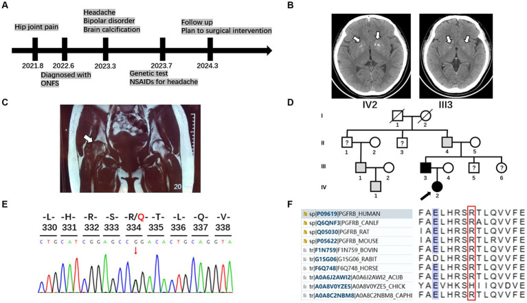
Primary familial brain calcification (PFBC) is a rare genetic neurodegenerative disorder characterized by bilateral calcifications in the brain. PFBC may manifest with a broad spectrum of motor, cognitive, and neuropsychiatric symptoms. Several causal genes have been identified in PFBC, which are inherited as both autosomal dominant and autosomal recessive traits. Herein, we present the case of a Chinese family diagnosed with PFBC. The family members carry a rare heterozygous variant (p. R334Q) in exon 7 of platelet-derived growth factor receptor β (PDGFRB) gene. The platelet-derived growth factor-B/PDGF receptor β (PDGF-B/PDGFRβ) signaling pathway plays a crucial role in pericyte development in various organs and tissues. Notably, this variant uniquely coexists with nontraumatic osteonecrosis of the femoral head. Additionally, we reviewed previous studies on PFBC-causing variants in PDGFRB.
Keywords: PDGFRB; case report; osteonecrosis of the femoral head; primary familial brain calcification; whole-exome sequencing.
Copyright © 2024 Cao, Luo and Wang.
Conflict of interest statement
The authors declare that the research was conducted in the absence of any commercial or financial relationships that could be construed as a potential conflict of interest.
Figures

Similar articles
-
Functional Characterization of Germline Mutations in PDGFB and PDGFRB in Primary Familial Brain Calcification.PLoS One. 2015 Nov 23;10(11):e0143407. doi: 10.1371/journal.pone.0143407. eCollection 2015. PLoS One. 2015. PMID: 26599395 Free PMC article.
-
Novel mutations of PDGFRB cause primary familial brain calcification in Chinese families.J Hum Genet. 2017 Jul;62(7):697-701. doi: 10.1038/jhg.2017.25. Epub 2017 Mar 16. J Hum Genet. 2017. PMID: 28298627
-
A novel PDGFRB sequence variant in a family with a mild form of primary familial brain calcification: a case report and a review of the literature.BMC Neurol. 2019 Apr 12;19(1):60. doi: 10.1186/s12883-019-1292-8. BMC Neurol. 2019. PMID: 30979360 Free PMC article. Review.
-
First Japanese family with primary familial brain calcification due to a mutation in the PDGFB gene: an exome analysis study.Psychiatry Clin Neurosci. 2015 Feb;69(2):77-83. doi: 10.1111/pcn.12238. Epub 2014 Oct 17. Psychiatry Clin Neurosci. 2015. PMID: 25211641
-
Genotype-Phenotype Relations in Primary Familial Brain Calcification: Systematic MDSGene Review.Mov Disord. 2021 Nov;36(11):2468-2480. doi: 10.1002/mds.28753. Epub 2021 Aug 25. Mov Disord. 2021. PMID: 34432325 Review.
References
-
- Ali N., Ayyub M., Khan S. A. (2014). High prevalence of protein C, protein S, antithrombin deficiency, and factor V Leiden mutation as a cause of hereditary thrombophilia in patients of venous thromboembolism and cerebrovascular accident. Pak. J. Med. Sci. 30, 1323–1326. doi: 10.12669/pjms.306.5878, PMID: - DOI - PMC - PubMed
Publication types
LinkOut - more resources
Full Text Sources
Miscellaneous