Review
. 2024 May 27:12:1397788.
doi: 10.3389/fcell.2024.1397788. eCollection 2024.

Non-coding RNAs and exosomal non-coding RNAs in lung cancer: insights into their functions

Affiliations
Review

Non-coding RNAs and exosomal non-coding RNAs in lung cancer: insights into their functions

Xiaolong Lv et al. Front Cell Dev Biol. .

Abstract

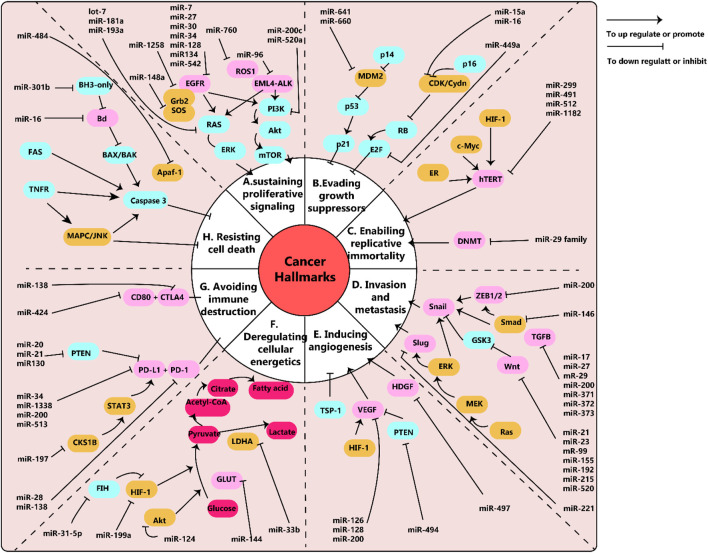
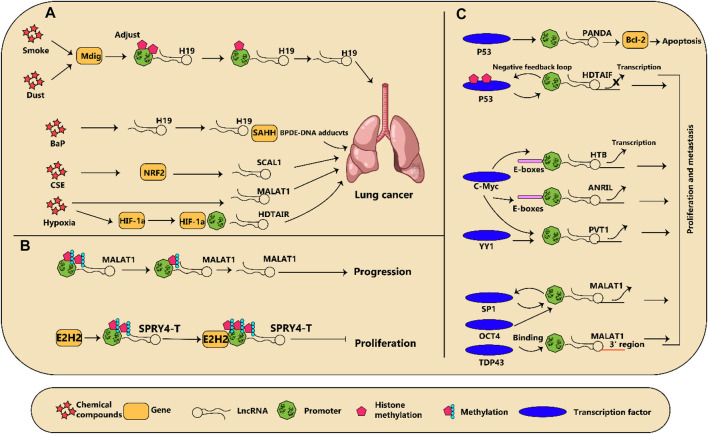
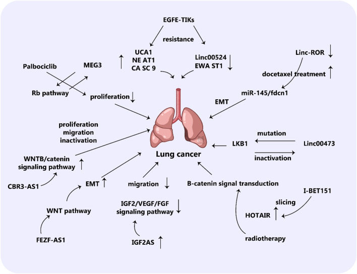
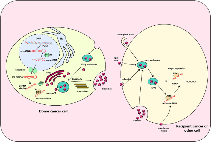
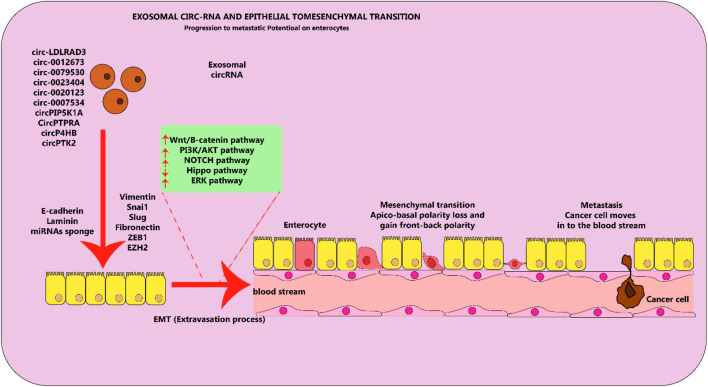
Lung cancer is the second most common form of cancer worldwide Research points to the pivotal role of non-coding RNAs (ncRNAs) in controlling and managing the pathology by controlling essential pathways. ncRNAs have all been identified as being either up- or downregulated among individuals suffering from lung cancer thus hinting that they may play a role in either promoting or suppressing the spread of the disease. Several ncRNAs could be effective non-invasive biomarkers to diagnose or even serve as effective treatment options for those with lung cancer, and several molecules have emerged as potential targets of interest. Given that ncRNAs are contained in exosomes and are implicated in the development and progression of the malady. Herein, we have summarized the role of ncRNAs in lung cancer. Moreover, we highlight the role of exosomal ncRNAs in lung cancer.

Keywords: circular RNA; long non-coding RNA; lung cancer; microRNA; non-coding RNA.


Conflict of interest statement

The authors declare that the research was conducted in the absence of any commercial or financial relationships that could be construed as a potential conflict of interest.

Figures

FIGURE 1
FIGURE 1
(A) schema of microRNAs and their pathways network in cancer. Epidermal growth factor (EGF) and its receptors, EGFR, work together to stimulate cell growth in lung cancer through the downstream RAS/ERK and PI3K/Akt/mTOR pathways. Similarly, EML4-ALK and ROS1 also trigger cancer cell growth through these pathways. Relevant miRNAs are depicted in the diagram. (B) Disruptions in tumor suppressors, RB and p53, enable cancer cells to avoid inhibition of growth. (C) The human telomerase reverse transcriptase (hTERT) is linked to the immortality of cancer cells. MiR-299, miR-491, miR-512, and miR-1182 have been found to target hTERT, but these findings have been studied in various cancer cells other than lung cancer. Additionally, the miR-29 family can control telomere length by targeting DNA methyltransferases (DNMT). (D) Snail, Slug, and Wnt are key factors in epithelial to mesenchymal transition (EMT), which is associated with cancer metastasis and invasion. The diagram shows the related miRNAs. (E) Vascular endothelial growth factors (VEGF) play a crucial role in promoting tumor angiogenesis. MiR-126, miR-128, and miR0-200 have been shown to target VEGF. (F) Cancer cells use aerobic glycolysis as their main metabolic pathway. For example, lung cancer cells with decreased levels of miR-144 exhibit increased expression of glucose transporter (GLUT1) and higher glucose uptake. (G) The interaction between programmed death-ligand 1 (PD-L1) and its receptor (PD-1) allows cancer cells to evade immune destruction. MiR-34, miR-138, miR-200, and miR-513 have been found to target PD-L1 and inhibit its expression. (H) Fas receptors (intrinsic pathway) and BH3-only proteins (extrinsic pathway) play important roles in preventing cell apoptosis. MiR-301b targets BH3-only proteins and miR-16 targets Bcl, both of which are involved in regulating cell apoptosis. Note: This diagram simplifies the major pathways involved in cancer, but there may also be cross-talk and interactions between different pathways that are not shown. For example, the EGFR pathway can not only promote cell proliferation but also enhance invasion, metastasis, angiogenesis, and resistance to apoptosis. This figure adapted from (Wu et al., 2019).
FIGURE 2
FIGURE 2
(A) diagram illustrating how regulation of dysregulated long non-coding RNAs (lncRNA) in lung cancer is influenced by various factors is shown. (A) Chemical compounds and reduction in oxygen levels regulate lncRNA expression which subsequently results in the advancement of cancer. (B) Modifications to the epigenetic makeup of lncRNAs can impact the growth of lung tumors. (C) Several transcription factors connect to lncRNAs and either incite or discourage their transcription, leading to changes in cancer development.
FIGURE 3
FIGURE 3
The role of lncRNAs in lung cancer. The lncRNA XLOC_008466 exhibits a high level of expression in patients with NSCLC. When XLOC_008466 expression is suppressed, there is a decrease in cell proliferation and invasion, along with a promotion of apoptosis. This lncRNA acts in a manner similar to a ceRNA, directly binding to and downregulating miR-874. As a result, the expression of miR-874 downstream targets, MMP2 and XIAP, is increased. Additionally, there is an axis involving lncRNA Gm15290, miR-615-5p, and targeted genes in lung cancer. AS5 expression is downregulated in lung cancer tissues and cells, while miR-135b expression is upregulated. The high expression of GAS5 and low expression of miR-135b have been shown to significantly reduce the survival rate of lung cancer cells under irradiation and improve radiotherapy sensitivity. Furthermore, this can also effectively inhibit tumor occurrence by suppressing the proliferation and invasion of tumor cells.
FIGURE 4
FIGURE 4
Biology of exosomal miRNAs. In animals, RNA polymerase II transcribes microRNA (miRNA) genes, producing primary miRNAs (pri-miRNAs), which are then modified by the Drosha complex into precursor miRNAs (pre-miRNAs). These pre-miRNAs are then transported to the cytoplasm by the exportin5 complex. After being digested by the Dicer complex, they become double-stranded miRNAs and are converted into single-stranded mature miRNAs with the help of a helicase. These mature miRNAs are then sorted into multivesicular bodies (MVBs), which are transported along microtubules to the plasma membrane and released as exosomes. These exosomes, containing specific miRNAs from the parent cell, can interact with recipient cells through various mechanisms including fusion through clathrin-dependent or independent endocytosis, caveolae-mediated endocytosis, or lipid raft-dependent endocytosis. Once inside the recipient cell, the exosomal miRNAs may repress their target genes.
FIGURE 5
FIGURE 5
Exosomal circRNA molecules play an important role in the EMT process in lung cancer cells. Circular RNA molecules found in exosomes, which are elevated in levels and contribute to cancer-causing actions, facilitate the process of EMT by stimulating or hindering various pathways in lung cancer. This figure adapted from (Hussen et al., 2022).

Similar articles

Cited by

References

    1. Adi Harel S., Bossel Ben-Moshe N., Aylon Y., Bublik D. R., Moskovits N., Toperoff G., et al. (2015). Reactivation of epigenetically silenced miR-512 and miR-373 sensitizes lung cancer cells to cisplatin and restricts tumor growth. Cell Death Differ. 22 (8), 1328–1340. 10.1038/cdd.2014.221 - DOI - PMC - PubMed
    1. Ajani J. A., Song S., Hochster H. S., Steinberg I. B. (2015). Cancer stem cells: the promise and the potential. Semin. Oncol. 42 (Suppl. 1), S3–S17. 10.1053/j.seminoncol.2015.01.001 - DOI - PubMed
    1. Arbour K. C., Riely G. J. (2019). Systemic therapy for locally advanced and metastatic non-small cell lung cancer: a review. Jama 322 (8), 764–774. 10.1001/jama.2019.11058 - DOI - PubMed
    1. Arcinas C., Tan W., Fang W., Desai T. P., Teh D. C. S., Degirmenci U., et al. (2019). Adipose circular RNAs exhibit dynamic regulation in obesity and functional role in adipogenesis. Nat. Metab. 1 (7), 688–703. 10.1038/s42255-019-0078-z - DOI - PubMed
    1. Bakir B., Chiarella A. M., Pitarresi J. R., Rustgi A. K. (2020). EMT, MET, plasticity, and tumor metastasis. Trends Cell Biol. 30 (10), 764–776. 10.1016/j.tcb.2020.07.003 - DOI - PMC - PubMed

LinkOut - more resources