. 2024 May 27:15:1385770.
doi: 10.3389/fneur.2024.1385770. eCollection 2024.

MRI parameters predict central lumbar spinal stenosis combined with redundant nerve roots: a prospective MRI study

Affiliations

MRI parameters predict central lumbar spinal stenosis combined with redundant nerve roots: a prospective MRI study

Jingli Qian et al. Front Neurol. .

Abstract

Background: To observe changes in the cauda equina nerve on lumbar MRI in patients with central lumbar spinal stenosis (LSS).

Methods: 878 patients diagnosed with LSS by clinical and MRI were divided into the redundant group (204 patients) and the nonredundant group (674 patients) according to the presence or absence of redundant nerve roots (RNRs). The anteroposterior diameter of the spinal canal (APDS) and the presence of multiple level stenosis, disc herniation, thickening of ligamentum flavum (LF) and increased epidural fat were assessed on MRI. Univariate and multivariate logistic regression analyses were performed to explore the predictors of LSS combined with RNRs.

Results: Patients with LSS combined with RNRs had thicker epidural fat, smaller APDS and more combined multifaceted stenosis. Female patients and older LSS patients were more likely to develop RNRs; there was no difference between two groups in terms of disc herniation (p > 0. 05). Age, APDS, multiple level stenosis, and increased epidural fat were significantly correlated with the formation of LSS combined with RNRs (p < 0.05).

Conclusion: A smaller APDS and the presence of multiple level stenosis, thickening of LF, and increased epidural fat may be manifestations of anatomical differences in patients with LSS combined with RNRs. Age, APDS, multiple level stenosis, and increased epidural fat play important roles. The lumbar spine was measured and its anatomy was observed using multiple methods, and cauda equina changes were assessed to identify the best anatomical predictors and provide new therapeutic strategies for the management of LSS combined with RNRs.

Keywords: epidural fat; lumbar spinal stenosis (LSS); magnetic resonance imaging (MRI); redundant nerve roots (RNRs); risk factors.


Conflict of interest statement

The authors declare that the research was conducted in the absence of any commercial or financial relationships that could be construed as a potential conflict of interest.

Figures

Figure 1
Figure 1
RNRs show thickening, buckling, and serpentine- or loop-shaped on their T2-weighted sagittal MRI slices.
Figure 2
Figure 2
APDS: It was measured at the disc level by drawing a line between the posterior border of discus and the ligamentum flavum at midline.
Figure 3
Figure 3
Measurement of thickness of ligamentum flavum: The measurement is at the midpoint of the medial-lateral line of the ligamentum flavum and perpendicular to its long axis.
Figure 4
Figure 4
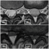
Axial appearance of epidural fat on T2-weighted MR images. Fat accumulation at each intervertebral disc level from L1–2 to L4–5 was classified as follows: absent (A), concave (B), flat (C), or convex (D).
Figure 5
Figure 5
The “Y” sign: when epidural fat was severely accumulated in the spinal canal, it compressed the dural sac into a Y-shape and named it the “Y” sign.
Figure 6
Figure 6
Sagittal grading of epidural fat on T1-weighted MR images. (A) Grade 1 was defined as epidural fat observed within the border between the anterosuperior edges of the upper and lower neighboring neural arches. (B) Grade 2 was defined as fat observed over the border at the middle but not at the edges of neural arches on both sides. (C) Grade 3 was defined as fat observed over the border at the edges of neural arches on at least one side.
Figure 7
Figure 7
Patient, male, 67 years old, with recurrent low back pain for 2 years, recurrence and exacerbation for 3 months, lumbosacral pressure and percussion. Magnetic resonance imaging: bilateral ligamentum flavum hypertrophy and spinal stenosis, and the cauda equina nerve in the sagittal position at T2WI showed flaccid, tortuous, and entangled high-signal shadows.
Figure 8
Figure 8
Stacked histograms of single factor analyses of LSS combined RNRs.
Figure 9
Figure 9
Patient, male, 54 years old, with lumbosacral distension, lumbar 3-sacral 1 paravertebral and interspinous tenderness, epidural fat sagittal grading of grade III, and varying degrees of indentation at the posterior edge of the dural sac, the Y-shaped sign(+), with the cauda equina showing dilated, curved and entangled in the sagittal plane of T2WI.
Figure 10
Figure 10
Predicted ROC curves for LSS combined with RNRs.

Similar articles

References

    1. Verbiest H. A radicular syndrome from developmental narrowing of the lumbar vertebral canal. J Bone Joint Surg Br. (1954) 36-B:230–7. doi: 10.1302/0301-620X.36B2.230, PMID: - DOI - PubMed
    1. Cressman MR, Pawl RP. Serpentine myelographic defect caused by a redundant nerve root. Case report. J Neurosurg. (1968) 28:391–3. doi: 10.3171/jns.1968.28.4.0391, PMID: - DOI - PubMed
    1. Nathani KR, Naeem K, Rai HH, Barakzai MD, Iftikhar H, Khan SA, et al. . Role of redundant nerve roots in clinical manifestations of lumbar spine stenosis. Surg Neurol Int. (2021) 12:218. doi: 10.25259/SNI_59_2021 - DOI - PMC - PubMed
    1. Papavero L, Ali N, Schawjinski K, Holtdirk A, Maas R, Ebert S. The prevalence of redundant nerve roots in standing positional MRI decreases by half in supine and almost to zero in flexed seated position: a retrospective cross-sectional cohort study. Neuroradiology. (2022) 64:2191–201. doi: 10.1007/s00234-022-03047-z, PMID: - DOI - PMC - PubMed
    1. Hur JW, Hur JK, Kwon TH, Park YK, Chung HS, Kim JH. Radiological significance of ligamentum flavum hypertrophy in the occurrence of redundant nerve roots of central lumbar spinal stenosis. J Korean Neurosurg Soc. (2012) 52:215–20. doi: 10.3340/jkns.2012.52.3.215, PMID: - DOI - PMC - PubMed

LinkOut - more resources