

Anesthetic Management and Considerations in a Rare Case of Parietal Bone Hemangioma
- PMID: 38860097
- PMCID: PMC11164296
- DOI: 10.7759/cureus.60098
Anesthetic Management and Considerations in a Rare Case of Parietal Bone Hemangioma
Abstract
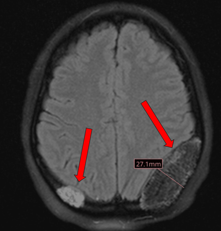
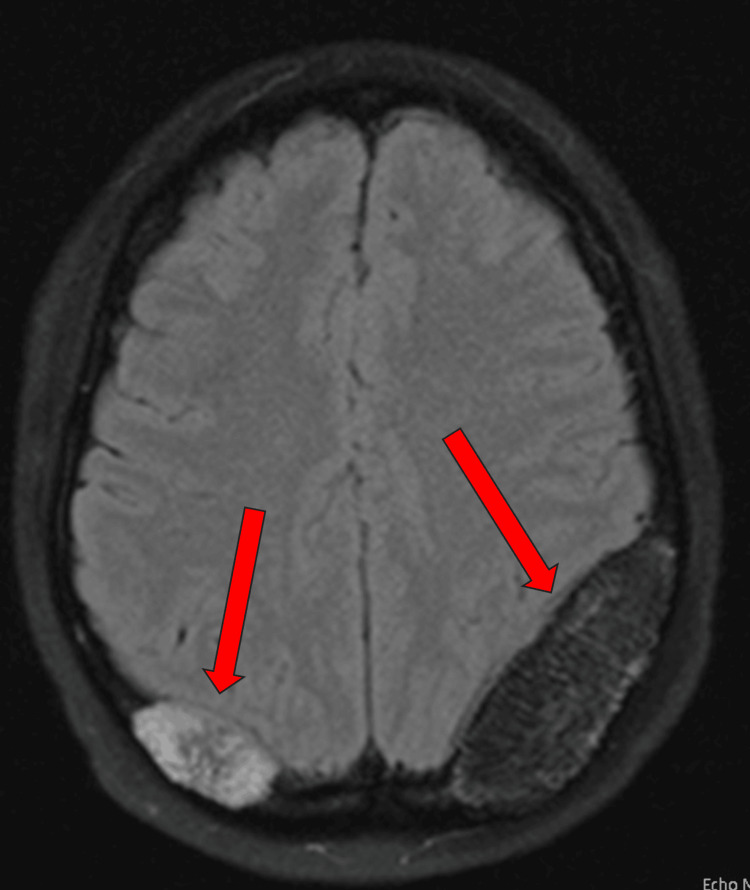
Parietal bone hemangiomas represent a minority of diagnosed brain tumors. These lesions require careful management under anesthesia due to their vascularity and cranial location. We discuss a 31-year-old female with chronic headaches who underwent surgery for the removal of a large parietal bone hemangioma, necessitating considerations for stable hemodynamics, intracranial pressure (ICP), and bleeding risks. There is no standard anesthetic for these cases, so a mixed anesthetic approach was used, combining intravenous anesthesia with sevoflurane, aimed at optimizing control during the procedure.
Keywords: anesthetic considerations; neuroanesthesia; parietal bone hemangiomas; total intravenous anesthetic; volatile anesthetic.
Copyright © 2024, Koenig et al.
Conflict of interest statement
The authors have declared that no competing interests exist.
Figures
References
-
- Primary intraosseous cavernous hemangioma of the cranium: a systematic review of the literature. Alexiou GA, Lampros M, Gavra MM, Vlachos N, Ydreos J, Boviatsis EJ. World Neurosurg. 2022;164:323–329. - PubMed
-
- Surgical treatment of aggressive intraosseous cavernous hemangioma in maxilla through surgical resection. Ribeiro IL, Vasconcelos ML, Panjwani CM, Silva HL, da Hora Sales PH, Andrade CS. J Craniofac Surg. 2020;31:0–8. - PubMed
-
- Occipital intraosseous hemangioma over torcula: unusual presentation with raised intracranial pressure. Rao KV, Beniwal M, Vazhayil V, Somanna S, Yasha TC. World Neurosurg. 2017;108:999–995. - PubMed
Publication types
LinkOut - more resources
Full Text Sources