

Magnetic Resonance Imaging of Radiation-Induced Brachial Plexopathy
- PMID: 38860104
- PMCID: PMC11163868
- DOI: 10.7759/cureus.60067
Magnetic Resonance Imaging of Radiation-Induced Brachial Plexopathy
Abstract
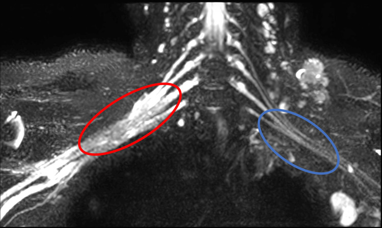
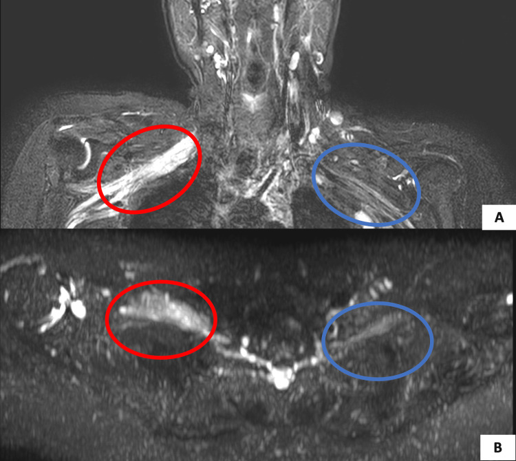
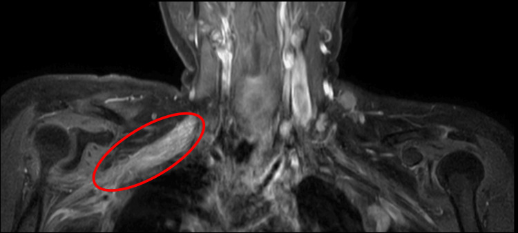
This report illustrates the case of a 37-year-old woman following chemoradiotherapy for invasive ductal carcinoma of the right breast. The patient underwent surgery and received a radiation dose of 50 gray to the chest wall and 45 gray to the regional lymph nodes in 25 total fractions. She developed motor and sensory weakness in the right upper limb eight years after treatment. Brachial plexus neuropathy in cancer patients may result from either trauma to the plexus during surgery, the spread of cancer, or radiation therapy, and distinguishing between them may be difficult. The case highlights the importance of recognizing the signs, symptoms, and possible differential diagnosis of radiation-induced brachial plexopathy in cancer patients post-radiation therapy. It emphasizes the role of magnetic resonance imaging in the careful assessment and diagnosis of such a case.
Keywords: brachial plexus injury; non traumatic plexopathy; oncoradiology; palliative radiation therapy; radiation-induced brachial plexopathy.
Copyright © 2024, Shetty et al.
Conflict of interest statement
The authors have declared that no competing interests exist.
Figures
Similar articles
-
A Case of Radiation-Induced Brachial Plexopathy Below the Tolerance Dose.Cureus. 2024 Jan 15;16(1):e52283. doi: 10.7759/cureus.52283. eCollection 2024 Jan. Cureus. 2024. PMID: 38357089 Free PMC article.
-
Treatment of radiation-induced brachial plexopathy with omentoplasty.Autops Case Rep. 2020 Sep 2;10(3):e2020202. doi: 10.4322/acr.2020.202. Autops Case Rep. 2020. PMID: 33344306 Free PMC article.
-
The role of surgery in the management of radiation-induced brachial plexopathy: a systematic review.J Hand Surg Eur Vol. 2024 Apr;49(4):490-498. doi: 10.1177/17531934231197794. Epub 2023 Sep 8. J Hand Surg Eur Vol. 2024. PMID: 37684017
-
Periscapular amputation as treatment for brachial plexopathy secondary to recurrent breast carcinoma: a case series and review of the literature.Eur J Surg Oncol. 2013 Dec;39(12):1325-31. doi: 10.1016/j.ejso.2013.10.005. Epub 2013 Oct 23. Eur J Surg Oncol. 2013. PMID: 24176674 Review.
-
Dose constraints to prevent radiation-induced brachial plexopathy in patients treated for lung cancer.Int J Radiat Oncol Biol Phys. 2012 Mar 1;82(3):e391-8. doi: 10.1016/j.ijrobp.2011.06.1961. Int J Radiat Oncol Biol Phys. 2012. PMID: 22284035 Free PMC article.
References
-
- Brachial plexopathy after breast cancer: a persistent late effect of radiotherapy. Harris SR. PM R. 2024;16:85–91. - PubMed
-
- Troublesome symptoms in cancer survivors: fatigue, insomnia, neuropathy, and pain. Pachman DR, Barton DL, Swetz KM, Loprinzi CL. J Clin Oncol. 2012;30:3687–3696. - PubMed
-
- Brachial plexus lesions in patients with cancer=100 cases. Kori SH, Foley KM, Posner JB. Neurology. 1981;31:45. - PubMed
-
- Radiation-induced brachial plexopathy: review. Complication without a cure. Schierle C, Winograd JM. J Reconstr Microsurg. 2004;20:149–152. - PubMed
Publication types
LinkOut - more resources
Full Text Sources