

Pioneering nanomedicine in orthopedic treatment care: a review of current research and practices
- PMID: 38860139
- PMCID: PMC11163052
- DOI: 10.3389/fbioe.2024.1389071
Pioneering nanomedicine in orthopedic treatment care: a review of current research and practices
Abstract
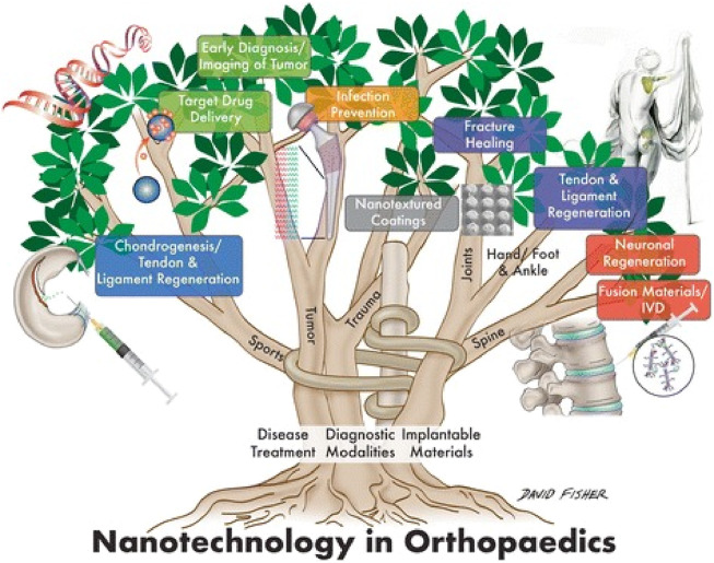
A developing use of nanotechnology in medicine involves using nanoparticles to administer drugs, genes, biologicals, or other materials to targeted cell types, such as cancer cells. In healthcare, nanotechnology has brought about revolutionary changes in the treatment of various medical and surgical conditions, including in orthopedic. Its clinical applications in surgery range from developing surgical instruments and suture materials to enhancing imaging techniques, targeted drug delivery, visualization methods, and wound healing procedures. Notably, nanotechnology plays a significant role in preventing, diagnosing, and treating orthopedic disorders, which is crucial for patients' functional rehabilitation. The integration of nanotechnology improves standards of patient care, fuels research endeavors, facilitates clinical trials, and eventually improves the patient's quality of life. Looking ahead, nanotechnology holds promise for achieving sustained success in numerous surgical disciplines, including orthopedic surgery, in the years to come. This review aims to focus on the application of nanotechnology in orthopedic surgery, highlighting the recent development and future perspective to bridge the bridge for clinical translation.
Keywords: bone regeneration; nanoparticle; nanotechnology; orthopaedic surgery; orthopedic.
Copyright © 2024 Liang, Zhou, Zhang, Bai, Long, Jiang, Liu, Xia, Jiang, Zhang and Zhao.
Conflict of interest statement
The authors declare that the research was conducted in the absence of any commercial or financial relationships that could be construed as a potential conflict of interest.
Figures
Similar articles
-
A mini-review on the emerging role of nanotechnology in revolutionizing orthopedic surgery: challenges and the road ahead.Front Bioeng Biotechnol. 2023 May 16;11:1191509. doi: 10.3389/fbioe.2023.1191509. eCollection 2023. Front Bioeng Biotechnol. 2023. PMID: 37260831 Free PMC article. Review.
-
Recent developments in nanomaterials for upgrading treatment of orthopedics diseases.Front Bioeng Biotechnol. 2023 Aug 9;11:1221365. doi: 10.3389/fbioe.2023.1221365. eCollection 2023. Front Bioeng Biotechnol. 2023. PMID: 37621999 Free PMC article. Review.
-
Targeting cancer cells with nanotherapeutics and nanodiagnostics: Current status and future perspectives.Semin Cancer Biol. 2021 Feb;69:52-68. doi: 10.1016/j.semcancer.2020.01.011. Epub 2020 Jan 31. Semin Cancer Biol. 2021. PMID: 32014609 Review.
-
Translation of nanotechnology-based implants for orthopedic applications: current barriers and future perspective.Front Bioeng Biotechnol. 2023 Aug 22;11:1206806. doi: 10.3389/fbioe.2023.1206806. eCollection 2023. Front Bioeng Biotechnol. 2023. PMID: 37675405 Free PMC article. Review.
-
Current developments and future perspectives of nanotechnology in orthopedic implants: an updated review.Front Bioeng Biotechnol. 2024 Mar 18;12:1342340. doi: 10.3389/fbioe.2024.1342340. eCollection 2024. Front Bioeng Biotechnol. 2024. PMID: 38567086 Free PMC article. Review.
References
-
- Alpuim P., Filonovich S., Costa C., Rocha P., Vasilevskiy M., Lanceros-Mendez S., et al. (2008). Fabrication of a strain sensor for bone implant failure detection based on piezoresistive doped nanocrystalline silicon. J. non-crystalline solids 354 (19-25), 2585–2589. 10.1016/j.jnoncrysol.2007.09.094 - DOI
Publication types
LinkOut - more resources
Full Text Sources