. 2024 Aug 1;327(2):G175-G187.
doi: 10.1152/ajpgi.00063.2024. Epub 2024 Jun 11.

FXR deletion attenuates intestinal barrier dysfunction in murine acute intestinal inflammation

Affiliations

FXR deletion attenuates intestinal barrier dysfunction in murine acute intestinal inflammation

MaKayla L O'Guinn et al. Am J Physiol Gastrointest Liver Physiol. .

Abstract

Accumulating literature suggests that the farnesoid-X receptor (FXR), a nuclear bile acid receptor best known for its role in bile acid homeostasis, is also a potent context-dependent regulator of inflammation. FXR may thus be relevant to several intestinal disease states including inflammatory bowel disease, necrotizing enterocolitis, and sepsis. In this study, we tested the effects of FXR deletion on acute murine intestinal inflammation. We found that FXR knockout (KO) mice were protected from intestinal injury and barrier dysfunction induced by lipopolysaccharide (LPS) injection, dithizone (DI)/Klebsiella, and cecal ligation/puncture models. In the LPS model, RNA sequencing and qPCR analysis showed that this protection correlated with substantial reduction in LPS-induced proinflammatory gene expression, including lower tissue levels of Il1a, Il1b, and Tnf. Examining functional effects on the epithelium, we found that LPS-induced tight junctional disruption as assessed by internalization of ZO-1 and occludin was ameliorated in FXR KO animals. Taken together, these data suggest a role for FXR in the intestinal barrier during inflammatory injury.NEW & NOTEWORTHY Intestinal barrier failure is a hallmark in gut-origin sepsis. We demonstrate that the intestinal barriers of farnesoid-X receptor (FXR) knockout (KO) animals are protected from inflammatory insult using multiple models of acute intestinal inflammation. This protection is due to decreased inflammatory cytokine production and maintenance of tight junctional architecture seen within the KO animals. This is the first report of FXR deletion being protective to the intestinal barrier.

Keywords: FXR; LPS; inflammation; intestinal barrier; tight junctions.


Conflict of interest statement

No conflicts of interest, financial or otherwise, are declared by the authors.

Figures

None
Graphical abstract
Figure 1.
Figure 1.
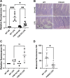
Farnesoid-X receptor (FXR) deletion in mice protects against intestinal barrier leakage. AD: wild-type (WT) and FXR knockout (KO) mice were injected intraperitoneally with lipopolysaccharide (LPS) and gavaged with fluorescein isothiocyanate (FITC)-dextran 4 kDa 16 h or 2 h before collection. A: relative FITC fluorescence in serum of 16-h group normalized to respective WT control. B: terminal ileum hematoxylin and eosin stains; arrowhead indicates villus injury and basement membrane separation. C: relative FITC fluorescence in serum 2 h after LPS normalized to respective WT control. D: measured FITC fluorescence in serum of 16-h group following 2 wk of bedding swap. Two-hour data are independent experiments from 16-h data. Each dot represents one animal. *P < 0.05; **P < 0.01; ***P < 0.001. Graphs displayed as means ± standard deviation.
Figure 2.
Figure 2.
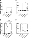
Farnesoid-X receptor (FXR) knockout (KO) mice are protected from acute inflammatory injury in dithizone (DI)/Klebsiella and cecal ligation puncture models of injury. Wild-type (WT) and FXR KO mice were given dithizone (DI) intraperitoneally and oral Klebsiella pneumoniae (Kleb) to represent a model of acute intestinal injury. A: standard injury grading of each of the controls (untreated control, Klebsiella, dithizone, dithizone + Klebsiella, and FXR KO dithizone). B: representative images of terminal ilium stained with hematoxylin and eosin used for histologic grading. Arrowhead, villus injury; arrow, basement membrane separation, both of which are absent in FXR KO. C: WT and FXR KO mice were subjected to cecal ligation puncture (CLP) and gavaged with fluorescein isothiocyanate (FITC) dextran 4 kDa. FITC fluorescence was measured from serum. D: hematoxylin and eosin histology of cecal ligation puncture. B and D: arrowhead, damage to villus; arrow, separation of basement membrane. Graphs displayed as means ± standard deviation.*P < 0.05; **P < 0.01.
Figure 3.
Figure 3.
Bulk RNA sequencing of wild-type (WT) and full body farnesoid-X receptor (FXR) knockout (FXR KO) mice given intraperitoneal lipopolysaccharide (LPS). A: principal component analysis (PCA) plot of RNA WT and FXR KO mice given LPS or normal saline (NS) control. B: heatmaps comparing gene expression between WT and FXR KO mice that have been treated with LPS or with normal saline control. C: heatmap comparing log2 fold change in gene expression between WT and FXR KO mice treated with LPS. Enrichment of Gene Ontology (GO) Biological Process terms in FXR KO mice compared with WT mice that have been injected with normal saline control (D) or LPS (E). Overrepresented and underrepresented terms were determined using statistically significant differentially expressed genes (adjusted P value <0.05) with positive or negative log2 fold changes, respectively.
Figure 4.
Figure 4.
Farnesoid-X receptor (FXR) loss does not affect MAPK phosphorylation. Wild-type (WT) and FXR knockout (KO) mice were given normal saline or lipopolysaccharide (LPS) at 16 h (A) or 2 h (B) before harvesting mucosal scrapings from terminal ileum. Lysates were then run on Western blot and probed for phospho- and total ERK. n ≥ 6. Graphs displayed as means ± standard deviation. *P < 0.05.
Figure 5.
Figure 5.
Farnesoid-X receptor (FXR) deletion affects pro- and anti-inflammatory cytokine in a time-sensitive manner. Mice [wild-type (WT) or FXR knockout (KO)] were injected with 200 µL of lipopolysaccharide (LPS) at 30 mg/kg diluted in normal saline or normal saline as a control. Mucosal scrapings from terminal ileum were collected at either 16 h or 2-h postinjection. AE: qPCR data analysis is normalized to the wild-type normal saline (WT NS) mouse using Hprt for reference. Each dot represents one animal. Graphs displayed as means ± standard deviation. *P < 0.05; **P < 0.01; ***P < 0.001.
Figure 6.
Figure 6.
Farnesoid-X receptor (FXR) deletion delays the expression of chemokines. Mice [wild-type (WT) or FXR knockout (KO]) were injected with 200 µL of lipopolysaccharide (LPS) at 30 mg/kg diluted in normal saline (NS) or normal saline as a control. Epithelial scrapings from terminal ileum were collected at either 16 h or 2-h postinjection. Cxcl1 at 16 h (A) and 2-h qPCR data analysis (B) is normalized to the wild-type normal saline (WT NS) group using Hprt for reference. Ccl2 at 16 h (C) and 2-h qPCR data analysis (D) is normalized to the wild-type (WT NS) group using Hprt for reference. Each dot represents one animal. Graphs displayed as means ± standard deviation. *P < 0.05; **P < 0.01.
Figure 7.
Figure 7.
Farnesoid-X receptor (FXR) deletion leads to maintained occludin distribution after lipopolysaccharide (LPS) treatment. Mice [wild-type (WT) or FXR knockout (KO)] were injected with 200 µL of LPS at 30 mg/kg diluted in normal saline (NS) or normal saline as a control. Segments of terminal ileum and mucosal scrapings from terminal ileum were collected at either 16 h or 2 h postinjection. A: relative amount of occludin by Western blot of wild-type normal saline (WT NS) and FXR KO normal saline (FXR KO NS) with representative blot; n = 6. B: representative immunofluorescence micrographs of mouse terminal ileum staining for occludin (green) and nuclei stained with DAPI (blue) from wild-type and FXR KO mice at 16 h and 2 h after being treated with or without LPS. The thin white arrow indicates the normal presentation of occludin. Thick white arrow indicates the lack of apical occludin after LPS. C: proportion of positive apical staining of occludin at 2 h. D: negative control with only secondary antibody (alexa-fluor 488) for reference. Graphs displayed as means ± standard deviation. *P < 0.05; **P < 0.01.
Figure 8.
Figure 8.
Farnesoid-X receptor (FXR) deletion leads to maintained zonula occludens-1 (ZO-1) distribution after lipopolysaccharide (LPS) treatment. Mice [wild-type (WT) or FXR knockout (KO)] were injected with 200 µL of LPS at 30 mg/kg diluted in normal saline (NS) or normal saline as a control. Segments of terminal ileum and mucosal scrapings from terminal ileum were collected at either 16 h or 2 h postinjection. A: relative amount of ZO-1 by Western blot of wild-type normal saline (WT NS) and FXR KO normal saline (FXR KO NS) with representative blot; n = 6. B: representative immunofluorescence micrographs of mouse terminal ileum staining for ZO-1 (red) and nuclei stained with DAPI (blue) from wild-type and FXR KO mice at 16 h and 2 h after being treated with or without LPS. The thin white arrow indicates normal presentation of ZO-1 with apical localization. Thick white arrow indicates the loss of apical ZO-1 after LPS. C: proportion of positive apical staining of ZO-1 at 2 h. D: negative control with only secondary antibody (alexa-fluor 568) for reference. Graphs displayed as means ± standard deviation. *P < 0.05; ***P < 0.001.
Figure 9.
Figure 9.
Farnesoid-X receptor (FXR) deletion does not alter E-cadherin distribution. Mice [wild-type (WT) or FXR knockout (KO)] were injected with 200 µL of lipopolysaccharide (LPS) at 30 mg/kg diluted in normal saline (NS) or normal saline as a control. Segments of terminal ileum and mucosal scrapings from terminal ileum were collected at either 16 h or 2 h postinjection. A: relative amount of E-cadherin by Western blot of wild-type normal saline (WT NS) and FXR KO normal saline (FXR KO NS) with representative blot; n = 6. B: representative immunofluorescence micrographs of mouse terminal ileum staining for E-cadherin (green) and nuclei stained with DAPI (blue) from wild-type and FXR KO mice at 16 h and 2 h after being treated with or without LPS. Scale bar is 20 µm and representative of all images. All images obtained with ×20 magnification. Graphs displayed as means ± standard deviation.

Comment in

Similar articles

Cited by

References

    1. Han X, Fink MP, Yang R, Delude RL. Increased iNOS activity is essential for intestinal epithelial tight junction dysfunction in endotoxemic mice. Shock 21: 261–270, 2004. doi: 10.1097/01.shk.0000112346.38599.10. - DOI - PubMed
    1. Guo S, Al-Sadi R, Said HM, Ma TY. Lipopolysaccharide causes an increase in intestinal tight junction permeability in vitro and in vivo by inducing enterocyte membrane expression and localization of TLR-4 and CD14. Am J Pathol 182: 375–387, 2013. doi: 10.1016/j.ajpath.2012.10.014. - DOI - PMC - PubMed
    1. Bein A, Zilbershtein A, Golosovsky M, Davidov D, Schwartz B. LPS induces hyper-permeability of intestinal epithelial cells. J Cell Physiol 232: 381–390, 2017. doi: 10.1002/jcp.25435. - DOI - PubMed
    1. Bain CC, Mowat AM. The monocyte-macrophage axis in the intestine. Cell Immunol 291: 41–48, 2014. doi: 10.1016/j.cellimm.2014.03.012. - DOI - PMC - PubMed
    1. Bain CC, Scott CL, Uronen-Hansson H, Gudjonsson S, Jansson O, Grip O, Guilliams M, Malissen B, Agace WW, Mowat AM. Resident and pro-inflammatory macrophages in the colon represent alternative context-dependent fates of the same Ly6Chi monocyte precursors. Mucosal Immunol 6: 498–510, 2013. doi: 10.1038/mi.2012.89. - DOI - PMC - PubMed

Substances

LinkOut - more resources