. 2024 Jul 22;52(13):8003-8016.
doi: 10.1093/nar/gkae479.

OptoLacI: optogenetically engineered lactose operon repressor LacI responsive to light instead of IPTG

Affiliations

OptoLacI: optogenetically engineered lactose operon repressor LacI responsive to light instead of IPTG

Meizi Liu et al. Nucleic Acids Res. .

Abstract

Optogenetics' advancement has made light induction attractive for controlling biological processes due to its advantages of fine-tunability, reversibility, and low toxicity. The lactose operon induction system, commonly used in Escherichia coli, relies on the binding of lactose or isopropyl β-d-1-thiogalactopyranoside (IPTG) to the lactose repressor protein LacI, playing a pivotal role in controlling the lactose operon. Here, we harnessed the light-responsive light-oxygen-voltage 2 (LOV2) domain from Avena sativa phototropin 1 as a tool for light control and engineered LacI into two light-responsive variants, OptoLacIL and OptoLacID. These variants exhibit direct responsiveness to light and darkness, respectively, eliminating the need for IPTG. Building upon OptoLacI, we constructed two light-controlled E. coli gene expression systems, OptoE.coliLight system and OptoE.coliDark system. These systems enable bifunctional gene expression regulation in E. coli through light manipulation and show superior controllability compared to IPTG-induced systems. We applied the OptoE.coliDark system to protein production and metabolic flux control. Protein production levels are comparable to those induced by IPTG. Notably, the titers of dark-induced production of 1,3-propanediol (1,3-PDO) and ergothioneine exceeded 110% and 60% of those induced by IPTG, respectively. The development of OptoLacI will contribute to the advancement of the field of optogenetic protein engineering, holding substantial potential applications across various fields.


Figures

Graphical Abstract
Graphical Abstract
Figure 1.
Figure 1.
Design and construction of the light-controlled repressor OptoLacI. (A) The design of the insertion sites of the photosensitive domain based on the structure of LacI (PDB ID:1LBG). Loop1 (D152-P155) and loop2 (Q311-N316) are in close proximity to the DNA-binding domain of LacIW220F, exerting an influence on the binding interaction between LacI and the operator. Loop3 (N333-P339) is positioned close to the oligomeric domain, affecting the oligomerization state of LacI. (B) The photosensitive domains used in this study. AsLOV2 domain contains the PAS core domain and the Ja helix at the N-terminus (PDB ID: 2V1A). cpLOV27 is a circular arrangement mutant of AsLOV2. The N-terminus of AsLOV2 is connected to the C-terminus via the GGGSGGS linker and is interrupted between the 525th and 526th residues (E525 and R526). (C) Schematic diagrams of the mechanisms for inducing target gene expression using OptoLacIL under blue light (top) and OptoLacID under dark condition (bottom), respectively. (D) OD600-normalized GFP fluorescence intensity of BL21(DE3) carrying wild type LacI (Control-1) induced by IPTG. OD600-normalized GFP fluorescence intensity of BL21(DE3) carrying variant LacIW220F (Control-2), and OptoLacI variants (LacIW220F, 153-cpLOV27, LacI W220F, 311-LOV2, LacI W220F, 152-cpLOV27 and LacIW220F, 335-LOV2, respectively) under blue light and dark conditions. Open circles represent individual data points. Error bars represent the standard deviation of at least three biological replicates.
Figure 2.
Figure 2.
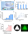
The OptoE.coliLight system excels in tunability, induced rigidity, and spatial rigidity. (A) Schematic diagrams of the mechanism of the OptoE.coliLight system. (B) Blue light induced GFP fluorescence of OptoBL21Light strain transformed with plasmids carrying different numbers of operator lacO1. (C) Blue light intensity-dependent induction of GFP expression by the OptoE.coliLight system. (D) GFP fluorescence intensity induced by various blue light pulses in the OptoE.coliLight system. The blue light pulse modes: full blue light exposure, 1 s off / 1000 s on, 10 s off /1000 s on, 100 s off /1000 s on, and full darkness. (E) Comparison of GFP fluorescence intensity induced by different IPTG doses (0, 0.001, 0.005, 0.01, 0.1, 0.5, and 1.0 mM) in a conventional BL21(DE3) strain with an expression vector (pET-28a). (F) The OptoE.coliLight system enables the production of high-contrast E. coli graphics. The triangular area used to expose to blue light (left). A triangular pattern generated by blue light induced GFP expression (right). The GFP fluorescence intensity was normalized to OD600. Open circles represent individual data points. Error bars represent the standard deviation of at least three biological replicates.
Figure 3.
Figure 3.
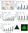
The OptoE.coliDark system exhibited the advantages in tunability, induced rigidity, and spatial rigidity. (A) The schematic diagrams of the mechanism of the OptoE.coliDark system. (B) Optimization of the number of operators lacO1 in the OptoE.coliDark system. (C) Substituting Lys84 with cysteine (C), serine (S), threonine (T), glutamate (E) or isoleucine (I) significantly enhanced GFP expression in the OptoE.coliDark system under dark condition. (D) Blue light intensity-dependent induction of GFP leakage expression by the OptoE.coliDark system. (E) The difference in GFP fluorescence intensity between the different OD600 was particularly significant within the first 4 h. The GFP fluorescence intensity was normalized to OD600. Open circles represent individual data points. Error bars represent the standard deviation of at least three biological replicates. (F) The OptoE.coliDark system allows the production of high-contrast E. coli graphics. The triangular photomask used to produce the E. coli picture (left), the triangular pattern generated by dark-induced GFP expression for 24 h (right).
Figure 4.
Figure 4.
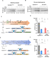
Application of the OptoE.coliDark system in protein production and control of metabolic pathways. (A, B) Production profiles of alkaline protease (A) and glucose dehydrogenase (B) using the OptoE.coliDark system. Inductions were initiated by switching the culture from blue light to darkness at their optimal cell density (OD600= 0.1). The IPTG-induced system served as a control, with induction triggered by adding 1 mM IPTG at the optimal cell density (OD600= 0.5). Samples were collected at 0, 2, 3, 4, 6, 9, and 12 h of induction to monitor target protein production. (C) Optogenetic control of the 1,3-propanediol (1,3-PDO) biosynthetic pathway using the OptoE.coliDark system. The OptoLacID protein regulates the expression of T7 RNA polymerase in the OptoBL21Dark strain, along with glycerol dehydrogenase (KpdhaB), glycerol dehydrogenase reactivase (KpgdrAB), and alcohol dehydrogenase (EcyqhD) on the plasmid. (D) Comparison of the OptoE.coliDark system with IPTG induction system for 1,3-PDO production. (E) Optogenetic control of the ergothioneine (EGT) biosynthetic pathway using the OptoE.coliDark system. The OptoLacID protein controls the expression of T7 RNA polymerase in the OptoBL21Dark strain, as well as S-adenosylmethionine-dependent methyltransferase (EgtD), C-S lyase (EgtE), and methyltransferase-sulfoxide synthase (Egt1) on the plasmid. (F) Comparison of the EGT production in the OptoE.coliDark system and the IPTG-induced system. Open circles represent individual data points. Error bars represent the standard deviation of at least three biological replicates.

Similar articles

Cited by

References

    1. Fang H., Li D., Kang J., Jiang P., Sun J., Zhang D Metabolic engineering of Escherichia coli for de novo biosynthesis of vitamin B12. Nat. Commun. 2018; 9:4917. - PMC - PubMed
    1. Wu Q.-Y., Huang Z.-Y., Wang J.-Y., Yu H.-L., Xu J.-H. Construction of an Escherichia coli cell factory to synthesize taxadien-5α-ol, the key precursor of anti-cancer drug paclitaxel. Bioresour. 2022; 9:82. - PMC - PubMed
    1. Zou X., Xiao X., Mo Z., Ge Y., Jiang X., Huang R., Li M., Deng Z., Chen S., Wang L. et al. . Systematic strategies for developing phage resistant Escherichia coli strains. Nat. Commun. 2022; 13:4491. - PMC - PubMed
    1. Lewis M., Chang G., Horton N.C., Kercher M.A., Pace H.C., Schumacher M.A., Brennan R.G., Lu P. Crystal structure of the lactose operon repressor and its complexes with DNA and inducer. Science. 1996; 271:1247–1254. - PubMed
    1. Jacob F., Monod J. Genetic regulatory mechanisms in the synthesis of proteins. J. Mole. Biol. 1961; 3:318–356. - PubMed

MeSH terms

LinkOut - more resources