. 2024 Jul 17;15(7):e0120924.
doi: 10.1128/mbio.01209-24. Epub 2024 Jun 11.

IFI207, a young and fast-evolving protein, controls retroviral replication via the STING pathway

Affiliations

IFI207, a young and fast-evolving protein, controls retroviral replication via the STING pathway

Eileen A Moran et al. mBio. .

Abstract

Mammalian AIM-2-like receptor (ALR) proteins bind nucleic acids and initiate production of type I interferons or inflammasome assembly, thereby contributing to host innate immunity. In mice, the Alr locus is highly polymorphic at the sequence and copy number level, and we show here that it is one of the most dynamic regions of the genome. One rapidly evolving gene within this region, Ifi207, was introduced to the Mus genome by gene conversion or an unequal recombination event a few million years ago. Ifi207 has a large, distinctive repeat region that differs in sequence and length among Mus species and even closely related inbred Mus musculus strains. We show that IFI207 controls murine leukemia virus (MLV) infection in vivo and that it plays a role in the STING-mediated response to cGAMP, dsDNA, DMXXA, and MLV. IFI207 binds to STING, and inclusion of its repeat region appears to stabilize STING protein. The Alr locus and Ifi207 provide a clear example of the evolutionary innovation of gene function, possibly as a result of host-pathogen co-evolution.IMPORTANCEThe Red Queen hypothesis predicts that the arms race between pathogens and the host may accelerate evolution of both sides, and therefore causes higher diversity in virulence factors and immune-related proteins, respectively . The Alr gene family in mice has undergone rapid evolution in the last few million years and includes the creation of two novel members, MndaL and Ifi207. Ifi207, in particular, became highly divergent, with significant genetic changes between highly related inbred mice. IFI207 protein acts in the STING pathway and contributes to anti-retroviral resistance via a novel mechanism. The data show that under the pressure of host-pathogen coevolution in a dynamic locus, gene conversion and recombination between gene family members creates new genes with novel and essential functions that play diverse roles in biological processes.

Keywords: AIM-2-like receptor; evolution of gene function; nucleic acid sensor; viral immunity.


Conflict of interest statement

The authors declare no conflict of interest.

Figures

Fig 1
Fig 1
Ifi207 structure. (A) Genome structure of the Alr locus in inbred mouse strains. Based on gene structure and phylogeny, the nine family members with variant copy numbers are indicated by the color blocks. Ifi208, Ifi207, Ifi204, and one copy of Ifi203 are encoded in a stable contig, and other regions in the Alr locus showed significant numbers of inversions, recombinations, and duplications. (B) Genome structure of the Alr locus in other rodent species and humans. Ifi207, MndaL, and Ifi202 are only found in the Mus genus. Some of the genes in Onychomys and Peromyscus were annotated tandemly, probably caused by low annotation quality. (C) IFI207 protein structure in different Mus genus members. Shown for comparison is the structure of IFI202, IFI204, MNDA, and IFI203 (see Tables S2 and S3 for sequences). Similar PYRIN domains are indicated by color, as are the HIN-A and HIN-B domains. The long linker sequence between these domains in IFI203 and IFI207 is indicated by a blue bar. (D) Comparison of C57BL/6 IFI207 (E9Q3L4) and IFI203 (E9QAN9) predicted protein structures. Structures were downloaded from AlphaFold (https://alphafold.ebi.ac.uk/). Domains are colored as follows: pyrin domain, pink; PYD-proximal repeat, brown; HIN-proximal repeat, teal; and HIN-B, blue.
Fig 2
Fig 2
IFI207 KO mice are more susceptible to MLV infection. (A) Pups of the indicated genotype were infected with 2,000 PFU of MLV (MLV or MLVgGag). At 16 dpi, splenic viral titers were determined on NIH3T3 cells by infectious center (IC) assays. Significance was determined by two-tail Mann-Whitney test (**, P < 0.003; ****, P < 0.0001). Right panel: RT-qPCR analysis to verify Ifi207 RNA levels in mice. Symbols represent individual mice. Significance was determined by one-way ANOVA (***, P < 0.002; ****, P < 0.0001). (B) BMDMs from the indicated mice were transfected with the indicated concentrations of ISD or cGAMP for 3 h. Ifnβ RNA levels were quantified by RT-qPCR. Shown are the averages of three independent experiments. (C) Primary fibroblasts from IFI207 KO and STINGmut mice were transfected with 8 µg/mL of ISD or cGAMP or 100 µg/mL DMXAA, and at 3 h post-transfection, Ifnβ RNA levels were measured. Shown is the average of three experiments (ISD and cGAMP) or two experiments (DMXAA). Significance was determined by two-way ANOVA (****, P < 0.0001).
Fig 3
Fig 3
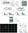
IFI207-deficient cells show decreased STING expression and diminished activation of the STING pathway. (A) NIH3T3 cells were transfected with the indicated siRNAs, and Western blots for endogenous STING were performed. Two concentrations of the 207 siRNA were used (10 and 30 pmol). Duplicate experimental replicates are shown. Quantification of Western blots from three independent experiments and Ifi207 knockdown (KD) verification by RT-qPCR are shown to the right (average + SD). One-way ANOVA was used (*, P ≤ 0.05; ****, P < 0.0001; ns, not significant). siC, control siRNA; U, untransfected. (B) BMDMs were transfected with ISD, and at 2, 4, and 8 h post-transfection, protein expression was analyzed by Western blot with the indicated antibodies. Blots were stripped and probed with anti-actin to control loading. (C and D) Fibroblasts (C) and macrophages (D) from IFI207 KO and wild-type mice were treated with 100 µg/mL DMXAA, and Western blots examining STING and phospho-STING levels were measured at the indicated times. Shown below the graph is the quantification of three independent experiments. Two-way ANOVA was used (*, P ≤ 0.03; **, P ≤ 0.001; ****, P < 0.0001; ns, not significant). (E) NIH3T3 cells were transfected with siRNAs targeting IFI207 or STING. Non-targeting (siC) siRNA was also used. At 48 h post-transfection, the cells were infected with MLVgGag for 2 h (MOI = 5). Protein expression was examined by Western blot with the indicated antibodies. Right panel: Ifi207 KD was verified by RT-qPCR. Average + SD from two independent experiments is shown. (F) Primary fibroblasts were transfected with 8 µg/mL ISD, and at 2 h post-transfection, the cells were fixed and stained with anti-STING and anti-LAMP1 (left) or anti-GM130 (right). Shown to the right of the graphs is the quantification of STING staining from untreated cells (NT) and treated cells. Different exposure times were used to quantify STING levels in the absence (4.5 s) and presence (2 s) of ISD. Shown to the bottom right are the fraction of cells with colocalized STING and LAMP1 or STING and GM130. The panels to the right show quantification of 15 fields for each cell type/condition.
Fig 4
Fig 4
IFI207 stabilizes STING. (A) HEK293T cells were transfected with full-length HA-tagged IFI207129, IFI207BL/6, IFI207DBA, IFI204, and FLAG-tagged STING expression constructs. Anti-HA was used to immunoprecipitate IFI207 in the transfected cell extracts, and Western blots with the indicated anti-HA and -FLAG (STING) as well as anti-tubulin antibodies were performed. (B) HEK293T cells were transfected with increasing amounts of the indicated full-length IFI207 (HA) (top) or STING (FLAG) (bottom) expression constructs. Protein expression was examined by Western blot with antibodies to the tags and to STING. Experiments were performed in triplicate and quantified by densitometry analysis in ImageJ (right panel). (C) HEK293T cells were co-transfected with HA-tagged full-length (FL) or repeat deletion (R1) IFI207129, IFI207B6, IFI207DBA (diagram), and FLAG-tagged STING expression plasmids. Protein expression was examined by Western blot with the antibodies to the tags and to STING. Experiments were performed in triplicate and were quantified by densitometry analysis in ImageJ (right panel). Green arrow, IFI207129 or IFI207DBA; purple arrow, IFI207B6; cyan arrow, R1 deletion constructs. (D) The same experiment was carried out with HA-tagged IFI203Iso1, IFI203Iso1R, IFI207B6, and IFI207DBA. (E) IFI207DBA FL or R1 lacking the PYD domain was examined for their ability to increase STING levels (left panel) and co-immunoprecipitate with STING (right panel). (F) IFI207, IFI203 HD, and IFI207 ΔNLS mutants and HIN constructs were tested for their ability to increase STING levels. All IFI207 plasmids are HA tagged, except the HIN construct, which contains a V5 tag. Protein expression was examined by Western blot with the indicated antibodies; STING was detected with anti-STING antibody. Right panel: triplicate experiments were quantified by densitometry analysis in ImageLab (BioRad). NS, non-specific; EV, empty vector. (G) HEK293T cells were co-transfected with the indicated IFI207, IFI203, IFI204, and FLAG-tagged STING expression plasmids. Immunoprecipitations were carried out with anti-HA (left panel) or V5 (right panel), and Western blots were examined for co-immunoprecipitation with STING. (H) NIH3T3 cells were transfected with the full-length IFI207BL/6 (HA) with or without STING (FLAG) expression constructs. Cells were fixed and stained with antibodies directed against expression construct tags. Images were acquired by fluorescence microscopy. Image brightness was manually adjusted to better visualize STING in the STING-alone panels because of the lower levels of STING. Images of cells transfected with IFI207 constructs without STING are in Figure S11B, and quantification of subcellular localization is in Figure S11C.

References

    1. Akira S, Uematsu S, Takeuchi O. 2006. Pathogen recognition and innate immunity. Cell 124:783–801. doi:10.1016/j.cell.2006.02.015 - DOI - PubMed
    1. Loo Y-M, Fornek J, Crochet N, Bajwa G, Perwitasari O, Martinez-Sobrido L, Akira S, Gill MA, García-Sastre A, Katze MG, Gale M. 2008. Distinct RIG-I and MDA5 signaling by RNA viruses in innate immunity. J Virol 82:335–345. doi:10.1128/JVI.01080-07 - DOI - PMC - PubMed
    1. Bhat N, Fitzgerald KA. 2014. Recognition of cytosolic DNA by cGAS and other STING-dependent sensors. Eur J Immunol 44:634–640. doi:10.1002/eji.201344127 - DOI - PMC - PubMed
    1. Schattgen SA, Fitzgerald KA. 2011. The PYHIN protein family as mediators of host defenses. Immunol Rev 243:109–118. doi:10.1111/j.1600-065X.2011.01053.x - DOI - PubMed
    1. Kawai T, Akira S. 2010. The role of pattern-recognition receptors in innate immunity: update on toll-like receptors. Nat Immunol 11:373–384. doi:10.1038/ni.1863 - DOI - PubMed