. 2024 Jul 1;21(7):3296-3309.
doi: 10.1021/acs.molpharmaceut.4c00046. Epub 2024 Jun 11.

Increasing the Dye Payload of Cetuximab-IRDye800CW Enables Photodynamic Therapy

Affiliations

Increasing the Dye Payload of Cetuximab-IRDye800CW Enables Photodynamic Therapy

Austin Nguyen et al. Mol Pharm. .

Abstract

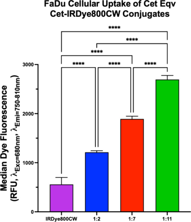
Cetuximab (Cet)-IRDye800CW, among other antibody-IRDye800CW conjugates, is a potentially effective tool for delineating tumor margins during fluorescence image-guided surgery (IGS). However, residual disease often leads to recurrence. Photodynamic therapy (PDT) following IGS is proposed as an approach to eliminate residual disease but suffers from a lack of molecular specificity for cancer cells. Antibody-targeted PDT offers a potential solution for this specificity problem. In this study, we show, for the first time, that Cet-IRDye800CW is capable of antibody-targeted PDT in vitro when the payload of dye molecules is increased from 2 (clinical version) to 11 per antibody. Cet-IRDye800CW (1:11) produces singlet oxygen, hydroxyl radicals, and peroxynitrite upon activation with 810 nm light. In vitro assays on FaDu head and neck cancer cells confirm that Cet-IRDye800CW (1:11) maintains cancer cell binding specificity and is capable of inducing up to ∼90% phototoxicity in FaDu cancer cells. The phototoxicity of Cet-IRDye800CW conjugates using 810 nm light follows a dye payload-dependent trend. Cet-IRDye800CW (1:11) is also found to be more phototoxic to FaDu cancer cells and less toxic in the dark than the approved chromophore indocyanine green, which can also act as a PDT agent. We propose that antibody-targeted PDT using high-payload Cet-IRDye800CW (1:11) could hold potential for eliminating residual disease postoperatively when using sustained illumination devices, such as fiber optic patches and implantable surgical bed balloon applicators. This approach could also potentially be applicable to a wide variety of resectable cancers that are amenable to IGS-PDT, using their respective approved full-length antibodies as a template for high-payload IRDye800CW conjugation.

Keywords: Cetuximab; IRDye800CW; antibody-conjugate; fluorescence image-guided surgery; photodynamic therapy.


Conflict of interest statement

The authors declare no competing financial interest.

Figures

Figure 1
Figure 1
Schematic of amide bond formation conjugation of IRDye800CW-NHS ester to Cetuximaba to form the 1:11 conjugate. (a3D representation of Cetuximab (IgG2a, Protein Data Bank ID: 1IGT)).
Figure 2
Figure 2
Characterization of various Cet-IRDye800CW conjugates and free IRDye800CW. (A) UV–vis spectra of free IRDye800CW, 1:2, 1:7, and 1:11 Cet-IRDye800CW conjugates in DPBS, normalized to the primary NIR absorption band (see Table 1). (B) Fluorescence emission spectra (λExc=773 nm) of 1 μM IRDye800CW equivalent free IRDye800CW, 1:2, 1:7, and 1:11 conjugates in DPBS. (C) Peak fluorescence emissions of 1:2, 1:7, 1:11, and free IRDye800CW.
Figure 3
Figure 3
Fluorescence of 1:11 can be proteolytically activated from its quenched state. (A) Fluorescence spectra of 1:11 conjugate incubated with trypsin, taken over various time points. (B) Fluorescence (λExc=773 nm, λEmi=798 nm) of 1:11 conjugate incubated with and without trypsin over 48 h.
Figure 4
Figure 4
Cet-IRDye800CW can produce various ROS when irradiated with a 41 mW·cm–2 810 nm light. (A) Singlet oxygen generation of conjugates measured by the decreasing absorbance of ADPA. (B) Area under the curve analysis of (A). It should be noted that, because ADPA decreases in absorbance when reacting with singlet oxygen, the AUC graph will appear differently compared to that of SOSG and HPF. (C) Singlet oxygen generation of conjugates measured by the increasing fluorescence emissions of SOSG at 530 nm. (D) Area under the curve analysis of (C). (E) Hydroxyl radical and peroxynitrite generation of conjugates measured by the increasing fluorescence emissions of HPF at 530 nm. (F) Area under the curve analysis of (E). (Data are mean ± SD, statistical significance was calculated using one-way ANOVA with Tukey’s multiple comparisons, *: P < 0.02; **: P < 0.01; ***: P < 0.001; ****: P < 0.0001).
Figure 5
Figure 5
ROS generation of 1:11 Cet-IRDye800CW conjugates is reduced in the presence of ROS scavengers. (A) Singlet oxygen generation of 1:11 conjugate in the presence of NaN3, measured by the decrease in absorbance of ADPA. (B) Area under the curve analysis of (A). It should be noted that, because ADPA decreases in absorbance when reacting with singlet oxygen, the AUC graph will appear differently compared to that of SOSG and HPF. (C) Singlet oxygen generation of 1:11 conjugate in the presence of NaN3, measured by the increasing fluorescence emission of SOSG at 530 nm. (D) Area under the curve analysis of (C). (E) Hydroxyl radical generation of 1:11 conjugate in the presence of mannitol, measured by the increasing fluorescence emission of HPF at 530 nm. (F) Area under the curve analysis of (E). (Data are mean ± SD. Statistical significance was calculated using unpaired t test, *: P < 0.02; ***: P < 0.001; ****: P < 0.0001).
Figure 6
Figure 6
Fluorescence and absorbance readings were taken between photoirradiations of Cet conjugates to measure photobleaching. (A) Photobleaching of 1:11 as measured by fluorescence (λExc=710 nm, λEmi=750–850 nm) over 300 J·cm–2 fluence. (B) Peak fluorescence values of 1:2, 1:7, 1:11, and IRDye800CW as a function of total fluence. Data are derived from (A) and Supp. Figure 5. (C) Peak absorbance value of the primary NIR absorption band of all conjugates as a function of total fluence. (D) Peak absorbance value of the secondary shoulder NIR absorption band of all conjugates as a function of the total fluence. (E) Rate of photobleaching of 1:11, 1:7, 1:2, and IRDye800CW as calculated by one-phase decay rate analysis of absorbance at 774 nm. (F) Rate of photobleaching of 1:11, 1:7, 1:2, and IRDye800CW as calculated by one-phase decay rate analysis of absorbance at 710 nm. (Data are mean ± SD. Statistical significance was calculated using one-way ANOVA with Tukey’s multiple comparisons, ***: P < 0.001; ****: P < 0.0001).
Figure 7
Figure 7
Fluorescence of 1:11 bound to F98 cells (EGFR-null) and FaDu cells (EGFR-positive) measured by flow cytometry. (A) Histogram of cell count vs their fluorescence emission at 780 nm. Both populations had a total cell count of 15,700 after gating. (B) Comparison of average median fluorescence of IRDye800CW between F98 and FaDu. Fluorescence corrections were done with untreated F98 and FaDu control groups to account for autofluorescence. (Data are mean ± SD. Statistical significance was calculated using unpaired t test, ****: P < 0.0001).
Figure 8
Figure 8
Fluorescence emission measured by flow cytometry of free IRDye800CW and various Cet-IRDye800CW conjugates uptaken by FaDu cells. Fluorescence emission corrections were done using untreated FaDu samples to account for autofluorescence. (Data are mean ± SD, statistical significance was calculated using one-way ANOVA with Tukey’s multiple comparisons, ****: P < 0.0001).
Figure 9
Figure 9
Metabolic activity of FaDu cells treated with various Cet conjugates and PDT determined through the MTT assay. (A) MTT assay using Cet equivalent concentrations of 1:2, 1:7, and 1:11 with 300 J·cm–2 of 810 nm light displayed significant reduction in cell activity for the high-payload conjugate. (B) Analysis of Figure 9A for 1 μM conjugate treatment groups. (C) MTT assay using dye equivalent concentrations of 1:2, 1:7, and 1:11 with 300 J·cm–2 of 810 nm light displayed a high reduction in cell activity for the 1:11 conjugate. (D) Analysis of Figure 9C for 11 μM dye equivalent treatment groups. (Data are mean ± SEM. Statistical significance was calculated using one-way ANOVA with Tukey’s multiple comparisons, **: P < 0.01, ***: P < 0.001, ****: P < 0.0001).

References

    1. Lamberts L. E.; Koch M.; De Jong J. S.; Adams A. L. L.; Glatz J.; Kranendonk M. E. G.; et al. Tumor-specific uptake of fluorescent bevacizumab-IRDye800CW microdosing in patients with primary breast cancer: A phase I feasibility study. Clin. Cancer Res. 2017, 23 (11), 2730–2741. 10.1158/1078-0432.CCR-16-0437. - DOI - PubMed
    1. Gao R. W.; Teraphongphom N.; de Boer E.; van den Berg N. S.; Divi V.; Kaplan M. J. Safety of panitumumab-IRDye800CW and cetuximab-IRDye800CW for fluorescence-guided surgical navigation in head and neck cancers. Theranostics 2018, 8 (9), 2488–2495. 10.7150/thno.24487. - DOI - PMC - PubMed
    1. Miller S. E.; Tummers W. S.; Teraphongphom N.; van den Berg N. S.; Hasan A.; Ertsey R. D.; et al. First-in-human intraoperative near-infrared fluorescence imaging of glioblastoma using cetuximab-IRDye800. J. Neurooncol. 2018, 139 (1), 135–143. 10.1007/s11060-018-2854-0. - DOI - PMC - PubMed
    1. Overholser J. P.; Prewett M. C.; Hooper A. T.; Waksal H. W.; Hicklin D. J. Epidermal growth factor receptor blockade by antibody IMC-C225 inhibits growth of a human pancreatic carcinoma xenograft in nude mice. Cancer. 2000, 89 (1), 74–82. 10.1002/1097-0142(20000701)89:1<74::AID-CNCR11>3.0.CO;2-K. - DOI - PubMed
    1. Saad M. A.; Zhung W.; Stanley M. E.; Formica S.; Grimaldo-Garcia S.; Obaid G.; et al. Photoimmunotherapy Retains Its Anti-Tumor Efficacy with Increasing Stromal Content in Heterotypic Pancreatic Cancer Spheroids. Mol. Pharmaceutics 2022, 19 (7), 2549–2563. 10.1021/acs.molpharmaceut.2c00260. - DOI - PMC - PubMed

LinkOut - more resources