

Comprehensive literature review on the application of the otological surgical planning software OTOPLAN® for cochlear implantation
- PMID: 38861031
- PMCID: PMC11618202
- DOI: 10.1007/s00106-023-01417-4
Comprehensive literature review on the application of the otological surgical planning software OTOPLAN® for cochlear implantation
Abstract
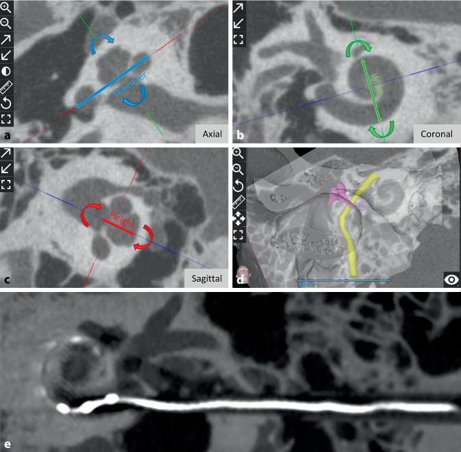
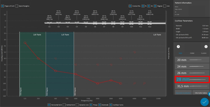
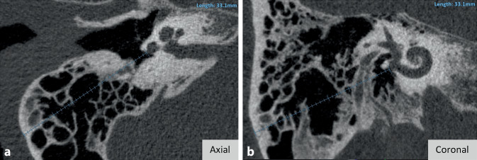
Background: The size of the human cochlear, measured by the diameter of the basal turn, varies between 7 and 11 mm. For hearing rehabilitation with cochlear implants (CI), the size of the cochlear influences the individual frequency map and the choice of electrode length. OTOPLAN® (CAScination AG [Bern, Switzerland] in cooperation with MED-EL [Innsbruck, Austria]) is a software tool with CE marking for clinical applications in CI treatment which allows for precise pre-planning based on cochlear size. This literature review aims to analyze all published data on the application of OTOPLAN®.
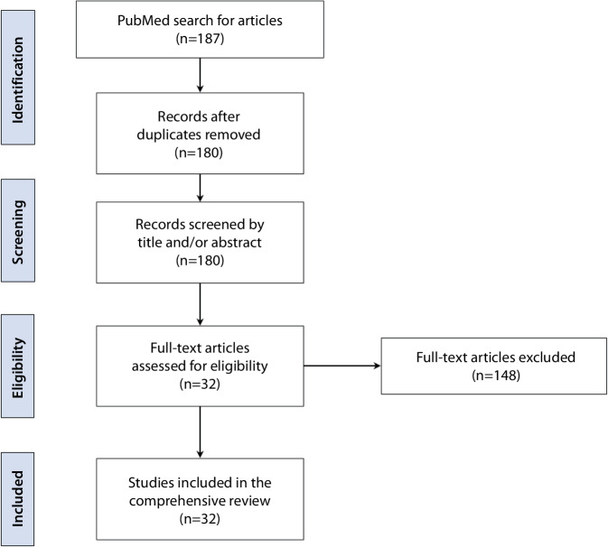
Materials and methods: The Preferred Reporting Items for Systematic Reviews and Meta-Analyses (PRISMA) guidelines were applied to identify relevant studies published in the PubMed search engine between January 2015 and February 2023 using the search terms "otoplan" [title/abstract] OR "anatomy-based fitting" [title/abstract] OR "otological software tool" [title/abstract] OR "computed tomography-based software AND cochlear" [title/abstract].
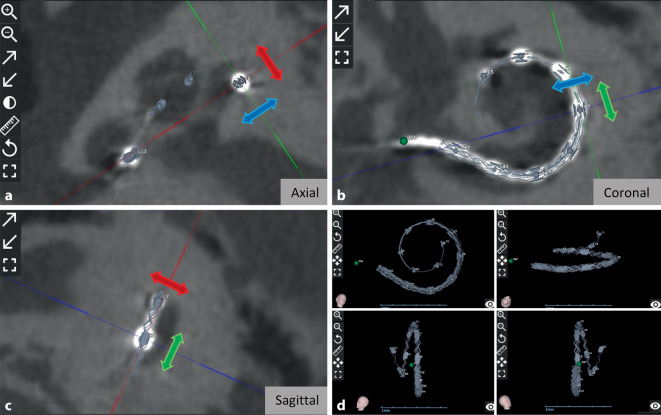
Results: The systematic review of the literature identified 32 studies on clinical use of OTOPLAN® in CI treatment. Most studies were reported from Germany (7 out of 32), followed by Italy (5), Saudi Arabia (4), the USA (4), and Belgium (3); 2 studies each were from Austria and China, and 1 study from France, India, Norway, South Korea, and Switzerland. In the majority of studies (22), OTOPLAN® was used to assess cochlear size, followed by visualizing the electrode position using postoperative images (5), three-dimensional segmentation of temporal bone structures (4), planning the electrode insertion trajectory (3), creating a patient-specific frequency map (3), planning of a safe drilling path through the facial recess (3), and measuring of temporal bone structures (1).
Conclusion: To date, OTOPLAN® is the only DICOM viewer with CE marking in the CI field that can process pre-, intra-, and postoperative images in the abovementioned applications.
Zusammenfassung: HINTERGRUND: Die Größe der menschlichen Cochlea, gemessen am Durchmesser der Basalwindung, schwankt zwischen 7 und 11 mm. Im Rahmen einer Hörrehabilitation durch ein Cochleaimplantat ist diese für die individuelle Zuordnung der Frequenzbänder und die Wahl der Elektrodenlänge von Bedeutung. OTOPLAN® (CAScination AG [Bern, Schweiz] in Kooperation mit MED-EL [Innsbruck, Österreich]) ist ein Softwaretool mit CE-Kennzeichnung für klinische Anwendungen in der Cochleaimplantat(CI)-Behandlung, welches die Vorplanung auf Grundlage der cochleären Größenparameter durchführt. Ziel dieser Literaturübersicht ist es, alle veröffentlichten Studien über die Anwendung von OTOPLAN® zu erfassen.
Materialien und methoden: Die PRISMA-Richtlinien (Preferred Reporting Items for Systematic Reviews and Meta-Analyses) wurden angewandt, um relevante Studien zu identifizieren, die zwischen Januar 2015 und Februar 2023 in der Suchmaschine PubMed veröffentlicht wurden (unter Verwendung der Suchbegriffe „otoplan“ [Titel/Abstract] OR „anatomy-based fitting“ [Titel/Abstract] OR „otological software tool“ [Titel/Abstract] OR „computed tomography-based software AND cochlear“ [Titel/Abstract]).
Ergebnisse: Bei der systematischen Durchsicht der Literatur wurden 32 Studien über den klinischen Einsatz von OTOPLAN® bei der CI-Behandlung gefunden. Die meisten Studien wurden von deutschen Arbeitsgruppen publiziert (7 von 32), gefolgt von Italien (5), Saudi-Arabien (4), USA (4) und Belgien (3). So stammten je 2 Studien aus Österreich und China, gefolgt von jeweils 1 Studie aus Frankreich, Indien, Norwegen, Südkorea und der Schweiz. In den meisten Studien (22) wurde OTOPLAN® zur Beurteilung der Cochleagröße verwendet, gefolgt von der Visualisierung der Elektrodenposition anhand postoperativer Bilder (5), der dreidimensionalen (3-D-)Segmentierung der Felsenbeinstrukturen (4), der Planung der Elektrodeneinführungstrajektorie (3), der Erstellung einer patientenspezifischen Frequenzbandzuordnung (3), der Planung eines sicheren Bohrpfads durch den Recessus facialis (3), und der Messung von Felsenbeinstrukturen (1).
Schlussfolgerung: OTOPLAN® ist bisher der einzige DICOM-Viewer mit CE-Kennzeichnung im CI-Bereich, der prä-, intra- und postoperative Bilder mit den genannten Anwendungen verarbeiten kann.
Keywords: Anatomy based fitting; Cochlear duct length; Computed tomography-based software ear/cochlear; Computer simulation; Imaging modalities (MRI, computer tomography [flat-panel volume CT]).
© 2024. The Author(s).
Conflict of interest statement
Declarations. Conflict of interest: F.-T. Müller-Graff, B. Spahn, D.P. Herrmann, A. Kurz, J. Völker, R. Hagen and K. Rak declare that they have no competing interests. For this article no studies with human participants or animals were performed by any of the authors. All studies mentioned were in accordance with the ethical standards indicated in each case. The supplement containing this article is not sponsored by industry.
Figures

References
-
- Almuhawas FA, Dhanasingh AE, Mitrovic D et al (2020) Age as a Factor of Growth in Mastoid Thickness and Skull Width. Otol Neurotol 41:709–714 - PubMed
-
- Andersen SAW, Bergman M, Keith JP et al (2021) Segmentation of Temporal Bone Anatomy for Patient-Specific Virtual Reality Simulation. Ann Otol Rhinol Laryngol 130:724–730 - PubMed
Publication types
MeSH terms
LinkOut - more resources
Full Text Sources
