. 2024 Jun 11;10(1):140.
doi: 10.1186/s40792-024-01945-3.

Laparoscopic left hepatectomy for a patient with an absence of portal bifurcation using real-time imaging: a case report

Affiliations

Laparoscopic left hepatectomy for a patient with an absence of portal bifurcation using real-time imaging: a case report

Shugo Mizuno et al. Surg Case Rep. .

Abstract

Background: Absence of portal bifurcation is an extremely rare anomaly that should be recognized preoperatively, especially prior to a major hepatectomy.

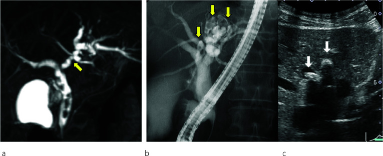
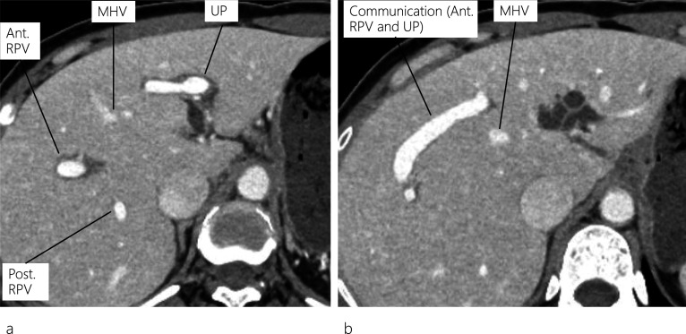
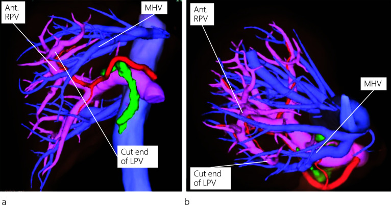
Case presentation: A 45-year-old woman presented with abdominal pain, and abdominal computed tomography (CT) revealed dilatation of both the common bile duct (CBD) and intrahepatic bile duct (IHBD). Endoscopic retrograde cholangiopancreatography (ERCP) showed CBD and IHBD stones (B2 and B4). The CBD stones were removed, but the IHBD stones could not be, yet there was no evidence of malignancy at the site of IHBD stenosis. Enhanced CT revealed a dilated IHBD, while three-dimensional CT images showed the left portal vein running through the ventral side of the middle hepatic vein, which was diagnosed as the absence of portal vein bifurcation (APB). Laparoscopic left hepatectomy was successfully performed using real-time indocyanine green (ICG) fluorescence imaging.

Conclusion: Surgeons should be aware of the possibility of APB, a rare portal vein anomaly, before performing major hepatectomy. Real-time ICG fluorescence imaging may be helpful to ensure the precise anatomy of the liver during laparoscopic surgery.

Keywords: Absence of portal vein bifurcation; ICG fluorescence real-time imaging; Laparoscopic left hepatectomy; Portal vein anomaly.


Conflict of interest statement

The authors declare that they have no competing interests associated with this manuscript.

Figures

Fig. 1
Fig. 1
Image findings. Magnetic resonance cholangiopancreatography (MRCP) (a) shows common bile duct (CBD) stones and stenosis at the confluence of B2 and B3. Endoscopic retrograde cholangiopancreatography (ERCP) (b) reveals intrahepatic bile duct (IHBD) stones in B2 and B4 (yellow arrows). Abdominal ultrasound scan (c) shows IHBD stones in B2 and B4 (white arrows)
Fig. 2
Fig. 2
Enhanced CT images. Enhanced CT scan reveals a dilated IHBD in the left lobe and a communication between the right portal vein and umbilical portion (a, b). CT: computed tomography; IHBD: intrahepatic bile duct; MHV: middle hepatic vein; Ant. RPV: anterior right portal vein; Post. RPV: posterior right portal vein, UP: umbilical portion
Fig. 3
Fig. 3
Three-dimensional reconstruction of the enhanced CT images. The left hepatic artery and bile duct are separated from the left portal vein (LPV) instead of being located in the left Glisson’s sheath as viewed from a left anterior oblique position (a). LPV, viewed from a neutral position, runs through the ventral side of the middle hepatic vein (MHV) (b). MHV: middle hepatic vein; Ant. RPV: anterior right portal vein; LPV: left portal vein
Fig. 4
Fig. 4
Intraoperative findings during laparoscopic left hepatectomy. A demarcation line (black arrows) appears after injecting indigo carmine (10 mg) into the LPV above the MHV under intraoperative ultrasound (US) guidance (a). The LPV and its branch are encircled (b). After clamping the LPV and its branch, negative staining using indocyanine green (ICG) fluorescence real-time imaging shows hepatic perfusion and a clear demarcation line (c)
Fig. 5
Fig. 5
Three-dimensional reconstruction of postoperative enhanced CT images. Both the MHV and Ant. RPV are preserved, as viewed from neutral (a) and left posterior oblique (b) positions. CT: computed tomography; MHV: middle hepatic vein; Ant. RPV: anterior right portal vein; LPV: left portal vein

References

    1. Marks C. Developmental basis of the portal venous system. Am J Surg. 1969;117:671–681. doi: 10.1016/0002-9610(69)90404-8. - DOI - PubMed
    1. Chaib E. Absence of bifurcation of the portal vein. Surg Radiol Anat. 2009;31:389–392. doi: 10.1007/s00276-008-0413-7. - DOI - PubMed
    1. Fraser-Hill MA, Atri M, Bret PM, et al. Intrahepatic portal venous system: variations demonstrated with duplex and color Doppler US. Radiology. 1990;177:523–526. doi: 10.1148/radiology.177.2.2217795. - DOI - PubMed
    1. Hardy KJ, Jones RM. Failure of the portal vein to bifurcate. Surgery. 1997;121:226–228. doi: 10.1016/S0039-6060(97)90295-6. - DOI - PubMed
    1. Teraoku H, Arakawa Y, Yoshikawa M, et al. Complication of portal vein thrombosis after right hemihepatectomy in a patient lacking the portal vein bifurcation. J Med Invest. 2016;63:315–318. doi: 10.2152/jmi.63.315. - DOI - PubMed

LinkOut - more resources