. 2024 Jun 11;26(1):23.
doi: 10.1007/s12017-024-08787-0.

Protein Disulfide Isomerase Endoplasmic Reticulum Protein 57 (ERp57) is Protective Against ALS-Associated Mutant TDP-43 in Neuronal Cells

Affiliations

Protein Disulfide Isomerase Endoplasmic Reticulum Protein 57 (ERp57) is Protective Against ALS-Associated Mutant TDP-43 in Neuronal Cells

Sonam Parakh et al. Neuromolecular Med. .

Abstract

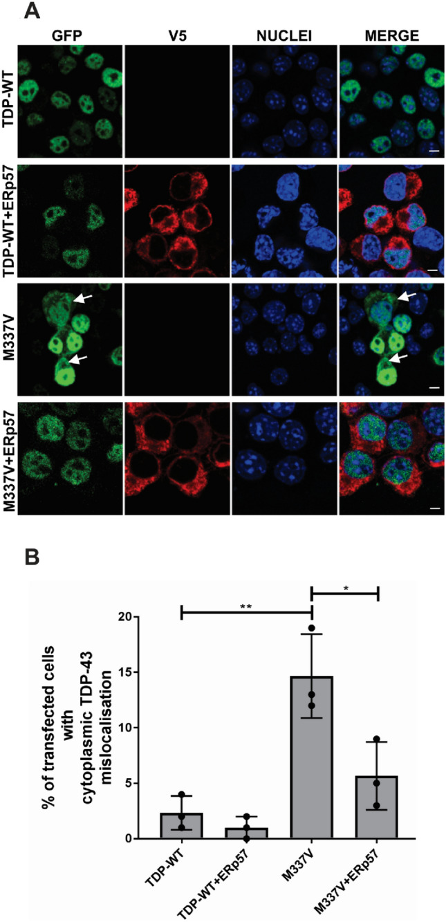
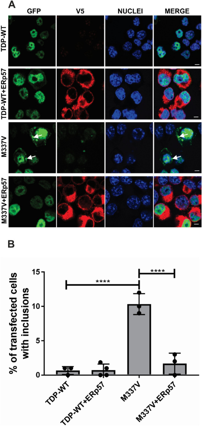
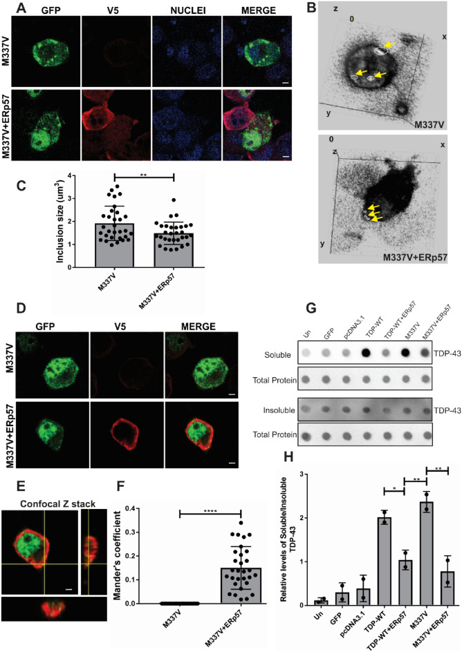
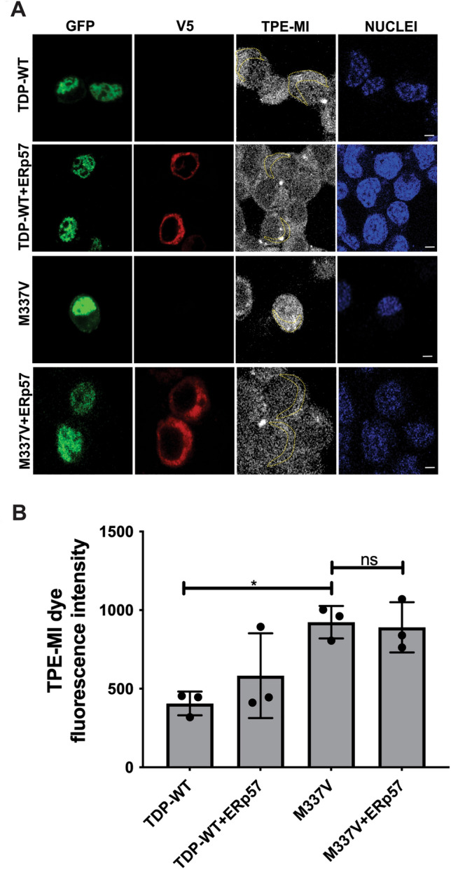
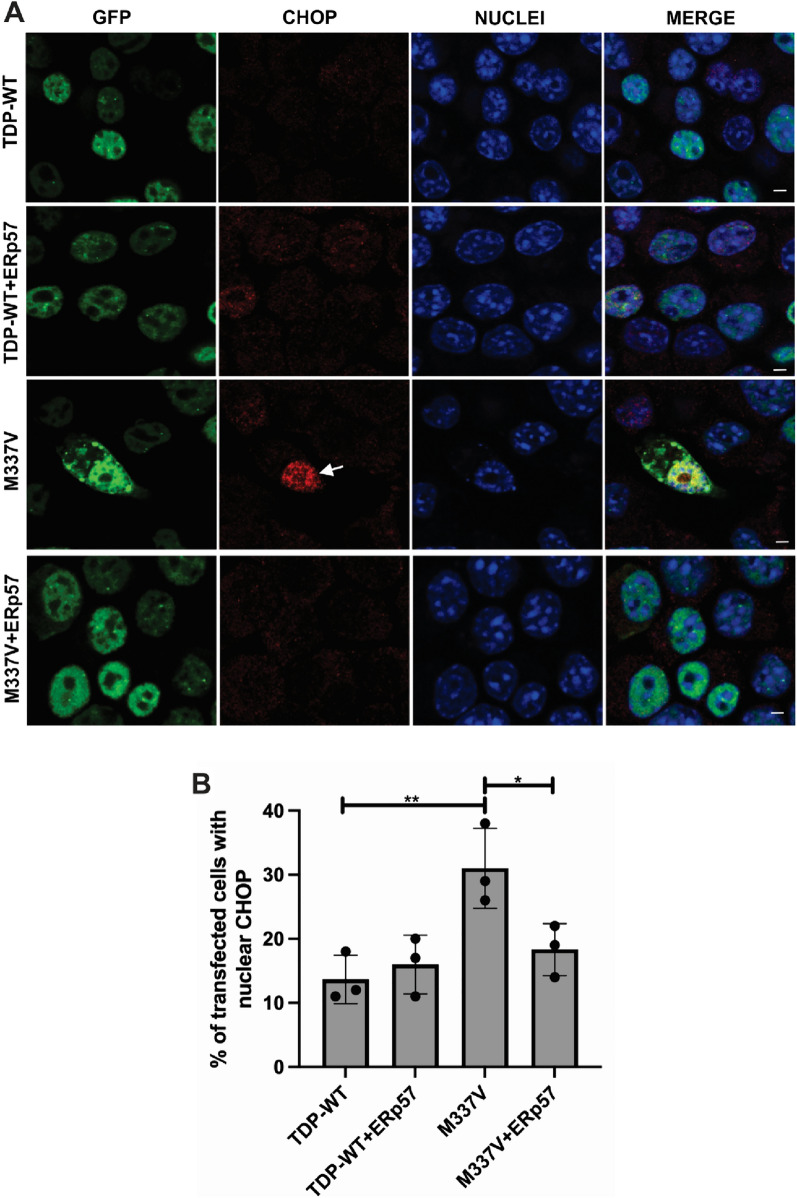
Amyotrophic Lateral Sclerosis (ALS) is a severe neurodegenerative disease affecting motor neurons. Pathological forms of Tar-DNA binding protein-43 (TDP-43), involving its mislocalisation to the cytoplasm and the formation of misfolded inclusions, are present in almost all ALS cases (97%), and ~ 50% cases of the related condition, frontotemporal dementia (FTD), highlighting its importance in neurodegeneration. Previous studies have shown that endoplasmic reticulum protein 57 (ERp57), a member of the protein disulphide isomerase (PDI) family of redox chaperones, is protective against ALS-linked mutant superoxide dismutase (SOD1) in neuronal cells and transgenic SOD1G93A mouse models. However, it remains unclear whether ERp57 is protective against pathological TDP-43 in ALS. Here, we demonstrate that ERp57 is protective against key features of TDP-43 pathology in neuronal cells. ERp57 inhibited the mislocalisation of TDP-43M337V from the nucleus to the cytoplasm. In addition, ERp57 inhibited the number of inclusions formed by ALS-associated variant TDP-43M337V and reduced the size of these inclusions. ERp57 was also protective against ER stress and induction of apoptosis. Furthermore, ERp57 modulated the steady-state expression levels of TDP-43. This study therefore demonstrates a novel mechanism of action of ERp57 in ALS. It also implies that ERp57 may have potential as a novel therapeutic target to prevent the TDP-43 pathology associated with neurodegeneration.

Keywords: ALS—Amyotrophic lateral sclerosis; ER stress; ERp57—Endoplasmic reticulum protein 57; PDI—Protein disulphide isomerase; TDP-43 pathology.


Conflict of interest statement

The authors declare no competing interests.

Figures

Fig. 1
Fig. 1
ERp57 protects against mislocalisation of mutant TDP-43 from the nucleus into the cytoplasm in neuronal cell lines A Immunocytochemistry and confocal microscopy of mislocalised cytoplasmic TDP-43 in Neuro-2a cells co-expressing TDP-43 and ERp57, at 72 h post transfection. Cells expressing Turbo-GFP tagged wild-type TDP-43 (TDP-WT, panel 1) with pcDNA3.1 empty vector or wild-type TDP-43 co-expressing ERp57 (panel 2) displayed mainly nuclear TDP-43 localisation, whereas more cells expressing mutant TDP-43M337V(M337V) exhibited cytoplasmic distribution, indicated with white arrows (panel 3). On co-expressing ERp57 with mutant TDP-43M337V, (panel 4) fewer cells displayed cytoplasmic TDP-43 compared to those expressing TDP-43M337V with vector alone. White arrows represent TDP-43 expression in the cytoplasm. Scale bar = 5 µm. B Quantification of cells in 1A, displaying cytoplasmic distribution of TDP-43. Results are expressed as mean ± SD, n = 3. Significant differences in the proportion of cells with cytoplasmic TDP-43 were detected between wild-type TDP-43 and mutant TDP-43M337V (**p < 0.01). Over-expression of ERp57 with mutant TDP-43M337V significantly decreased the proportion of cells displaying cytoplasmic TDP-43, compared to TDP-43M337V cells expressing empty vector only (*p < 0.05)
Fig. 2
Fig. 2
ERp57 inhibits the formation of mutant TDP-43M337V inclusions in neuronal cell lines A Immunocytochemistry and confocal microscopy of GFP-positive inclusions present in Neuro-2a cells expressing wild-type TDP-43 (TDP-WT) or mutant TDP-43M337V (M337V, green) with V5 tagged ERp57 (red), at 72 h post transfection. Nuclei are visualised by Hoechst staining (blue). A small proportion of cells expressing wild-type TDP-43 formed inclusions (panel 1), but this was not altered by co-expression with ERp57 (panel 2). More inclusion-positive cells were present in populations expressing mutant TDP-43M337V with vector alone (panel 3) represented by white arrows. In contrast, fewer cells formed inclusions when ERp57 was co-expressed with mutant TDP-43M337V (panel 4). White arrows represent TDP-43 inclusions in the cytoplasm. Scale bar = 5 µm. B Quantification of the percentage of transfected Neuro-2a cells bearing inclusions represented in 2A. Results are expressed as mean ± SD, n = 3. Significant fewer TDP-43 inclusions were present in wild-type TDP-43 compared to mutant TDP-43M337V populations (****p < 0.0001). Significantly fewer cells formed inclusions when ERp57 was co-expressed with TDP-43M337V (****p < 0.0001) compared with cells transfected with pcDNA3.1 empty vector alone
Fig. 3
Fig. 3
ERp57 decreases the size of mutant TDP-43M337V inclusions and co-localises with TDP-43 in neuronal cell lines A Immunocytochemistry and confocal microscopy of GFP-positive inclusions in Neuro-2a cells expressing mutant TDP-43M337V (M337V, green) with V5 tagged ERp57 (red), 72 h post transfection. Nuclei are visualised by Hoechst staining (blue). Scale bar = 5 µm. B 3D images using ImageJ depicting inclusion size variation shown with white circles and yellow arrows in ERp57 cells compared to TDP-43M337V with vector alone. C Quantification using Z stack images (A) demonstrated that ERp57 significantly reduced inclusion size (**p < 0.01) compared to TDP-43M337V with vector alone. D Immunofluorescence detection of GFP-positive TDP-43M337V in Neuro-2a cells expressing empty vector (panel 1) with V5 tagged ERp57 (panel 2) demonstrated partially co-localized ERp57 with TDP-43-positive inclusions in neuronal cells. E Orthogonal section of the confocal image in (D), showing co-localization of TDP-43 inclusions with ERp57. F The degree of co-localization between TDP-43 inclusions with ERp57 in neuronal cells was quantified using Mander’s coefficient, revealing significant overlap (****p < 0.0001). Results are expressed as mean ± SD. G Protein lysates were subjected to vacuum filtration through a 96-well dot blot apparatus with a nitrocellulose membrane. Immunoblotting was then performed with anti-TDP-43 and anti-phospho-TDP-43 antibodies. Ponceau S staining was used as a reference for total protein levels (lane 2 and 4). Soluble TDP-43 levels (lane 1) and insoluble TDP-43 levels (lane 3) are shown. H Densitometric quantitation of TDP-43 protein (soluble/insoluble) levels normalized to total protein from the dot blot shown in (G) confirms that the ratio of soluble:insoluble wild-type or TDP-43M337V was reduced in ERp57 expressing cells (**p < 0.01, *p < 0.05)
Fig. 4
Fig. 4
ERp57 has no effect on mutant TDP-43 unfolding in neuronal cell lines A Protein unfolding detection using TPE-MI  dye fluorescence in Neuro-2a cells. TDP-WT with empty vector (panel 1) and with V5 tagged ERp57 (panel 2), TDP-43M337V with empty vector alone (panel 3), or co-expressing ERp57 (panel 4). Scale bar = 5 µm. Yellow dotted area representing TPE-MI levels in the cytoplasm. B TPE-MI fluorescence in the cytoplasm was significantly lower in cells expressing TDP-WT compared to TDP-43M337V (*p < 0.05). However, no statistical difference was observed when ERp57 was co-expressed with TDP-43M337V compared to controls, ns, non-significant
Fig. 5
Fig. 5
ERp57 inhibits ER stress induced by mutant TDP-43M337V in neuronal cell lines A Immunocytochemistry and confocal microscopy of Neuro-2a cells expressing wild-type TDP-43 (TDP-WT) or mutant TDP-43M337V (M337V, green) with V5 tagged ERp57, and nuclear CHOP immunoreactivity (red), were examined at 72 h post transfection. Nuclei were visualised by Hoechst staining (blue). A small proportion of cells expressing wild-type TDP-43 displayed nuclear CHOP (panel 1), and this was not altered by co-expression with ERp57 (panel 2). More nuclear CHOP immunoreactivity was present in populations expressing mutant TDP-43M337V with vector alone (panel 3), represented by white arrows. In contrast, fewer cells formed inclusions when ERp57 was co-expressed with mutant TDP-43M337V (panel 4). White arrows represent nuclear CHOP. Scale bar = 5 µm. B Quantification of the percentage of transfected Neuro-2a cells showing nuclear CHOP immunoreactivity in 5A. Results are expressed as mean ± SD, n = 3. Significant fewer cells expressing nuclear CHOP in wild-type TDP-43 compared to mutant TDP-43M337V populations (**p < 0.01). Significantly fewer cells displayed nuclear CHOP when ERp57 was co-expressed with TDP-43M337V (*p < 0.05) compared with cells transfected with pcDNA3.1 empty vector alone
Fig. 6
Fig. 6
ERp57 protects against mutant TDP-43 induced cell death in neuronal cell lines A Neuro-2a cells were co-expressed with wild-type TDP-43 (TDP-WT) or TDP-43M337V (M337V, green) and V5 tagged ERp57 (red), examined by confocal microscopy at 72 h post transfection. Nuclei are shown by Hoechst stain (blue). Arrow represents condensed or fragmented nuclei, indicating apoptosis is underway. Few cells expressing TDP-WT (panel 1) or TDP-WT co-expressing ERp57 (panel 2) contained fragmented nuclei and hence were apoptotic (< 1%) but more cells expressing TDP-43M337V (panel 3) displayed Hoechst-stained condensed nuclei, indicating apoptosis, indicated by white arrows (middle panel). However, fewer cells co-expressing TDP-43M337V with ERp57 (panel 3) were undergoing apoptosis compared to those transfected with empty vector, scale bar = 5 µm. B Quantification of apoptotic nuclei in cells in 5A expressing TDP-43 and ERp57. Results are expressed as mean ± SD, n = 3. A significant difference in apoptosis was observed between wild-type TDP-43 and TDP-43M337V cells (****p < 0.0001). Over-expression of ERp57 with TDP-43M337V resulted in significantly fewer cells undergoing apoptosis compared to cells transfected with empty vector only (***p < 0.001). C Activated caspase-3 immunoreactivity, confirming induction of apoptosis, in cells expressing TDP-43M337V and ERp57. Neuro-2a cells were co-expressed with either wild-type TDP-43 or TDP-43M337V (green) and ERp57 for 72 h, followed by immunocytochemistry using anti-activated caspase-3 antibodies (red), visualized using confocal microscopy. Nuclei are shown by Hoechst stain (blue). White arrow represents caspase-3 activation, indicating apoptosis is underway. As expected, fewer cells expressing wild-type TDP-43 (row 1) displayed caspase-3 activation, compared to cells expressing TDP-43M337V (row 3). However, fewer cells expressing TDP-43M337V with ERp57 (row 4) displayed caspase-3 activation, compared to those TDP-43M337V cells transfected with empty vector. D Quantification of transfected cells visualized in 6C, immunostained using anti-activated caspase-3 antibodies. Results are expressed as mean ± SD, n = 3. Over-expression of ERp57 with TDP-43M337V significantly decreased the proportion of cells with activated caspase-3, indicating apoptotic cell death is underway, compared to cells expressing empty vector only (**p < 0.01, ***p<0.001 )

Similar articles

Cited by

References

    1. Acewicz, A., Stępień, T., Felczak, P., Tarka, S., & Wierzba-Bobrowicz, T. (2022). Incidence and morphology of secondary TDP-43 proteinopathies: Part 1. Folia Neuropathologica,60(3), 267–276. 10.5114/fn.2022.120314 - PubMed
    1. Bargsted, L., Hetz, C., & Matus, S. (2016). ERp57 in neurodegeneration and regeneration. Neural Regeneration Research,11(2), 232–233. 10.4103/1673-5374.177722 - PMC - PubMed
    1. Barmada, S. J., Serio, A., Arjun, A., Bilican, B., Daub, A., Ando, D. M., Tsvetkov, A., Pleiss, M., Li, X., Peisach, D., Shaw, C., Chandran, S., & Finkbeiner, S. (2014). Autophagy induction enhances TDP43 turnover and survival in neuronal ALS models. Nature Chemical Biology,10(8), 677–685. 10.1038/nchembio.1563 - PMC - PubMed
    1. Barmada, S. J., Skibinski, G., Korb, E., Rao, E. J., Wu, J. Y., & Finkbeiner, S. (2010). Cytoplasmic mislocalization of TDP-43 is toxic to neurons and enhanced by a mutation associated with familial amyotrophic lateral sclerosis. Journal of Neuroscience,30(2), 639–649. 10.1523/jneurosci.4988-09.2010 - PMC - PubMed
    1. Bilches Medinas, D., Malik, S., Yildiz-Bölükbasi, E., Borgonovo, J., Saaranen, M. J., Urra, H., Pulgar, E., Afzal, M., Contreras, D., Wright, M. T., Bodaleo, F., Quiroz, G., Rozas, P., Mumtaz, S., Díaz, R., Rozas, C., Cabral-Miranda, F., Piña, R., Valenzuela, V., … Tolun, A. (2022). Mutation in protein disulfide isomerase A3 causes neurodevelopmental defects by disturbing endoplasmic reticulum proteostasis. EMBO Journal,41(2), e105531. 10.15252/embj.2020105531 - PMC - PubMed

Publication types