. 2024 Aug 5;221(8):e20231518.
doi: 10.1084/jem.20231518. Epub 2024 Jun 11.

A viral E3 ubiquitin ligase produced by herpes simplex virus 1 inhibits the NLRP1 inflammasome

Affiliations

A viral E3 ubiquitin ligase produced by herpes simplex virus 1 inhibits the NLRP1 inflammasome

Pooja Parameswaran et al. J Exp Med. .

Abstract

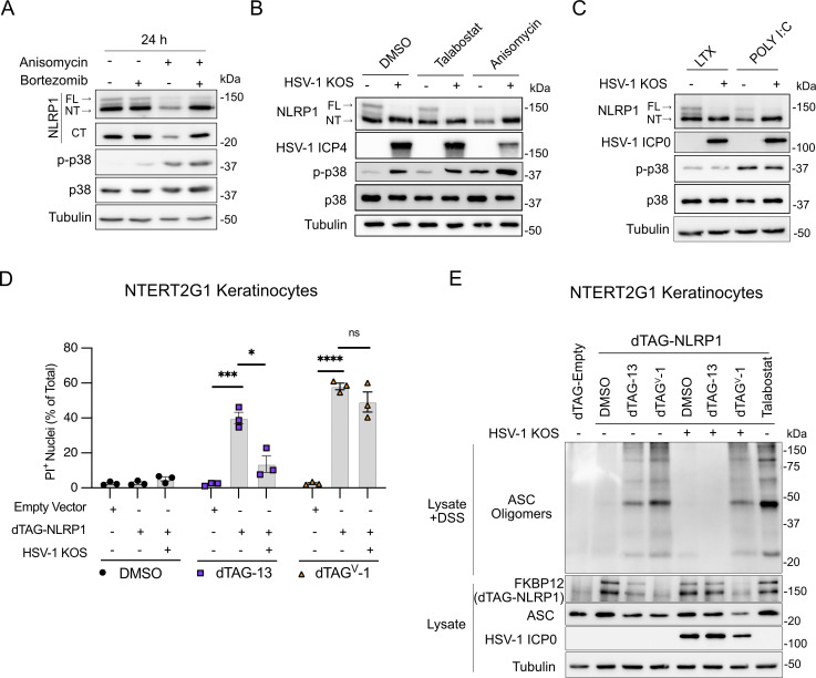
Guard proteins initiate defense mechanisms upon sensing pathogen-encoded virulence factors. Successful viral pathogens likely inhibit guard protein activity, but these interactions have been largely undefined. Here, we demonstrate that the human pathogen herpes simplex virus 1 (HSV-1) stimulates and inhibits an antiviral pathway initiated by NLRP1, a guard protein that induces inflammasome formation and pyroptotic cell death when activated. Notably, HSV-1 infection of human keratinocytes promotes posttranslational modifications to NLRP1, consistent with MAPK-dependent NLRP1 activation, but does not result in downstream inflammasome formation. We identify infected cell protein 0 (ICP0) as the critical HSV-1 protein that is necessary and sufficient for inhibition of the NLRP1 pathway. Mechanistically, ICP0's cytoplasmic localization and function as an E3 ubiquitin ligase prevents proteasomal degradation of the auto-inhibitory NT-NLRP1 fragment, thereby preventing inflammasome formation. Further, we demonstrate that inhibiting this inflammasome is important for promoting HSV-1 replication. Thus, we have established a mechanism by which HSV-1 overcomes a guard-mediated antiviral defense strategy in humans.


Conflict of interest statement

Disclosures: The authors declare no competing interests exist.

Figures

Figure 1.
Figure 1.
HSV-1 infection activates receptor proximal steps in the NLRP1 inflammasome pathway but does not form a functional inflammasome. (A) Immunoblots of lysates examining NLRP1 abundance in primary human keratinocytes transduced with control or shNLRP1-expressing lentiviruses. Images are representative of n = 3 biological replicates. (B) Immunoblots of lysates from mock-infected or WT HSV-1 strains KOS or 17syn+-infected primary human keratinocytes (MOI 10) at 24 h.p.i. Images are representative of n = 3 biological replicates. (C) Immunoblots of lysates from HSV-1 (MOI 10) or anisomycin (10 μM) -treated primary human keratinocytes harvested at indicated time points. Images are representative of n = 3 biological replicates. (D) Immunoblots of ASC oligomerization status in DSS-treated lysates from mock-infected, HSV-1 KOS–infected (MOI 10), or talabostat-treated (30 μM) primary human keratinocytes for 24 h. A portion of the lysates was not treated with DSS and run in parallel to examine total ASC abundance under the indicated experimental conditions. Images are representative of n = 3 biological replicates. (E) Quantification of PI+ primary human keratinocyte nuclei following infection with HSV-1 KOS (MOI 10) or treatment with talabostat (30 μM). PI+ nuclei were quantified hourly and plotted as a percentage of total nuclei detected at each time point imaged. Data are presented as the mean of three independent experiments ± SEM. The area under the curve was calculated for each condition and statistical significance was determined by Student’s t test (**P < 0.01). Source data are available for this figure: SourceData F1.
Figure S1.
Figure S1.
HSV-1 induces the proteasome-dependent loss of FL-NLRP1. (A) Immunoblots of lysates from mock-infected or HSV-1 KOS strain–infected primary human keratinocytes (MOI 10) at 6, 12, 24 h.p.i. Images are representative of n = 3 biological replicates. (B) Immunoblots of lysates from DMSO or bortezomib (10 μM) -treated primary human keratinocytes infected with HSV-1 strain KOS (MOI 10) and harvested at indicated time points. Images are representative of n = 3 biological replicates. Source data are available for this figure: SourceData FS1.
Figure 2.
Figure 2.
HSV-1 infection inhibits the NLRP1 inflammasome. (A–D) Quantification of membrane permeability in mock- or HSV-1–infected (MOI 10) primary human keratinocytes treated with (A) talabostat (30 μM), (B) poly(I:C) (1 μg/ml), (C) anisomycin (1 μM), or (D) LPS (1 μg/ml). Lipofectamine (LTX) was used as the transfection control. PI+ nuclei were quantified hourly and results are presented as the mean of three independent experiments ± SEM. The area under the curve was calculated for each treatment condition and statistical significance was determined by Student’s t test (***P < 0.001, ****P < 0.0001). (E) Immunoblots of lysates from HSV-1–infected primary human keratinocytes (MOI 10) treated with talabostat (30 μM) for 24 h. Immunoblots of DSS-treated lysates and protein extracted from cell culture supernatants were also performed to assess ASC oligomerization and caspase-1 cleavage, respectively. Images are representative of n = 3 biological replicates. (F) ELISA for IL-1β in the supernatants of HSV-1–infected (MOI 10) keratinocytes treated with talabostat (30 μM) for 24 h. Data are presented as the mean of three independent experiments ± SEM. Statistical significance was calculated by Student’s t test (****P < 0.0001). (G) ASC speck detection by indirect immunofluorescence in HSV-1–infected (MOI 10) keratinocytes treated with talabostat (30 μM) for 24 h. Arrows indicate ASC specks. Images are representative of n = 3 biological replicates. Scale bar = 10 μm. (H) Quantification of ASC specks in G. Specks were quantified by counting the number of ASC+ cells as a percentage of total Hoechst+ cells. Data are presented as the mean of three independent experiments ± SEM. Statistical significance was calculated by Student’s t test (*P < 0.05). (I) Immunoblots from DSS-treated lysates to assess ASC oligomerization in HSV-1–infected primary human keratinocytes (MOI 10) treated with talabostat (30 μM) or anisomycin (10 μM). Images are representative of n = 3 biological replicates. (J) Immunoblots from DSS-treated lysates to assess ASC oligomerization in HSV-1–infected primary human keratinocytes (MOI 10) treated with poly(I:C) (1 μg/ml) or anisomycin (10 μM). Images are representative of n = 3 biological replicates.
Figure S2.
Figure S2.
HSV-1 inhibits talabostat-induced ASC speck formation in A549-ASC(GFP)-NLRP1 cells. (A) Micrographs visualizing ASC speck formation in HSV-1–infected (MOI 10) A549-ASC(GFP)-NLRP1 cells followed by treatment with talabostat (30 μM) or DMSO. Images are representative of n = 3 biological replicates. Scale bars = 20 μm. (B) Quantification of specks in A549-ASC(GFP)-NLRP1 cells infected with HSV-1 (MOI 10) followed by treatment with DMSO or talabostat (30 μM). Data are presented as the mean ± SEM of three independent experiments. Statistical significance was calculated for individual time points by Student’s t test (*P < 0.05, ***P < 0.001). (C) Immunoblot of lysates from TNFα-treated (2.5 μg/ml) A549-ASC(GFP)-NLRP1 cells infected with HSV-1 (MOI 10) for 6 h. Images are representative of n = 3 biological replicates. Source data are available for this figure: SourceData FS2.
Figure S3.
Figure S3.
Talabostat-induced PI uptake in primary human keratinocytes is proteasome and neddylation dependent. Quantification of membrane permeability of primary human keratinocytes treated with talabostat (30 μM) in the presence or absence of bortezomib (10 μM) or MLN4924 (2.5 μM). Data are presented as the mean ± SEM of three independent experiments.
Figure 3.
Figure 3.
HSV-1 inhibits proteasome-dependent NT-NLRP1 degradation. (A) Immunoblots of lysates from primary human keratinocytes treated with anisomycin (1 μM) and/or bortezomib (10 μM) for 24 h. Images are representative of n = 3 biological replicates. (B) Immunoblots of lysates from primary human keratinocytes infected with HSV-1 KOS (MOI 10) for 4 h followed by treatment with talabostat (30 μM) or anisomycin (1 μM) for 24 h. Images are representative of n = 3 biological replicates. (C) Immunoblots of lysates from primary keratinocytes infected with HSV-1 KOS (MOI 10) for 4 h followed by transfection with poly(I:C) (1 μg/ml) for 24 h. Images are representative of n = 3 biological replicates. (D) NTERT2G1 keratinocytes expressing an empty vector or dTAG-NLRP1 were infected with HSV-1 KOS for 4 h followed by treatment with dTAG-13 or dTAGV-1. Membrane permeability was assessed at 8 h after treatment. Data are presented as the mean of three independent experiments ± SEM. Statistical significance was calculated by Student’s t test (*P < 0.05, ***P < 0.001, ****P < 0.0001). (E) Immunoblot of ASC oligomers and lysates from NTERT2G1 keratinocytes expressing dTAG-NLRP1 infected with HSV-1 for 4 h followed by treatment with DMSO, dTAG-13, or dTAGV-1 for 4 h. Images are representative of n = 3 biological replicates. Source data are available for this figure: SourceData F3.
Figure S4.
Figure S4.
HSV-1 ICP0 is required to inhibit the NLRP1 inflammasome. (A) Viral yields at 24 h.p.i. from PAA-treated (125 μg/ml) primary human keratinocytes infected with HSV-1 (MOI 10). Data are presented as the mean ± SEM of three independent experiments. Statistical significance was calculated by Student’s t test (****P < 0.0001). (B) Kinetics of membrane permeability in HSV-1–infected primary human keratinocytes treated with talabostat (30 μM) in the presence or absence of PAA. Data are presented as the mean ± SEM of three independent experiments. Statistical significance was calculated by Student’s t test using the area under the curve for each treatment condition (***P < 0.001). (C) Kinetics of membrane permeability in primary human keratinocytes infected with HSV-1 ∆ICP0 virus 7134 and the corresponding rescue 7134R at MOI 10 followed by treatment with anisomycin (10 μM). Data are representative of two independent experiments and are presented as the mean ± SEM of three technical replicates. (D) Quantification of ASC specks in A549-ASC(GFP)-NLRP1 cells. Cells were infected with HSV-1 ∆ICP0 virus 7134 or the 7134R rescue virus at MOI 10 followed by treatment with talabostat (30 μM). Data represent the mean of three independent experiments ± SEM. Statistical significance was calculated by Student’s t test (*P < 0.05, **P < 0.01, ***P < 0.001). (E) Immunoblots of lysates from primary human keratinocytes infected with HSV-1 ∆ICP0 virus 7134 or the 7134R rescue virus at MOI 10. Lysates were harvested at 6, 12, and 24 h.p.i. Images are representative of n = 3 biological replicates. (F) Immunoblots of lysates from primary human keratinocytes infected with non-replicative viruses HSV-1 d106 and d109 (MOI 10) for 24 h. Images are representative of n = 3 biological replicates. Source data are available for this figure: SourceData FS4.
Figure 4.
Figure 4.
HSV-1 ICP0 is required to inhibit the NLRP1 inflammasome. (A) Kinetics of membrane permeability in primary keratinocytes infected with HSV-1 ∆ICP0 virus 7134 and the corresponding rescue 7134R at MOI 10 followed by treatment with talabostat (30 μM). Data are presented as the mean of three independent experiments ± SEM. Area under the curves was calculated for each treatment and statistical significance was calculated by Student’s t test (*P < 0.05, **P < 0.01). (B) Immunoblots of lysates from primary human keratinocytes infected with HSV-1 ∆ICP0 virus 7134 or the 7134R rescue virus at MOI 10 for 4 h followed by treatment with talabostat (30 μM) for 24 h. Immunoblots were also run from DSS-treated lysates to assess ASC oligomerization and from protein extracted from supernatants to test for caspase-1 cleavage. Images are representative of n = 3 biological replicates. (C) Immunoblots of lysates from primary human keratinocytes infected with HSV-1 ∆ICP0 virus 7134 or the 7134R rescue virus at MOI 10 for 4 h followed by treatment with anisomycin (1 μM) for 24 h. Images are representative of n = 3 biological replicates. (D) Quantification of membrane permeability from control NTERT2G1 keratinocytes (empty vector) or cells expressing dTAG-NLRP1 infected with HSV-1 7134 or 7134R (MOI 10) for 4 h followed by treatment with dTAG-13 or dTAGV-1 for 8 h. Data are presented as the mean of three independent experiments ± SEM. Statistical significance was calculated by Student’s t test (*P < 0.05, ***P < 0.001, ****P < 0.0001). (E) Kinetics of membrane permeability in primary human keratinocytes infected with non-replicative viruses HSV-1 d106 and d109 (MOI 10) for 4 h followed by treatment with talabostat (30 μM). Data are presented as the mean of three independent experiments ± SEM. Area under the curve was calculated for each treatment and statistical significance was calculated by Student’s t test (**P < 0.01, ***P < 0.001, ****P < 0.0001). (F) Immunoblots of lysates from primary human keratinocytes infected with HSV-1 d106 or d109 at MOI 10 for 4 h followed by treatment with anisomycin (1 μM) for 24 h. Images are representative of n = 3 biological replicates. (G) Quantification of membrane permeability from control NTERT2G1 keratinocytes (empty vector) or cells expressing dTAG-NLRP1 infected with HSV-1 d106 or d109 for 4 h followed by treatment with dTAG-13 or dTAGV-1 for 8 h. Data are presented as the mean of four independent experiments ± SEM. Statistical significance was calculated by Student’s t test (***P < 0.001, ****P < 0.0001) Source data are available for this figure: SourceData F4.
Figure 5.
Figure 5.
Cytoplasmic ICP0’s E3 ubiquitin ligase activity is required to inhibit the NLRP1 inflammasome. (A) Kinetics of membrane permeability in primary human keratinocytes infected with HSV-1 ICP0 RING finger mutant (RFm) and RING finger rescue (RFr) at a MOI of 10 for 4 h followed by treatment with talabostat (30 μM). Data are presented as the mean ± SEM of three independent experiments. Area under the curves were calculated for each treatment and statistical significance was calculated by Student’s t test (**P < 0.01). (B) Immunoblots of ASC oligomers in DSS-treated primary human keratinocytes infected with HSV-1 RFm or RFr at a MOI of 10 for 4 h followed by treatment with talabostat (30 μM). Images are representative of n = 3 biological replicates. (C) Immunoblots of lysates from primary human keratinocytes infected with HSV-1 RFm or RFr rescue virus at a MOI of 10 for 4 h followed by treatment with Anisomycin (1 μM) for 24 h. Images are representative of n = 3 biological replicates. (D) Quantification of membrane permeability from control or dTAG-NLRP1 NTERT2G1 keratinocytes infected with HSV-1 RFm or RFr (MOI 10) for 4 h followed by treatment with dTAG-13 or dTAGV-1 for 8 h. Data are presented as the mean ± SEM of three independent experiments. Statistical significance was calculated by Student’s t test (***P < 0.001, ****P < 0.0001). (E) Diagram of WT and mutant ICP0 constructs generated for this study and used in panels F–H. (F) Micrographs of A549-ASC(GFP)-NLRP1 cells transfected with indicated ICP0 constructs followed by TNF and talabostat (30 μM) treatment (scale bar = 10 μm). Images are representative of n = 3 biological replicates. (G) Quantification of ASC speck formation in micrographs from F. Data are presented as the mean ± SEM of three experiments. Statistical significance was calculated by Student’s t test (*P < 0.05, **P < 0.01). (H) Immunoblots from HEK293T cells transfected with dTAG-NLRP1 and/or pICP0ΔNLS and pICP0ΔNLS C116G followed by treatment with dTAG-13 or dTAGV-1 for 24 h. Images are representative of n = 3 biological replicates. Source data are available for this figure: SourceData F5.
Figure S5.
Figure S5.
HSV-1 ICP0 E3 ubiquitin ligase activity is required to inhibit the NLRP1 inflammasome in A549-ASC(GFP)-NLRP1 cells. Quantification of ASC specks in A549-ASC(GFP)-NLRP1 cells. Cells were infected with HSV-1 RFm or the RFr rescue virus at MOI 10 followed by treatment with talabostat (30 μM). Data represent the mean of three independent experiments ± SEM. Statistical significance was calculated by Student’s t test (*P < 0.05, **P < 0.01, ***P < 0.001).
Figure 6.
Figure 6.
NLRP1 inflammasome activation inhibits replication of ICP0-deficient HSV-1. (A) Viral yields from primary human keratinocytes infected with HSV-1 7134 or 7134R (MOI 1) followed by treatment with DMSO or talabostat (30 μM). Samples were collected for plaque assays at 6, 12, 24, and 36 h.p.i. Data are presented as the mean ± SEM of three independent experiments. Statistical significance was calculated for individual time points by Student’s t test (**P < 0.01, ***P < 0.001). (B) Viral yields from primary human keratinocytes infected with HSV-1 7134 or 7134R (MOI 1) followed by treatment with DMSO or talabostat (30 μM) in the presence or absence of the pan-caspase inhibitor zVAD-FMK (20 μM). Samples were collected for plaque assays at 36 h.p.i. Data are presented as the mean ± SEM of three independent experiments. Statistical significance was calculated for individual time points by Student’s t test (*P < 0.05). (C) Kinetics of membrane permeability in response to talabostat in primary human keratinocytes transduced with non-targeting (shNT) control or shNLRP1 expressing lentiviruses. Data are presented as the mean ± SEM of three independent experiments. Area under the curves were calculated for each treatment condition and statistical significance was calculated by Student's t test (***P < 0.001). (D) Viral yields from non-targeting (shNT) control or shNLRP1 expressing primary human keratinocytes infected with HSV-1 7134 or 7134R (MOI 1) followed by treatment with DMSO or talabostat (30 μM). Samples were collected for plaque assays at 36 h.p.i. Data are presented as the mean ± SEM of three independent experiments. Statistical significance was calculated for individual time points by Student’s t test (**P < 0.01).
Figure 7.
Figure 7.
Model figure. HSV-1 infections in primary human keratinocytes induce ICP0-dependent phosphorylation of the MAPKs p38 and JNK and the addition of posttranslational modifications to the N-terminus of NLRP1 indicative of NLRP1 activation. Instead of the formation of a functional inflammasome in these infected cells that will limit HSV-1 replication, the virus blocks the NLRP1 inflammasome pathway by inhibiting Cullin RING E3 ubiquitin ligase-dependent degradation of NT-NLRP1. This inhibitory function is also mediated by ICP0, suggesting a single virulence factor produced by HSV-1 activates and inhibits the NLRP1 inflammasome pathway. Created with https://BioRender.com.

Similar articles

Cited by

References

    1. Ball, D.P., Taabazuing C.Y., Griswold A.R., Orth E.L., Rao S.D., Kotliar I.B., Johnson D.C., and Bachovchin D.A.. 2019. Human caspase-1 autoproteolysis is required for ASC-dependent and -independent inflammasome activation. bioRxiv. 10.1101/681304 (Preprint posted June 24, 2019). - DOI
    1. Ball, D.P., Tsamouri L.P., Wang A.E., Huang H.C., Warren C.D., Wang Q., Edmondson I.H., Griswold A.R., Rao S.D., Johnson D.C., and Bachovchin D.A.. 2022. Oxidized thioredoxin-1 restrains the NLRP1 inflammasome. Sci. Immunol. 7:eabm7200. 10.1126/sciimmunol.abm7200 - DOI - PMC - PubMed
    1. Barnett, K.C., Li S., Liang K., and Ting J.P.. 2023. A 360° view of the inflammasome: Mechanisms of activation, cell death, and diseases. Cell. 186:2288–2312. 10.1016/j.cell.2023.04.025 - DOI - PMC - PubMed
    1. Bauernfried, S., Scherr M.J., Pichlmair A., Duderstadt K.E., and Hornung V.. 2020. Human NLRP1 is a sensor for double-stranded RNA. Science. 371:eabd0811. 10.1126/science.abd0811 - DOI - PubMed
    1. Cai, W., and Schaffer P.A.. 1992. Herpes simplex virus type 1 ICP0 regulates expression of immediate-early, early, and late genes in productively infected cells. J. Virol. 66:2904–2915. 10.1128/jvi.66.5.2904-2915.1992 - DOI - PMC - PubMed

MeSH terms