. 2024 Jul;30(7):1361-1373.
doi: 10.3201/eid3007.240689. Epub 2024 Jun 11.

Sialic Acid Receptor Specificity in Mammary Gland of Dairy Cattle Infected with Highly Pathogenic Avian Influenza A(H5N1) Virus

Sialic Acid Receptor Specificity in Mammary Gland of Dairy Cattle Infected with Highly Pathogenic Avian Influenza A(H5N1) Virus

Rahul K Nelli et al. Emerg Infect Dis. 2024 Jul.

Abstract

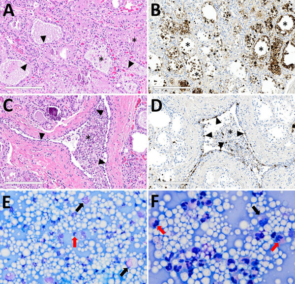
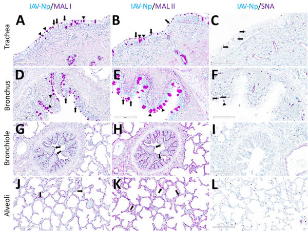
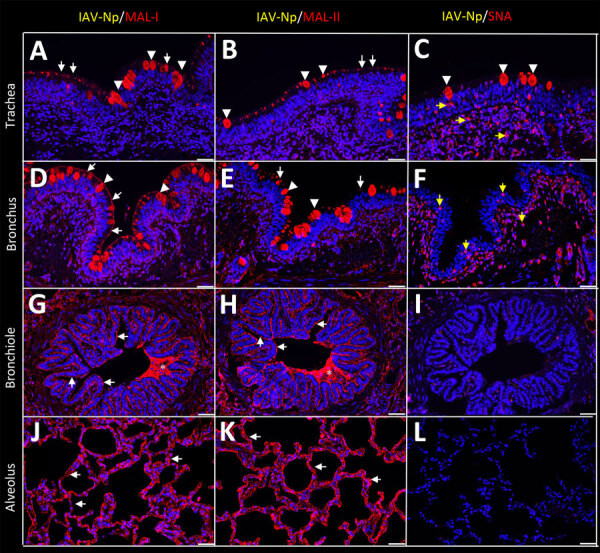
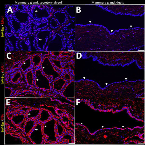
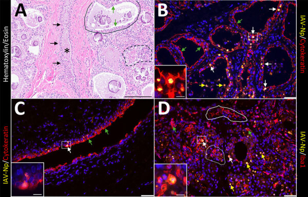
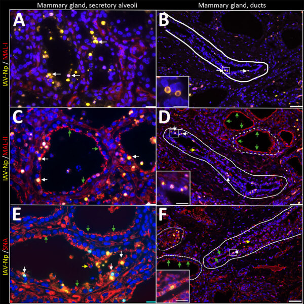
In March 2024, the US Department of Agriculture's Animal and Plant Health Inspection Service reported detection of highly pathogenic avian influenza (HPAI) A(H5N1) virus in dairy cattle in the United States for the first time. One factor that determines susceptibility to HPAI H5N1 infection is the presence of specific virus receptors on host cells; however, little is known about the distribution of the sialic acid (SA) receptors in dairy cattle, particularly in mammary glands. We compared the distribution of SA receptors in the respiratory tract and mammary gland of dairy cattle naturally infected with HPAI H5N1. The respiratory and mammary glands of HPAI H5N1-infected dairy cattle are rich in SA, particularly avian influenza virus-specific SA α2,3-gal. Mammary gland tissues co-stained with sialic acids and influenza A virus nucleoprotein showed predominant co-localization with the virus and SA α2,3-gal. HPAI H5N1 exhibited epitheliotropism within the mammary gland, and we observed rare immunolabeling within macrophages.

Keywords: H5N1; Neu5Ac; United States; dairy cattle; epithelial cell; highly pathogenic avian influenza; influenza; macrophage; mammary gland; respiratory tract; sialic acid; viruses; zoonoses; α2,3-gal; α2,6-gal.


Figures

Figure 1
Figure 1
Microscopic mammary lesions of an index case in US dairy cattle naturally infected with highly pathogenic avian influenza A(H5N1) virus clade 2.3.4.4b. A) Mammary gland alveoli show epithelial attenuation and vacuolation (arrows), leading to degeneration with intraluminal sloughing and neutrophilic intraluminal inflammation (asterisk). Hematoxylin and eosin stain. B) Mammary glad alveoli show degenerative epithelial cells (asterisks) and strong intracytoplasmic and nuclear immunoreactivity to influenza A virus nucleoprotein. C) Cuboidal epithelium lining of the interlobular duct was markedly attenuated (arrowheads) with abundant intraluminal sloughing and neutrophilic inflammation (asterisk). Hematoxylin and eosin stain. D) Interlobular duct shows of attenuating and degenerative epithelium by intranuclear and cytoplasmic immunoreactivity (brown labeling) with immunopositive intraluminal debris and inflammation (asterisk). Scale bars indicate 200 μm. E, F) Modified Wright's stained representative cytology images of milk from a dairy cow with highly pathogenic avian influenza A(H5N1) virus infection, demonstrating moderate to marked neutrophilic inflammation (red arrows) with low numbers of vacuolated macrophages (black arrows) among large numbers of lipid vacuoles. A 100-cell count was performed, and nucleated cells were found to consist of 83% neutrophils, 12% macrophages, and 5% lymphocytes. Original magnifications ×500 for panel E and ×1,000 for panel F.
Figure 2
Figure 2
Respiratory tract tissues from a US dairy cow infected with highly pathogenic avian influenza A(H5N1) virus, showing IAV-Np (teal chromogen), individually duplexed with MAL-I (magenta chromogen), MAL-II (magenta chromogen), and SNA (magenta chromogen) using chromogenic staining. Representative images are shown for IAV-Np/MAL-I (A, D, G, J), IAV-Np/MAL-II (B, E, H, K), and IAV-Np/SNA (C, F, I, L) are shown. No IAV-Np was observed in the unaffected respiratory tissue sections. Intense granular to punctate labeling for MAL-I (A) and MAL-II (B) were observed within goblet cells (arrowheads), along the apical ciliated margin (arrows), and glands of the trachea. SNA labeling (C) was confined to intraepithelial round cells (arrows), endothelium, and glands of the trachea. Similar labeling for MAL-I (D) and MALII (E) was observed within the bronchial lumen within goblet cells (arrowheads) and along the apical cell margin (arrows). SNA (F) labeling was only observed in rare goblet cells (arrowheads) and lamina proprial round cells (arrows) in the bronchus. Bronchioles had diffuse, fine, fibrillary to apical membranous labeling (arrows) MAL-I (G) and MALII (H). No substantial labeling was detected within the mucosal epithelial cells of bronchioles with SNA (I). Diffuse, fine, apical membranous labeling of pneumocytes lining alveoli was observed with MAL-I (J) and MAL-II (K) (arrows). SNA (L) labeling within the alveolar portions of the lung was confined to endothelium and interstitial round cells. Scale bars indicate 200 μm. IAV-Np, influenza A virus nucleoprotein; MAL, Maackia amurensis lectin; SNA, Sambucus nigra lectin.
Figure 3
Figure 3
Respiratory tract tissues from a US dairy cow infected with highly pathogenic avian influenza A(H5N1) virus, labeled with IAV-Np (yellow pseudocolor, DyLight 594), individually duplexed with MAL-I (red pseudocolor, Alexa Fluor 647), MAL-II (red pseudocolor, Alexa Fluor 647), and SNA (red pseudocolor, Alexa Fluor 647) using fluorescent staining. Representative merged images are shown for IAV-Np and MAL-I (A, D, G, J), IAV-Np and MAL-II (B, E, H, K), and IAV-Np and SNA (C, F, I, L). IAV-Np labeling was not detected within the respiratory tissue sections. Intense, granular to punctate, cytoplasmic MAL-I, MAL-II, and SNA labeling was observed in goblet cells (arrowheads) and glands of the trachea (A–C). Similar goblet cell labeling (arrowheads) for MAL-I (D) and MAL-II (E) was observed in the bronchi with weak SNA labeling (F). Multifocal, moderate, fibrillar, apical, membranous MAL-I (A) and MAL-II (B) labeling (white arrows) was observed on the tracheal epithelium. The respiratory epithelium of the bronchi, bronchioles, and alveoli had diffuse, moderate to intense, apical, fibrillar MAL-I labeling (white arrows) (D, G, J). The respiratory epithelium of the bronchi had multifocal, moderate, fibrillar, apical MAL-II labeling (white arrows) (E). The respiratory epithelium of the bronchioles and alveoli had diffuse MAL-II labeling (white arrows) (H, K). Intraluminal secretory material (asterisks) in the bronchi and bronchioles were intensely labeled with MAL-I and MAL-II (G, H). Membranous, granular SNA labeling (yellow arrows) was observed in intraepithelial and lamina proprial round cells in the trachea and bronchi (C, F). Scale bars indicate 50 μm. IAV-Np, influenza A virus nucleoprotein; MAL, Maackia amurensis lectin; SNA, Sambucus nigra lectin.
Figure 4
Figure 4
Unaffected region of the mammary gland from a US dairy cow infected with highly pathogenic avian influenza A(H5N1) virus, showing IAV-Np (teal chromogen), individually duplexed with MAL-I (magenta chromogen), MAL-II (magenta chromogen), and SNA (magenta chromogen) using chromogenic staining. Representative images of IAV-Np/MAL-I (A, B), IAV-Np/MAL-II (C, D), and IAV-Np/SNA (E, F) showing no IAV-Np labeling within unaffected mammary gland tissue sections. No MAL-I was detected in the mammary glandular (A) or interlobular duct epithelium (B). Within the alveolar gland epithelium, intense, granular, fibrillar labeling (arrows) of the apical portion labeling of MAL-II (C) was noted, with no epithelial labeling within the interlobular duct (D). Multifocal, strong, punctate, apical labeling (arrows) with SNA was observed within the mammary glandular epithelium (E). Scant apical labeling (arrow) was observed within the interlobular ductal epithelium with SNA (F). Scale bars indicate 200 μm. IAV-Np, influenza A virus nucleoprotein; MAL, Maackia amurensis lectin; SNA, Sambucus nigra lectin.
Figure 5
Figure 5
Unaffected region of the mammary gland from a US dairy cow infected with highly pathogenic avian influenza A(H5N1) virus, labeled with IAV-Np (yellow pseudocolor, DyLight 594), individually duplexed with MAL-I (red pseudocolor, Alexa Fluor 647), MAL-II (red pseudocolor, Alexa Fluor 647), and SNA (red pseudocolor, Alexa Fluor 647) using fluorescent staining. Representative merged images of IAV-Np/MAL-I (A, B), IAV-Np/MAL-II (C, D), and IAV-Np/SNA (E, F). IAV-Np labeling was not detected within unaffected mammary gland tissue sections. MAL-I labeling was neither observed in the epithelial cells (white arrows) lining the secretory alveoli (A) nor on the epithelial cells (white arrowheads) lining the interlobular ducts (B). Intense, apical, fibrillary MAL-II labeling was observed in the epithelial cells (white arrows) lining the secretory alveoli of unaffected mammary glands (C). MAL-II labeling was not observed in the epithelial cells (arrowheads) lining the interlobular ducts (D). Intense, apical, granular, membranous to cytoplasmic SNA labeling in the epithelial cells lining (white arrows) the secretory alveoli (E) and lining the interlobular ducts (arrowheads) (F) was observed. Scale bars indicate 50 μm. IAV-Np, influenza A virus nucleoprotein; MAL, Maackia amurensis lectin; SNA, Sambucus nigra lectin.
Figure 6
Figure 6
Infected region of the mammary gland from a US dairy cow infected with highly pathogenic avian influenza A(H5N1) virus, labeled with IAV-Np (yellow pseudocolor, DyLight 594), individually duplexed either with epithelial marker pan-cytokeratin (red pseudocolor, Alexa Fluor 647) (B, C) or macrophage or monocytic marker ionized calcium binding adaptor molecule 1 (Iba1) (red pseudocolor, Alexa Fluor 647) (D) using fluorescent labeling. A representative image from a hematoxylin and eosin stain section highlights the cellular architecture of the affected mammary gland (A). Unaffected secretory alveoli were observed in the section (solid black outline). Secretory alveoli variably contain eosinophilic proteinaceous material and corpora amylacea. The secretory alveoli were lined by cuboidal epithelial cells (green arrows) that were variably vacuolated. A few secretory alveoli were disrupted by inflammation (macrophages and neutrophils) and epithelial necrosis (dashed black outline). A single interlobular duct (black asterisk) lined by a bilayer of low cuboidal epithelial cells (black arrows) was observed. The duct contains eosinophilic proteinaceous fluids with scattered inflammatory cells (macrophages and neutrophils) and sloughed epithelium (A). Epithelial cells lining secretory alveoli (B) and interlobular ducts (C) were labeled with pancytokeratin as expected. IAV-Np intranuclear co-labeling (white arrows) in epithelial cells lining the secretory alveoli (B) and ducts (C). Intraluminal cells within secretory alveoli had intranuclear IAV-Np labeling (yellow arrows) that variably co-labeled with pan-cytokeratin. Likewise, Iba1 labeling (green arrows) was observed in the lumens of secretory alveoli or interlobular (alveolus labeling highlighted by white dotted outline) and interstitium (highlighted by the solid white outline) (D). Iba1 labeling was intense, diffuse, and cytoplasmic. IAV-Np intranuclear and intracytoplasmic labeling was less commonly co-labeled within Iba1 labeled cells (white arrows) (D). IAV-Np labeling was not observed in Iba1-labeled cells in the interstitium solid white outline). Insets highlight intranuclear labeling in panels C, B, and D, and the white boxes in the corresponding images represent the origin of the inset image. Scale bars indicate 200 μm (A), 50 μm (B, C, D), and 20 μm (insets). IAV-Np, influenza A virus nucleoprotein; Iba1, ionized calcium binding adaptor molecule 1.
Figure 7
Figure 7
Infected region of the mammary gland from a US dairy cow infected with highly pathogenic avian influenza A(H5N1) virus, labeled with IAV-Np (yellow pseudocolor, DyLight 594), individually duplexed with MAL-I (red pseudocolor, Alexa Fluor 647), MAL-II (red pseudocolor, Alexa Fluor 647), and SNA (red pseudocolor, Alexa Fluor 647) using fluorescent staining. Representative merged images of IAV-Np/MAL-I (A, B), IAV-Np/MAL-II (C, D), and IAV-Np/SNA (E, F) are shown. Moderate to intense intranuclear and intracytoplasmic IAV-Np labeling (yellow, white arrows) was observed in epithelial cells lining the secretory alveolus of the mammary gland; however, no MAL-I labeling was detected (A). Rare, moderate intranuclear IAV-Np labeling (yellow, white arrows), but no MAL-I labeling was observed in epithelial cells lining the interlobular duct (solid white outline) (B). Intense, apical, fibrillary MAL-II labeling (green arrows) was observed in the epithelial cells lining the secretory alveoli of infected mammary glands (C). IAV-Np labeling was co-labeled with MAL-II in individual epithelial cells (white arrows). IAV-Np labeling (white arrows) was observed in epithelial cells of interlobular ducts (solid white outline) (D). MAL-II labeling was not observed in interlobular ducts but was detected in the unaffected secretory alveoli (dashed white outline) (D). Intraluminal cells with cytoplasmic MAL-II labeling were observed (yellow arrow) (D). Intense, apical, granular, membranous to cytoplasmic SNA labeling (green arrows) was observed in the epithelial cells lining the secretory alveoli of infected mammary glands (E). IAV-Np intranuclear labeling was co-labeled with SNA in individual epithelial cells (white arrows) in the secretory alveoli (E). SNA (green arrows) and IAV-Np (white arrows) were observed in epithelial cells of the interlobular ducts (dashed or solid white outline) and were co-labeled to individual ductal epithelial cells (F). Sloughed intraluminal cells had membranous SNA labeling (yellow arrows). Adjacent secretory alveoli had prominent SNA labeling (dashed white outline). The dotted white outline highlights a severely affected secretory gland (F). Insets highlight white boxed areas in panels B, D, and F. Scale bar indicate 20 μm (A, C, E), 50 μm (B, D, F), and 20 μm (insets). IAV-Np, influenza A virus nucleoprotein; MAL, Maackia amurensis lectin; SNA, Sambucus nigra lectin.
Figure 8
Figure 8
Infected region of the mammary gland from a US dairy cow infected with highly pathogenic avian influenza A(H5N1) virus, showing IAV-Np (teal chromogen), individually duplexed with MAL-I (magenta chromogen), MAL-II (magenta chromogen), and SNA (magenta chromogen) using chromogenic staining. Representative images IAV-Np/MAL-I (A, B), IAV-Np/MAL-II (C, D), and IAV-Np/SNA (E, F) in infected mammary gland and interlobular duct are shown. Strong intracytoplasmic and intranuclear immunoreactivity with IAV-Np was observed within the glandular epithelium of the mammary alveolus and interlobular ducts (arrowheads) with no substantial MAL-I labeling (A, B). Scant, multifocal, apical, punctate MAL-II labeling was observed in degenerative mammary gland epithelial cells (arrows) with strong, intranuclear, and intracytoplasmic immunoreactivity for IAV-Np (arrowheads) (C). Interlobular ductal epithelium exhibits strong, multifocal intranuclear and intracytoplasmic immunoreactivity to IAV-Np in attenuated and sloughed cells (arrowheads) with no substantial epithelial MAL-II detected (D). Strong, multifocal, punctate, apical labeling with SNA in attenuated or degenerative epithelial cells (arrows) with strong, intranuclear, and intracytoplasmic immunoreactivity for IAV-Np (arrows) was observed within mammary gland epithelium (E). Scant, multifocal, delicate, apical labeling for SNA (arrows) within ductal epithelium with strong, intranuclear, and intracytoplasmic immunoreactivity within sloughed, intraluminal epithelial cells for IAV-Np (arrowheads) was observed in an interlobular duct (F). Scale bars indicate 200 μm. IAV-Np, influenza A virus nucleoprotein; MAL, Maackia amurensis lectin; SNA, Sambucus nigra lectin.

Similar articles

Cited by

References

    1. Burrough ER, Magstadt DR, Petersen B, Timmermans SJ, Gauger PC, Zhang J, et al. Highly pathogenic avian influenza A(H5N1) clade 2.3.4.4b virus infection in domestic dairy cattle and cats, United States, 2024. Emerg Infect Dis. 2024;30:30. 10.3201/eid3007.240508 - DOI - PMC - PubMed
    1. US Department of Agriculture, Animal and Plant Health Inspection Service. Federal and state veterinary, public health agencies share update on HPAI detection in Kansas, Texas dairy herds. 2024. [cited 2024 May 5]. https://www.aphis.usda.gov/news/agency-announcements/federal-state-veter...
    1. Gilbertson B, Subbarao K. Mammalian infections with highly pathogenic avian influenza viruses renew concerns of pandemic potential. J Exp Med. 2023;220:e20230447. 10.1084/jem.20230447 - DOI - PMC - PubMed
    1. US Department of Agriculture, US Department of Health and Human Services. USDA, HHS announce new actions to reduce impact and spread of H5N1. 2024. [cited 2024 May 5]. https://www.usda.gov/media/press-releases/2024/05/10/usda-hhs-announce-n...
    1. Bevins SN, Shriner SA, Cumbee JC Jr, Dilione KE, Douglass KE, Ellis JW, et al. Intercontinental movement of highly pathogenic avian influenza A(H5N1) clade 2.3.4.4 virus to the United States, 2021. Emerg Infect Dis. 2022;28:1006–11. 10.3201/eid2805.220318 - DOI - PMC - PubMed

MeSH terms