

Pathogenic differences of cynomolgus macaques after Taï Forest virus infection depend on the viral stock propagation
- PMID: 38861571
- PMCID: PMC11195944
- DOI: 10.1371/journal.ppat.1012290
Pathogenic differences of cynomolgus macaques after Taï Forest virus infection depend on the viral stock propagation
Abstract
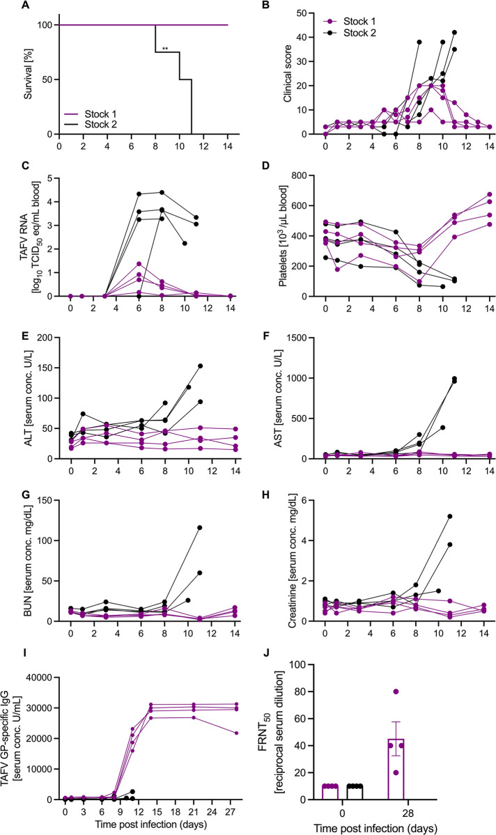
Taï Forest virus (TAFV) is a negative-sense RNA virus in the Filoviridae family. TAFV has caused only a single human infection, but several disease outbreaks in chimpanzees have been linked to this virus. Limited research has been done on this human-pathogenic virus. We sought to establish an animal model to assess TAFV disease progression and pathogenicity at our facility. We had access to two different viral stock preparations from different institutions, both originating from the single human case. Type I interferon receptor knockout mice were inoculated with TAFV stock 1 or stock 2 by the intraperitoneal route. Inoculation resulted in 100% survival with no disease regardless of viral stock preparation or infectious dose. Next, cynomolgus macaques were inoculated with TAFV stock 1 or stock 2. Inoculation with TAFV stock 1 resulted in 100% survival and robust TAFV glycoprotein-specific IgG responses including neutralizing antibodies. In contrast, macaques infected with TAFV stock 2 developed disease and were euthanized 8-11 days after infection exhibiting viremia, thrombocytopenia, and increased inflammatory mediators identified by transcriptional analysis. Histopathologic analysis of tissue samples collected at necropsy confirmed classic filovirus disease in numerous organs. Genomic differences in both stock preparations were mapped to several viral genes which may have contributed to disease severity. Taken together, we demonstrate that infection with the two TAFV stocks resulted in no disease in mice and opposing disease phenotypes in cynomolgus macaques, highlighting the impact of viral stock propagation on pathogenicity in animal models.
Copyright: This is an open access article, free of all copyright, and may be freely reproduced, distributed, transmitted, modified, built upon, or otherwise used by anyone for any lawful purpose. The work is made available under the Creative Commons CC0 public domain dedication.
Conflict of interest statement
The authors have declared that no competing interests exist.
Figures

Similar articles
-
Single-dose VSV-based vaccine protects cynomolgus macaques from disease after Taï Forest virus infection.Emerg Microbes Infect. 2023 Dec;12(2):2239950. doi: 10.1080/22221751.2023.2239950. Emerg Microbes Infect. 2023. PMID: 37470396 Free PMC article.
-
Taï Forest Virus Does Not Cause Lethal Disease in Ferrets.Microorganisms. 2021 Jan 21;9(2):213. doi: 10.3390/microorganisms9020213. Microorganisms. 2021. PMID: 33494199 Free PMC article.
-
Pathogenicity of simian-human immunodeficiency virus SHIV-89.6P and SIVmac is attenuated in cynomolgus macaques and associated with early T-lymphocyte responses.J Virol. 2005 Jul;79(14):8878-85. doi: 10.1128/JVI.79.14.8878-8885.2005. J Virol. 2005. PMID: 15994781 Free PMC article.
-
Influenza virus A/Anhui/1/2013 (H7N9) replicates efficiently in the upper and lower respiratory tracts of cynomolgus macaques.mBio. 2014 Aug 12;5(4):e01331-14. doi: 10.1128/mBio.01331-14. mBio. 2014. PMID: 25118237 Free PMC article.
-
Establishment of a cynomolgus macaque model of influenza B virus infection.Virology. 2010 Nov 25;407(2):178-84. doi: 10.1016/j.virol.2010.08.006. Epub 2010 Sep 6. Virology. 2010. PMID: 20822788
Cited by
-
An update on nonhuman primate usage for drug and vaccine evaluation against filoviruses.Expert Opin Drug Discov. 2024 Oct;19(10):1185-1211. doi: 10.1080/17460441.2024.2386100. Epub 2024 Aug 18. Expert Opin Drug Discov. 2024. PMID: 39090822 Review.
References
-
- Marzi A, Banadyga L. Ebola Virus (Filoviridae). Encyclopedia of Virology2021. p. 232–44.
-
- Formenty P, Hatz C, Guenno BL, Stoll A, Rogenmoser P, Widmer A. Human Infection Due to Ebola Virus, Subtype Coˆte d’Ivoire: Clinical and Biologic Presentation. The Journal of Infectious Diseases. 1999;179:S48–53. - PubMed
-
- Formenty P, Boesch C, Wyers M, Steiner C, Donati F, Dind F, et al.. Ebola Virus Outbreak among Wild Chimpanzees Living in a Rain Forest of Cote d’Ivoire. The Journal of Infectious Diseases. 1999;179:S120–S6. - PubMed
MeSH terms
Substances
LinkOut - more resources
Full Text Sources
