. 2024 Jun 18;121(25):e2316143121.
doi: 10.1073/pnas.2316143121. Epub 2024 Jun 11.

Vibrio MARTX toxin processing and degradation of cellular Rab GTPases by the cytotoxic effector Makes Caterpillars Floppy

Affiliations

Vibrio MARTX toxin processing and degradation of cellular Rab GTPases by the cytotoxic effector Makes Caterpillars Floppy

Alfa Herrera et al. Proc Natl Acad Sci U S A. .

Abstract

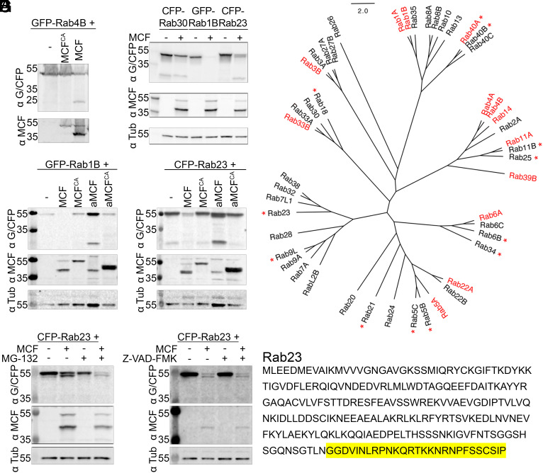
Vibrio vulnificus causes life-threatening wound and gastrointestinal infections, mediated primarily by the production of a Multifunctional-Autoprocessing Repeats-In-Toxin (MARTX) toxin. The most commonly present MARTX effector domain, the Makes Caterpillars Floppy-like (MCF) toxin, is a cysteine protease stimulated by host adenosine diphosphate (ADP) ribosylation factors (ARFs) to autoprocess. Here, we show processed MCF then binds and cleaves host Ras-related proteins in brain (Rab) guanosine triphosphatases within their C-terminal tails resulting in Rab degradation. We demonstrate MCF binds Rabs at the same interface occupied by ARFs. Moreover, we show MCF preferentially binds to ARF1 prior to autoprocessing and is active to cleave Rabs only subsequent to autoprocessing. We then use structure prediction algorithms to demonstrate that structural composition, rather than sequence, determines Rab target specificity. We further determine a crystal structure of aMCF as a swapped dimer, revealing an alternative conformation we suggest represents the open, activated state of MCF with reorganized active site residues. The cleavage of Rabs results in Rab1B dispersal within cells and loss of Rab1B density in the intestinal tissue of infected mice. Collectively, our work describes an extracellular bacterial mechanism whereby MCF is activated by ARFs and subsequently induces the degradation of another small host guanosine triphosphatase (GTPase), Rabs, to drive organelle damage, cell death, and promote pathogenesis of these rapidly fatal infections.

Keywords: MARTX toxin; Rab GTPases; Vibrio vulnificus; effector; host-pathogen interaction.


Conflict of interest statement

Competing interests statement:The authors declare no competing interest.

Figures

Fig. 1.
Fig. 1.
Model of MCF activation. The MARTX toxin is secreted as a single polypeptide from the bacterium. Using its N- and C-terminal repeats it forms a pore at the eukaryotic membrane to translocate its effectors, in this case the Domain of Unknown Function 1 (DUF1, green box), the Rho GTPase-inactivation domain (RID, red box), the Alpha/Beta Hydrolase domain (ABH, yellow box), and MCF (purple, box) as well as a CPD into the host cytoplasm. Inside, CPD is activated by IP6 to cleave and release effectors individually. However, it does not cleave between MCF and the effector in front of it resulting in an effector module. GTP-bound ARFs (ARF1, shown) stimulate MCF autoprocessing to release aMCF from the effector it is tethered to. Following release aMCF binds phosphatidylinositol-5-phosphate at membranes and is N-terminally acetylated. This figure was made with BioRender.com.
Fig. 2.
Fig. 2.
The cysteine protease activity of the MCF toxin induces variable degradation of Rab GTPases. (A and B) Fluorescently tagged Rabs ectopically coexpressed in HEK 293T cells with MCF, MCFCA, or empty vector control (p3xFlag-CMV-7.1). Representative western blots shown of 35 μg of total protein from whole cell lysates recovered from cells (n = 3). Rabs detected by anti-GFP/CFP (α G/CFP), MCF by anti-MCF (α MCF), and tubulin as a loading control by anti-α-Tubulin (α Tub) antibodies. (C) Unrooted amino acid phylogenetic cladogram of Rab isoforms tested in the screen. Names in red indicate Rabs significantly degraded when coexpressed in HEK 293T cells with MCF, established by degradation factor determined by densitometry of bands on western blots from three independent experiments (Material and Methods). Names with a red asterisk denote Rabs cleaved when coexpressed with MCF, as manually assessed. Western blots of independent transfections used to make the designations can be found in SI Appendix, Fig. S2 and analysis in SI Appendix, Fig. S3. (D) HEK 293T cells were co‐transfected with MCF, MCFCA, aMCF, aMCFCA, or p3xFlag-CMV-7.1 and either GFP-Rab1B or CFP-Rab23 (n = 3). Western blots on cell lysates recovered from these cells were performed as in (A). (E and F) Coexpression experiments and western blot analysis were completed as in (B) with CFP-Rab23 and MCF in the presence of either (E) 5 μM proteasome inhibitor MG-132 or (F) 5 μM pancaspase inhibitor Z-VAD-FMK. (G) Protein sequence of Rab23 designating residues cleaved off (shaded in yellow) during coexpression with MCF in HEK 293T cells.
Fig. 3.
Fig. 3.
MCF coprecipitates (IP) with and cleaves Rabs. (A) Recombinant aMCF incubated with either GST-Rab1B or GST-Rab23 at 37 °C and reactions subsequently separated by SDS-PAGE gel and stained with Coomassie. Arrows indicate cleaved Rab bands. (B) Purified (Left) GST‐Rab1B or (Right) GST-Rab23 incubated with either MCF, aMCF, or residues 85 to 324 of aMCF (aMCFdomain II) at 37 °C. Purified recombinant ACD was used as a control for nonspecific binding. Western blot on the incubated samples (input), flow through following incubation with GST beads (unbound), and sample following anti‐GST IP (elution) using anti-MCF (α MCF), anti‐GST (α GST), and anti-ACD (α ACD) antibodies. Representative gels (n = 3). (C) Cleavage of Rab1B and Rab23 by MCF determined by densitometry of bands on Coomassie-stained gels (% Cleaved Rab = (cleaved Rab band/(cleaved Rab band + uncleaved Rab band)) × 100) from three independent reactions. Significance was calculated using parametric t tests, P value: 0.1234 (ns), 0.0332 (*), 0.0021 (**), 0.0002 (***), < 0.0001 (****). (D) Recombinant MCF or aMCF were incubated with either ARF1 or GST-Rab11A for 2 h prior to the addition of the other GTPase followed by overnight incubation at 37 °C. GTPase added after 2 h is in bolded font. Anti-GST IP and western blot on input, unbound, and elution samples were completed as above in (B) including anti-ARF1 (α ARF1) antibodies.
Fig. 4.
Fig. 4.
Structure of unbound aMCF toxin and predicted complex structure of aMCF with Rabs. (A) Three views of predicted complex ribbon structure of Rab1B (cornflower blue) bound to surface representation of aMCF (purple) overlaid onto the ribbon structure of ADP Ribosylation Factor 3 mutated to mimic the active state (ARF3Q71L) (cyan) bound to aMCFCS (not shown) (PDB code 6ii6) (5). Residues important for proteolytic activity of aMCF (Gln118, Arg132, Cys133, Asp134, His245, and Asp264) highlighted in red. (B) Predicted ribbon structure of Rab23 (blue) in complex with surface representation of aMCF (purple). Residues removed during cotransfection with MCF in HEK 293T cells are highlighted in yellow. (C–E) Ribbon structure of (C) closed aMCFCS derived from the complex structure with ADP Ribosylation Factor 3 mutated to mimic the active state (ARF3Q71L) (PDB code 6ii6) (5), (D) closed unbound aMCFCS (PDB code 6ii0) (5), and (E) open unbound aMCFCA (PDB code 8sfg) with surface representation of swapped dimer in light gray, in a rainbow spectrum from the N-terminus in blue to the C-terminus in red. α4 and α7 helices (according to PDB code 6ii6) (5) important for flexibility and binding denoted and distinct domains of unbound aMCF outlined. Residues important for proteolytic activity of aMCF (Gln118, Arg132, Cys133, Asp134, His245, and Asp264) are highlighted in magenta on structures (4, 5, 7). (F and G) Length in number of amino acid residues of the (F) fifth α-helix and (G) hypervariable tail in Rabs screened determined from structure prediction models. Significance was calculated using a nonparametric Kruskal–Wallis test, P value: 0.1234 (ns), 0.0332 (*), 0.0021 (**), 0.0002 (***), <0.0001 (****).
Fig. 5.
Fig. 5.
MCF mis-localizes endogenous Rab1B and decreases Rab1B intensity in infected tissues. (A) Immunofluorescence microscopy was completed on Cos7 cells transfected with empty vector control (pEGFP-N3) or MCF-EGFP (green) for 18 h, fixed, and stained for DAPI (blue), and endogenous Rab1B (red). (B) ICR mice were intoxicated with CMCP6 ΔvvhA rtxA1::bla or CMCP6 ΔvvhA rtxA1::mcf-bla at 3.3 × 10⁸ to 1 × 10⁹ CFU/mL intragastrically and experiment allowed to continue for up 24 h. At time of death or at the end of the experiment the small intestine were excised and Rab1B signal intensity quantified in the tissue (n = 6). Significance was calculated using parametric t tests, P value: 0.1234 (ns), 0.0332 (*), 0.0021 (**), 0.0002 (***), <0.0001 (****).
Fig. 6.
Fig. 6.
Model of MCF mediated Golgi fragmentation. aMCF binds many membrane-bound Rabs, with their hypervariable tail exposed (undetermined whether dependent on the nucleotide bound status of Rabs). aMCF induces widespread degradation and cleavage of Rabs which ultimately results in Golgi fragmentation. Golgi modified from BioRender.com.
Fig. 7.
Fig. 7.
Proposed models of Rab1B binding based on solved structures of open and closed autoprocessed MCF. N-terminal domain I (residues 1 to 82), linker (residues 83 to 118), and the core domain (residues 119 to 361) of open aMCF in magenta, hot pink, and purple, respectively, and Rab1B in cornflower blue. Models shown as surface structures with the exception of α4 of closed aMCF as it transitions into α4, β0, and α4’ of open aMCF presented as a ribbon structure. In model 1, closed unbound aMCF (PDB code 6ii0) (5) transitions to a structure that resembles its ARF3-bound conformation (PDB code 6ii6) (5) to bind Rab1B, as is predicted in Fig. 4A. In model 2, Rab1B binds to open aMCF (PDB code 8sfg), as overlaid onto the (2A) predicted Rab1B-aMCF (Fig. 4A) or the (2B) predicted Rab1B-aMCF119-361 costructure (SI Appendix, Fig. S14).

Update of

Similar articles

Cited by

References

    1. Satchell K. J. F., Multifunctional-autoprocessing repeats-in-toxin (MARTX) Toxins of Vibrios. Microbiol. Spectr. 3, (2015), 10.1128/microbiolspec.ve-0002-2014. - DOI - PMC - PubMed
    1. Gavin H. E., Satchell K. J., MARTX toxins as effector delivery platforms. Pathog. Dis. 73, ftv092 (2015). - PMC - PubMed
    1. Kling K., et al. , Genetic divergence of Vibrio vulnificus clinical isolates with mild to severe outcomes. mBio 13, e0150022 (2022), 10.1128/mbio.01500-22. - DOI - PMC - PubMed
    1. Herrera A., et al. , N-terminal autoprocessing and acetylation of multifunctional-autoprocessing repeats-in-toxins (MARTX) Makes Caterpillars Floppy-like effector is stimulated by adenosine diphosphate (ADP)-Ribosylation Factor 1 in advance of Golgi fragmentation. Cell Microbiol. 22, e13133 (2020). - PMC - PubMed
    1. Lee Y., et al. , Makes caterpillars floppy-like effector-containing MARTX toxins require host ADP-ribosylation factor (ARF) proteins for systemic pathogenicity. Proc. Natl. Acad. Sci. U.S.A. 116, 18031–18040 (2019). - PMC - PubMed

LinkOut - more resources