. 2024 Jun 18;121(25):e2322588121.
doi: 10.1073/pnas.2322588121. Epub 2024 Jun 11.

Single-tissue proteomics in Caenorhabditis elegans reveals proteins resident in intestinal lysosome-related organelles

Affiliations

Single-tissue proteomics in Caenorhabditis elegans reveals proteins resident in intestinal lysosome-related organelles

Chieh-Hsiang Tan et al. Proc Natl Acad Sci U S A. .

Abstract

The nematode intestine is the primary site for nutrient uptake and storage as well as the synthesis of biomolecules; lysosome-related organelles known as gut granules are important for many of these functions. Aspects of intestine biology are not well understood, including the export of the nutrients it imports and the molecules it synthesizes, as well as the complete functions and protein content of the gut granules. Here, we report a mass spectrometry (MS)-based proteomic analysis of the intestine of the Caenorhabditis elegans and of its gut granules. Overall, we identified approximately 5,000 proteins each in the intestine and the gonad and showed that most of these proteins can be detected in samples extracted from a single worm, suggesting the feasibility of individual-level genetic analysis using proteomes. Comparing proteomes and published transcriptomes of the intestine and the gonad, we identified proteins that appear to be synthesized in the intestine and then transferred to the gonad. To identify gut granule proteins, we compared the proteome of individual intestines deficient in gut granules to the wild type. The identified gut granule proteome includes proteins known to be exclusively localized to the granules and additional putative gut granule proteins. We selected two of these putative gut granule proteins for validation via immunohistochemistry, and our successful confirmation of both suggests that our strategy was effective in identifying the gut granule proteome. Our results demonstrate the practicability of single-tissue MS-based proteomic analysis in small organisms and in its future utility.

Keywords: lysosome-related organelle; microproteomics; tissue-specific mass-spectrometry; yolk protein.


Conflict of interest statement

Competing interests statement:The authors declare no competing interest.

Figures

Fig. 1.
Fig. 1.
MS–based proteomic analysis of extracted nematode tissues. (A) A young adult stage hermaphrodite C. elegans with the intestine (blue shade) and gonad (red shade) colored for identification. (B) An extracted gonad arm severed at the spermatheca. (C) Extracted intestinal tissue. (D) Schematic representation of the workflow. Intestinal tissues and gonads were extracted from young adult worms, snap-frozen, heated, sonicated, and digested prior to MS and data analysis. The figure was created with BioRender.com.
Fig. 2.
Fig. 2.
The intestinal and gonadal proteomes of C. elegans. (A) A total of 5,962 proteins were detected in our wild-type gonad and intestine samples. 5,243 were identified in the gonad, and 4,848 were identified in the intestine. 4,129 proteins were identified in both the intestine and the gonad, while 1,114 were identified only in the gonad, and 719 were identified only in the intestine. (B–D) Overview of proteins identified in the pooled tissue samples. Tissues were collected from five animals in each of the four samples used per tissue type. (B) The data from the four pooled samples for each tissue were analyzed together. From these pooled tissue samples, 5,464 proteins were detected. 3,532 of those were shared in both tissues, while 1,188 were identified only in the gonad and 744 only in the intestine. (C and D) The tissue samples of the same type show a high degree of similarity in both the identified proteins and their abundances while being clearly distinctive from those of the other tissue type. (C) PCA of the pooled tissue samples. Samples were divided into two distinct groups separated on the PC1. (D) Heatmap displaying the proteins of the pooled-tissue samples. The Z-score, representing the distance in SDs from the mean within each row, was computed by subtracting the mean protein abundance from each individual abundance and then dividing by the SD across all samples in that row. (E) Gene Ontology biological processes enrichment analysis of proteins identified at least twice in the same tissue types. 5,297 proteins were detected more than once in the same tissue type. From these, 3,501 were shared in both tissue types, with an overrepresentation of proteins involved in essential housekeeping processes such as ribosome assembly. 1,060 were detected only in the gonad, with an overrepresentation of proteins involved in processes expected to be predominant in the germ cells, such as cell cycle. 726 were detected only in the intestine, with an overrepresentation of proteins involved in processes expected to be predominant in the intestinal cells, such as immune response, metal transport, and lipid metabolism. The Euler diagrams are area-proportional. Lists of proteins described in this figure can be found in Dataset S1.
Fig. 3.
Fig. 3.
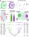
Single-tissue proteomic analysis. (A) A total of 4,012 proteins were identified from six samples of gonadal tissue, each isolated from a single animal, with up to 3,316 (82.7%) identified in a single gonad sample. (B) A total of 3,732 proteins were identified from six samples of intestinal tissue each extracted from a single animal, with up to 2,889 (77.4%) identified in a single intestinal tissue sample. (C and D) The single-tissue samples of the same type show a high degree of similarity and were clearly distinctive from that of the other tissue type. (C) PCA of the single-tissue samples. Samples were divided into two distinct groups separated on the PC1. (D) Heatmap of the single-tissue samples. The Z-score, representing the distance in SDs from the mean within each row, was computed by subtracting the mean protein abundance from each individual abundance and then dividing by the SD across all samples in that row. (E) The vast majority of the identified proteins were repeatedly identified in tissues isolated from different animals, with nearly half of them identified in every animal. (F) A high-confidence set of proteins present in each tissue, 2,368 in the gonad and 1,713 in the intestine, were identified based on consistent identification of the proteins across different animals and experimental sets. Of these proteins, 450 and 418 were consistently enriched in the gonad and intestine, respectively. (G) Center: A volcano plot showing the relative abundance of the proteins between the gonad and the intestine. Points representing proteins in the high-confidence sets found to be enriched in the gonad (68) and intestine (143) are highlighted with the corresponding colors (green and purple, respectively), and some of the most abundant proteins are labeled. The vitellogenins, which are highly abundant in both tissues, are also labeled in yellow. Flanking the volcano plot on either side are lists of highly abundant proteins selected from those highly enriched (maxed out fold-change) in either the gonad (382) or the intestine (275) and cannot be placed in the plot. Two intestinal-enriched proteins, LRO-1 (plot) and MRP-3 (side), are also labeled and will be discussed more in later sections. The values used in this panel were based on one of the two experimental sets, but the highlighted (colored) proteins were those that were consistently enriched in the respective tissue. Green: gonad; purple: intestine; Yellow: vitellogenins. The Euler diagrams are area-proportional. Lists of proteins described in this figure can be found in Dataset S2.
Fig. 4.
Fig. 4.
Proteome–transcriptome comparison reveals gonadal proteins with an intestinal origin. (A and B) The majority of the genes encoding the proteins enriched in the gonad (A) and the intestine (B) were also highly enriched as mRNA in the same tissues. (C) We identified 99 proteins that are likely gonadal proteins transcribed as mRNA in the intestine by comparing proteins that are consistently found in both the gonad and the intestine and not intestinally enriched to the list of intestinally enriched mRNAs (50) (D) Enrichment analysis of the list of 99 likely transported from the intestine to the gonad features proteins involved in lipid transport and localization as well as other metabolic processes. The Euler diagrams are area-proportional. The lists of proteins analyzed in this figure are in Dataset S5.
Fig. 5.
Fig. 5.
Identification of LRO–associated proteins through MS–based genetic analysis. (A) Proteins identified in glo-1(-) and wild-type intestines. 11 intestines of each genotype were assayed individually in two independent experiments. (B) Proteins repeatedly identified in the intestines of respective genotypes. (C) Heatmap comparing protein abundances of the glo-1(-) and wild-type intestines. The intestine proteome of the two genotypes is similar but distinctive. The Euler diagrams are area-proportional. Lists of proteins described in this figure can be found in Dataset S6.
Fig. 6.
Fig. 6.
MRP-3 and LRO-1 are gut granule proteins. Anti-FLAG staining of gonad and intestine tissues dissected from wild-type control animals (WT), and from worms with a C-terminal 3XFLAG epitope inserted into the endogenous loci encoding either MRP-3 or LRO-1. (A and B) Images of gonad and intestine tissues that were dissected from worms shown in DIC (differential interference contrast microscopy), staining with the DNA dye DAPI, anti-FLAG antibody, and merged. (A) MRP-3 and LRO-1 are abundant in the intestine but not in the gonad. Exposure was kept constant across experiments. Scale bar: 100 µm (B) MRP-3 and LRO-1 are gut granule proteins. MRP-3 and LRO-1 are subcellularly localized to granule-like organelles in the glo-1(+) background and were largely absent (MRP-3) or scattered with reduced abundance (LRO-1) in the glo-1(-) background, in which gut granule synthesis is impaired. Exposure was kept constant across experiments. (Scale bar: 20 µm.)

Similar articles

Cited by

References

    1. Sulston J. E., Horvitz H. R., Post-embryonic cell lineages of the nematode, Caenorhabditis elegans. Dev. Biol. 56, 110–156 (1977). - PubMed
    1. Hedgecock E. M., White J. G., Polyploid tissues in the nematode Caenorhabditis elegans. Dev. Biol. 107, 128–133 (1985). - PubMed
    1. McKay R. M., McKay J. P., Avery L., Graff J. M., C elegans: A model for exploring the genetics of fat storage. Dev. Cell 4, 131–142 (2003). - PMC - PubMed
    1. Ashrafi K., et al. , Genome-wide RNAi analysis of Caenorhabditis elegans fat regulatory genes. Nature 421, 268–272 (2003). - PubMed
    1. Rajagopal A., et al. , Haem homeostasis is regulated by the conserved and concerted functions of HRG-1 proteins. Nature 453, 1127–1131 (2008). - PMC - PubMed

MeSH terms

LinkOut - more resources