. 2024 Jun 18;121(25):e2400601121.
doi: 10.1073/pnas.2400601121. Epub 2024 Jun 11.

Progesterone inactivation in decidual stromal cells: A mechanism for inflammation-induced parturition

Affiliations

Progesterone inactivation in decidual stromal cells: A mechanism for inflammation-induced parturition

Angela DeTomaso et al. Proc Natl Acad Sci U S A. .

Erratum in

Abstract

The process of human parturition involves inflammation at the interface where fetal chorion trophoblast cells interact with maternal decidual stromal (DS) cells and maternal immune cells in the decidua (endometrium of pregnancy). This study tested the hypothesis that inflammation at the chorion-decidua interface (CDI) induces labor by negating the capacity for progesterone (P4) to block labor and that this is mediated by inactivation of P4 in DS cells by aldo-keto reductase family 1 member C1 (AKR1C1). In human, Rhesus macaque, and mouse CDI, AKR1C1 expression increased in association with term and preterm labor. In a human DS cell line and in explant cultures of term human fetal membranes containing the CDI, the prolabor inflammatory cytokine, interleukin-1ß (IL-1ß), and media conditioned by LPS-stimulated macrophages increased AKR1C1 expression and coordinately reduced nuclear P4 levels and P4 responsiveness. Loss of P4 responsiveness was overcome by inhibition of AKR1C1 activity, inhibition of AKR1C1 expression, and bypassing AKR1C1 activity with a P4 analog that is not metabolized by AKR1C1. Increased P4 activity in response to AKR1C1 inhibition was prevented by the P4 receptor antagonist RU486. Pharmacologic inhibition of AKR1C1 activity prevented parturition in a mouse model of inflammation-induced preterm parturition. The data suggest that inflammatory stimuli at the CDI drive labor by inducing AKR1C1-mediated P4 inactivation in DS cells and that inhibiting and/or bypassing of AKR1C1-mediated P4 inactivation is a plausible therapeutic strategy to mitigate the risk of inflammation-associated preterm birth.

Keywords: inflammation; parturition; pregnancy; progesterone metabolism.


Conflict of interest statement

Competing interests statement:The authors declare no competing interest.

Figures

Fig. 1.
Fig. 1.
AKR1C1 expression in human chorion-decidua increases in association with labor. (A) Immunohistochemical detection of AKR1C1 (brown staining) in the decidual compartment of the CDI at term (representative of N = 8). (B) AKR1C1 and (C) IL1B expression in preterm (N = 3/group) and term [(N = 9 or 10/group) ± labor] CDI (mean ± SEM; P calculated by Student’s t test). (D) Correlation (Pearson’s) of AKR1C1 and IL1B expression in human CDI (N = 25) (P calculated by Pearson’s correlation test).
Fig. 2.
Fig. 2.
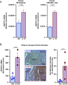
Prolabor stimuli increase AKR1C1 in DS cells. (A) Effect of IL-1ß (1 ng/mL) or PBS for 12 h on the AKR1C1 expression in FM explants (Left) and hFM-DEC cells (Right) (mean ± SEM; P calculate by Student’s t test). (B) AKR1C1 expression (Left) [normalized RNA reads obtained from GSE181054 (9)] (N = 3/group) and representative immunohistochemical localization (indicated by brown staining) of AKR1C1 in the CDI in Rhesus macaques 48 h after inoculation with E. coli or vehicle (Veh) into the amniotic fluid at 80% gestation (N = 3 or 5/group). Mean ± SEM; P calculated by Student’s t test.
Fig. 3.
Fig. 3.
LPS-stimulated immune cells increase AKR1C1 expression in DS cells. (A) Effect of LPS (1 µg/mL) or PBS for 16 h on expression of AKR1C1, IL1B, CXCL8, and IL6 in hFM-DEC cells. (B) Effect of LPS (1 ng/mL) or PBS for 16 h on AKR1C1 expression in term, not in in labor FM explants. (C) Effect of LPS (1 µg/mL), IL-1ß (1 ng/mL), and media from THP-1 cells exposed for 16 h to LPS (1 µg/mL) or vehicle (veh; PBS) on AKR1C1 expression in hFM-DEC cells. (D) hFM-DEC cells were treated as in C ± the IL1R signaling antagonist, anakinra (ana; 2.5 µg/mL). Mean ± SEM; P values calculated by Student’s t test (A and B) and one-way ANOVA (C and D). Groups that are not significantly different from one another (P > 0.05) are denoted by a (C and D).
Fig. 4.
Fig. 4.
AKR1C1 inhibits P4/PR signaling in DS cells. Effect of BPSA (100 nM), RU486 (50 nM), and R5020 (100 nM) on IL-1ß-induced CXCL8 expression (A and B) and IL15 expression (C) in FM explants (term not in labor). (D) Effect of AKR1C1 knockdown by RNAi (SI Appendix, Fig. S3) in hFM-DEC cells on IL-1ß-induced CXCL8 expression. (E) Effect of IL-1ß (1 ng/mL for 16 h; Left or 22 h; Right) and AKR1C1 knockdown by RNAi (Right) on the level of P4 in nuclear lysates from hFM-DEC cells. Mean ± SEM; P values calculated by Student’s t test.
Fig. 5.
Fig. 5.
Inhibition of AKR1C1 prevents inflammation-induced parturition in mice. Mice were treated with LPS on GD14.5. (A) Effect of LPS (100 µg) on Il1b (Right) and Akr1c18 (Left) expression in the decidua of mice at 1 h and 3 h after treatment (N = 5/group). Mean ± SEM; P values calculated by one-way ANOVA. (B) Effect of BPSA on LPS-induced preterm birth (Materials and Methods); N = 7 or 8 mice/group; P values calculated by Chi-square test.
Fig. 6.
Fig. 6.
Working model. We propose that for most of pregnancy P4/PR signaling in DS cells block labor in part by promoting IL-15-mediated tissue-level immune quiescence. In response to net inflammatory load (e.g., intrauterine infection; fetal development; maternal stress) immune cells at the CDI become activated leading to inflammation and the production of IL-1ß, which induces AKR1C1-mediated local P4/PR withdrawal (i.e., prevention of ligand activation of PRs) in DS cells leading to loss of the P4/PR block to labor and further amplification of tissue-level inflammation that transforms the adjacent myometrium and cervix to the labor state.

References

    1. Young A., et al. , Immunolocalization of proinflammatory cytokines in myometrium, cervix, and fetal membranes during human parturition at term. Biol. Reprod. 66, 445–449 (2002). - PubMed
    1. Keelan J. A., et al. , Cytokines, prostaglandins and parturition–A review. Placenta 24, S33–S46 (2003). - PubMed
    1. Osman I., Young A., Jordan F., Greer I. A., Norman J. E., Leukocyte density and proinflammatory mediator expression in regional human fetal membranes and decidua before and during labor at term. J. Soc. Gynecol. Invest. 13, 97–103 (2006). - PubMed
    1. Norman J. E., Bollapragada S., Yuan M., Nelson S. M., Inflammatory pathways in the mechanism of parturition. BMC Pregnancy Childbirth. 7, S7 (2007). - PMC - PubMed
    1. Galinsky R., Polglase G. R., Hooper S. B., Black M. J., Moss T. J., The consequences of chorioamnionitis: Preterm birth and effects on development. J. Pregnancy 2013, 412831 (2013). - PMC - PubMed

Substances