. 2024 Aug 29;144(9):977-987.
doi: 10.1182/blood.2024024523.

In vivo CAR T-cell generation in nonhuman primates using lentiviral vectors displaying a multidomain fusion ligand

Affiliations

In vivo CAR T-cell generation in nonhuman primates using lentiviral vectors displaying a multidomain fusion ligand

Christopher J Nicolai et al. Blood. .

Abstract

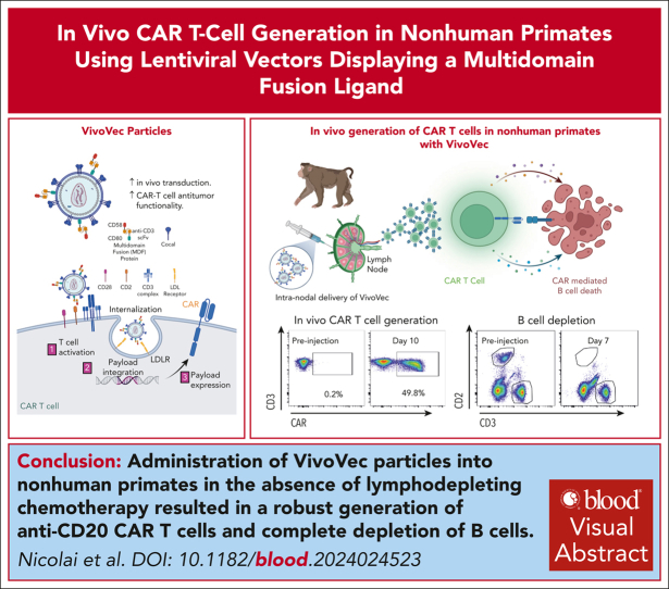
Chimeric antigen receptor (CAR) T-cell therapies have demonstrated transformative efficacy in treating B-cell malignancies. However, high costs and manufacturing complexities hinder their widespread use. To overcome these hurdles, we have developed the VivoVec platform, a lentiviral vector capable of generating CAR T cells in vivo. Here, we describe the incorporation of T-cell activation and costimulatory signals onto the surface of VivoVec particles (VVPs) in the form of a multidomain fusion protein and show enhanced in vivo transduction and improved CAR T-cell antitumor functionality. Furthermore, in the absence of lymphodepleting chemotherapy, administration of VVPs into nonhuman primates resulted in the robust generation of anti-CD20 CAR T cells and the complete depletion of B cells for >10 weeks. These data validate the VivoVec platform in a translationally relevant model and support its transition into human clinical testing, offering a paradigm shift in the field of CAR T-cell therapies.


Conflict of interest statement

Conflict-of-interest disclosure: All authors, except for H.-P.K., are paid employees of Umoja Biopharma and hold equity in the company. H.-P.K. is a member of the scientific advisory board at Umoja Biopharma.

Figures

None
Graphical abstract
Figure 1.
Figure 1.
VVPs containing CD80 and CD58 costimulatory domains enhance T-cell activation and generate greater numbers of CAR T cells with increased in vitro and in vivo antitumor functionality. (A) VivoVec–induced CD4 and CD8 T-cell activation, as measured by CD25, 3 days after VVP addition, n = 3 PBMC donors and representative of 5 individual experiments. (B) Total CAR+ CD4 and CD8 T cells generated by VVPs in vitro, 7 days after vector addition. Data combined from 2 separate experiments with n = 3 PBMC donors for each. (C) CAR T-cell cytokine production (interferon gamma [IFN-γ], IL-2, and tumor necrosis factor-α [TNF-α]) upon Nalm6 coculture for 24 hours. Representative of 2 individual experiments, n = 3 PBMC donors. (D) CAR T-cell–Nalm6 in vitro serial stimulation assay, representative of 2 individual experiments, n = 3 PBMC donors. Arrows indicate restimulation with fresh Nalm6 cells. (E) Human T-cell activation, as measured by CD25 and CD71, in humanized NSG MHC I/II KO model 4 days after VivoVec administration at several particle doses, n = 5 to 9. (F) Percentage of CD19 CAR+ of human CD3+ cells and total human CD19 CAR+ cells per μL blood at day 11 after VivoVec particle administration, n = 5 to 9. (G) Representative flow plots for panel F. (H) Tumor burden over time as measured by Nalm6 bioluminescence, n = 5 to 9. Two-way analysis of variance (ANOVA) with Sidak multiple comparisons for panels A-B. Two-way ANOVA for panels C-D. Two-tailed unpaired student t test for panels E-F. ∗∗∗∗P < .0001; ∗∗∗P < .0002; ∗∗P < .002; and ∗P < .03. +Costim, VVP displaying anti-CD3scFv, CD80, and CD58 costimulatory ligands; MOI, multiplicity of infection; ns, not significant; TU, transducing units.
Figure 2.
Figure 2.
MDF protein comprising an anti-CD3 scFv fused to the CD58 extracellular domain and CD80 exhibit potentiated T-cell activation and transduction-enhancing properties. (A) VivoVec T-cell–binding assay. A total of 20 × 106 PBMCs cultured with VivoVec at MOI = 10 for 1 hour followed by flow cytometry for T-cell–associated cocal glycoprotein, n = 3 PBMC donors and representative of 2 individual experiments. (B) VivoVec–induced CD4 and CD8 T-cell activation, as measured by CD25, 3 days after vector addition, n = 3 PBMC donors and representative of 3 individual experiments. (C) Percent of CD4 and CD8 T cells expressing anti-CD19 CAR 7 days after VivoVec addition to PBMCs, n = 3 PBMC donors and representative of 3 individual experiments. (D) CAR T-cell cytokine production (IFN-γ, IL-2, and TNF-α) upon Nalm6 coculture for 24 hours, representative of 2 individual experiments, n = 3 PBMC donors. (E) CellTrace Violet (CTV) dilution on VivoVec–generated anti-CD19 CAR T cells 5 days after Nalm6 stimulation, representative of 2 independent experiments, n = 3 PBMC donors. (F) CAR T-cell–Nalm6 in vitro serial stimulation assay, representative of 2 individual experiments, n = 3 PBMC donors. Arrows indicate restimulation with fresh Nalm6 cells. Two-tailed unpaired student t test for panels A,E. Two-way ANOVA with Sidak multiple comparisons for panels B-C. Two-way ANOVA for panels D,F. ∗∗∗∗P < .0001; ∗∗∗P < .0002; and ∗P < .03.
Figure 3.
Figure 3.
MDF VVPs exhibit enhanced in vivo functionality in a humanized NSG MHC I/II KO Nalm6 mouse model. (A) Human T-cell activation, as measured by CD25 and CD71, in humanized NSG MHC I/II KO model 4 days after VivoVec administration. (B) Percent CD19 CAR+ of human CD3+ cells and total human CD19 CAR+ cells per microliter blood (left) and representative flow plots (right) on day 11 after VivoVec administration. (C) Tumor burden over time as measured by Nalm6 bioluminescence. (D) Overall survival. For all figures, n = 7 to 8 for VivoVec groups and n = 3 for vehicle control. Two-tailed unpaired student t test for panels A-B. Two-way ANOVA with multiple comparisons for panel C. Log-rank Mantel Cox test for panel D. ∗∗∗∗P < .0001; ∗∗∗P < .0002; ∗∗P < .002; and ∗P < .03.
Figure 4.
Figure 4.
MDF VVPs administered intranodally induce potent and prolonged B-cell depletion in NHPs. (A) Activation of NHP T cells in the blood 7 days after VivoVec administration, as measured by CD25. (B) Anti-CD20 CAR expression (Flag-tag) on NHP CD3+ T cells in the blood 10 days after VivoVec administration. (C) Percent of NHP T cells expressing CAR (top), overall numbers of CAR positive T cells (middle), and total B cells in the blood (bottom) over time. (D) Representative flow plots of B-cell depletion in Z20106. (E) Biodistribution of VivoVec CAR payload in various tissues 139 days after particle administration in Z20106, measured by ddPCR. VCN, vector copy number.

Comment in

References

    1. Neelapu SS, Locke FL, Bartlett NL, et al. Axicabtagene ciloleucel CAR T-cell therapy in refractory large B-cell lymphoma. N Engl J Med. 2017;377(26):2531–2544. - PMC - PubMed
    1. Maude SL, Laetsch TW, Buechner J, et al. Tisagenlecleucel in children and young adults with B-cell lymphoblastic leukemia. N Engl J Med. 2018;378(5):439–448. - PMC - PubMed
    1. Schuster SJ, Svoboda J, Chong EA, et al. Chimeric antigen receptor T cells in refractory B-cell lymphomas. N Engl J Med. 2017;377(26):2545–2554. - PMC - PubMed
    1. Zhao W-H, Wang B-Y, Chen L-J, et al. Four-year follow-up of LCAR-B38M in relapsed or refractory multiple myeloma: a phase 1, single-arm, open-label, multicenter study in China (LEGEND-2) J Hematol Oncol. 2022;15(1):86. - PMC - PubMed
    1. Schmidts A, Ormhøj M, Choi BD, et al. Rational design of a trimeric APRIL-based CAR-binding domain enables efficient targeting of multiple myeloma. Blood Adv. 2019;3(21):3248–3260. - PMC - PubMed