. 2024 Jun;14(6):230448.
doi: 10.1098/rsob.230448. Epub 2024 Jun 12.

Structural and functional insights into the C-terminal signal domain of the Bacteroidetes type-IX secretion system

Affiliations

Structural and functional insights into the C-terminal signal domain of the Bacteroidetes type-IX secretion system

Danuta Mizgalska et al. Open Biol. 2024 Jun.

Abstract

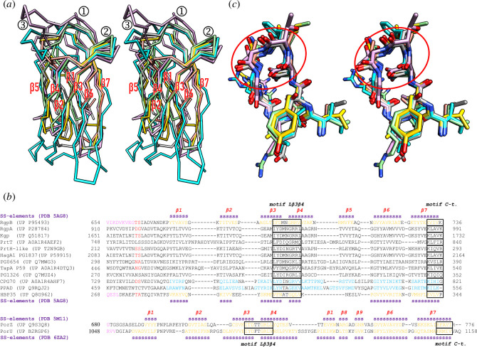
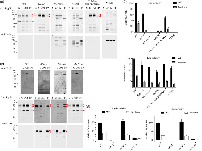
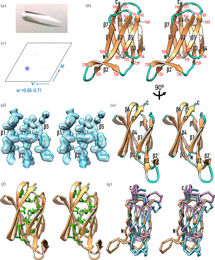
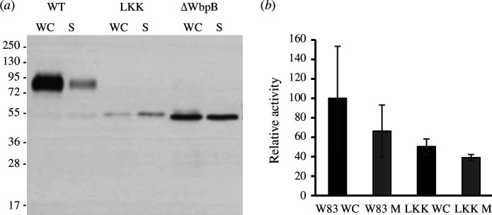
Gram-negative bacteria from the Bacteroidota phylum possess a type-IX secretion system (T9SS) for protein secretion, which requires cargoes to have a C-terminal domain (CTD). Structurally analysed CTDs are from Porphyromonas gingivalis proteins RgpB, HBP35, PorU and PorZ, which share a compact immunoglobulin-like antiparallel 3+4 β-sandwich (β1-β7). This architecture is essential as a P. gingivalis strain with a single-point mutant of RgpB disrupting the interaction of the CTD with its preceding domain prevented secretion of the protein. Next, we identified the C-terminus ('motif C-t.') and the loop connecting strands β3 and β4 ('motif Lβ3β4') as conserved. We generated two strains with insertion and replacement mutants of PorU, as well as three strains with ablation and point mutants of RgpB, which revealed both motifs to be relevant for T9SS function. Furthermore, we determined the crystal structure of the CTD of mirolase, a cargo of the Tannerella forsythia T9SS, which shares the same general topology as in Porphyromonas CTDs. However, motif Lβ3β4 was not conserved. Consistently, P. gingivalis could not properly secrete a chimaeric protein with the CTD of peptidylarginine deiminase replaced with this foreign CTD. Thus, the incompatibility of the CTDs between these species prevents potential interference between their T9SSs.

Keywords: T9SS; X-ray crystal structure; bacterial virulence factor; infectious disease; periodontal disease; protein secretion.


Conflict of interest statement

We declare we have no competing interests.

Figures

Structural comparison of P. gingivalis CTDs.
Figure 1.
Structural comparison of P. gingivalis CTDs. (a) Overall superposition in cross-eye stereo of the Cα-traces of the non-cleaved CTDs of PorU (Protein Data Bank (PDB) access code 6ZA2; A1054–Q1158, for UP codes, see (b); cyan) and PorZ (PDB 5M11; E688–R776; plum), as well as of the cleaved CTDs of RgpB (PDB 5AG8; K672–K736; light green), HBP35 (PDB 5Y1A; P287–P344; salmon), CPG70 (predicted model; Q746–G821; gold) and PPAD (predicted model; K476–K556; dark grey). The seven consensus strands are labelled β1–β7, as well as the two essential structural motifs for T9SS function (①, motif C-t. and ②, motif Lβ3β4) and the non-functional β-hairpin within Lβ5β6 (③). (b) Structure-based sequence alignments of the cleaved CTDs of selected cargoes (top alignment) and the non-cleaved CTDs of PorZ and PorU (bottom alignment). The respective UP access codes and flanking residue numbers are provided. Residues in β-strand conformation (‘SS-elements’) according to the respective PDB entries for RgpB and PorZ (above the respective alignment block) and HBP35 and PorU (below the respective alignment block) are displayed in orange. They are earmarked with a magenta ‘s’ and labelled β1–β7. PorU and PorZ have an extra β-ribbon (strands β8+β9) inserted after the fifth strand. Predicted strand residues of the two homology models (CPG70 and PPAD) are in blue. The last residues of the respective upstream domains of structurally analysed proteins are shown in magenta. Determined or putative cleavage sites by PorU are flanked by residues in red [50,53,55]. Motifs Lβ3β4 and C-t. are boxed and are similar to regions B and E of [46]. (c) Superposition in stereo of motif Lβ3β4 (red ellipse, end-on view) from PorU (residues I1090–V1098, carbons in cyan), PorZ (I720–L728, plum), RgpB (I694–V702, light green), HBP35 (V287–V295, tan), CPG70 (L773–V781, gold) and PPAD (L505–V513, grey). The residue numbers correspond to the respective UP entries in (b). The orientation results from that of (a) after successive vertical and horizontal rotations of 120°C and 40°C, respectively.
Cell-fraction analysis and proteolytic activity of RgpB and PorU mutant strains.
Figure 2.
Cell-fraction analysis and proteolytic activity of RgpB and PorU mutant strains. (a) Western blotting analysis of RgpB-mutant strains in the supernatant (S), whole-cell extract (C), outer-membrane (OM) fraction and periplasmic/cytoplasmic fraction (PP) employing polyclonal antibodies against RgpA and RgpB (top row, pAb GP1) or the CTD of RgpB (bottom row). Red square brackets pinpoint membrane-type RgpB and asterisks denote the isolated RgpA catalytic domain. The latter is missing in the RgpA-C strain and the mutants generated with a RgpA-C template. The absence of gingipain activity results from deficient proteolytic processing of the respective zymogens, which require the entire prodomain to be removed and degraded to release activity [56]. (b) RgpB (top panel) and Kgp (bottom panel) activity relative to the wild-type (100%) of the RgpB-mutant strains of (a) in whole-cell (WC) cultures and the culture medium after cell centrifugation. Experiments were performed in triplicate. (c) Western blotting analysis of PorU-mutant strains employing the monoclonal antibody 7G9 against PorU (top row) and the polyclonal antibodies of (a) against RgpB and its CTD (middle and bottom rows). Red square brackets pinpoint membrane-type RgpB; asterisks denote the isolated RgpA catalytic domain; open triangles hallmark the unprocessed, full-length 80 kDa RgpB zymogen including the intact N-terminal prodomain and the CTD; and full triangles designate the N-terminally truncated RgpB zymogen. (d) RgpB (left panel) and Kgp (right panel) activity of the PorU-mutant strains of (c) in whole cultures (WC) and the culture medium after cell centrifugation.
Crystallographic studies of T. forsythia LKK.
Figure 3.
Crystallographic studies of T. forsythia LKK. (a) Trigonal crystal of space group P31 of selenomethionine-derivatized LKK diffracting to 1.6 Å resolution. (b) Native Patterson map section (axes u, v, w) viewed down the w axis for fraction 0.65–0.71. A peak of 72% the height of the origin peak at fractional coordinates 0.331, 0.663 and 0.678 accounts for strong translational non-crystallographic symmetry, which obscures hemihedral twinning following twin law h,–h–k,–l (estimated twin fraction α = 0.423). (c) Ribbon-type plot in cross-eye stereo of LKK, which encompasses eight β-strands as salmon arrows (β1–β7 plus β2’) connected by coil regions in cyan. The flanking residues of each strand are numbered in red, and the N- and the C-terminus are labelled in black. In this view, the molecule has been rotated vertically ~180° with respect to figure 1a for clarity. (d) Detail of the final σA-weigthed (2mFobsDFcalc)-type Fourier map, contoured at 1σ above the threshold, centred on the three β-strands (β1, β2 and β5) of the front sheet of molecule A. The final refined model is shown for segments L706–S709, T718–L721 and P759–Q762, approximately in the view of (c). (e) Same as (c) after a vertical 90° rotation. (f) Variant of (e) depicting the side chains that form the hydrophobic core of the LKK moiety, with carbons in light green. The participating residues are L706, L708, A713, V717, L719, L721, Y740, I742, I744, F754, T756, F761, I763, P764, M765, L768, Y773, V775, V777, K779, Y784, L788 and K790. (g) Superposition of LKK as Cα-plot in sandy brown in the view of (c) onto the structures of the PorU (cyan; PDB 6ZA2) and PorZ (plum; PDB 5M11) CTDs.
Cross-species CTD exchange between P. gingivalis and T. forsythia.
Figure 4.
Cross-species CTD exchange between P. gingivalis and T. forsythia. (a) Western blotting analysis of PPAD export in WC cultures and the culture medium (S) for the wild-type protein (labelled WT) and a PPAD chimaera with LKK as CTD (labelled LKK), as well as for a P. gingivalis mutant strain (ΔWbpB), which does not attach lipopolysaccharide and thus does not anchor substrates to the OM. The chimaera mimics the phenotype of the latter strain, though with a substantially lower yield, which indicates that the protein is not properly secreted. (b) PPAD activity in whole-cell extract and medium for the wild-type protein and the chimaera.

Similar articles

Cited by

References

    1. Economou A, Christie PJ, Fernandez RC, Palmer T, Plano GV, Pugsley AP. 2006. Secretion by numbers: protein traffic in prokaryotes. Mol. Microbiol. 62, 308–319. (10.1111/j.1365-2958.2006.05377.x) - DOI - PMC - PubMed
    1. Christie PJ. 2019. The rich tapestry of bacterial protein translocation systems. Protein J. 38, 389–408. (10.1007/s10930-019-09862-3) - DOI - PMC - PubMed
    1. Trivedi A, Gosai J, Nakane D, Shrivastava A. 2022. Design principles of the rotary type 9 secretion system. Front. Microbiol. 13, 845563. (10.3389/fmicb.2022.845563) - DOI - PMC - PubMed
    1. Paillat M, Lunar Silva I, Cascales E, Doan T. 2023. A journey with type IX secretion system effectors: selection, transport, processing and activities. Microbiology (Reading) 169, 001320. (10.1099/mic.0.001320) - DOI - PMC - PubMed
    1. Lasica AM, et al. . 2016. Structural and functional probing of PorZ, an essential bacterial surface component of the type-IX secretion system of human oral-microbiomic Porphyromonas gingivalis. Sci. Rep. 6, 37708. (10.1038/srep37708) - DOI - PMC - PubMed

MeSH terms

Substances

LinkOut - more resources