. 2024 Jun;14(6):240025.
doi: 10.1098/rsob.240025. Epub 2024 Jun 12.

Kinetoplastid kinetochore proteins KKT14-KKT15 are divergent Bub1/BubR1-Bub3 proteins

Affiliations

Kinetoplastid kinetochore proteins KKT14-KKT15 are divergent Bub1/BubR1-Bub3 proteins

Daniel Ballmer et al. Open Biol. 2024 Jun.

Abstract

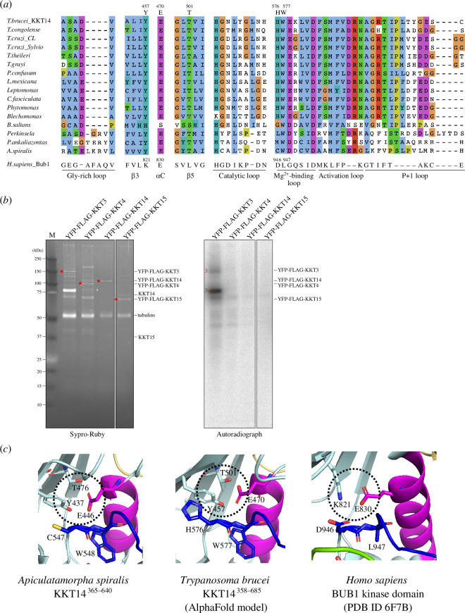
Faithful transmission of genetic material is crucial for the survival of all organisms. In many eukaryotes, a feedback control mechanism called the spindle checkpoint ensures chromosome segregation fidelity by delaying cell cycle progression until all chromosomes achieve proper attachment to the mitotic spindle. Kinetochores are the macromolecular complexes that act as the interface between chromosomes and spindle microtubules. While most eukaryotes have canonical kinetochore proteins that are widely conserved, kinetoplastids such as Trypanosoma brucei have a seemingly unique set of kinetochore proteins including KKT1-25. It remains poorly understood how kinetoplastids regulate cell cycle progression or ensure chromosome segregation fidelity. Here, we report a crystal structure of the C-terminal domain of KKT14 from Apiculatamorpha spiralis and uncover that it is a pseudokinase. Its structure is most similar to the kinase domain of a spindle checkpoint protein Bub1. In addition, KKT14 has a putative ABBA motif that is present in Bub1 and its paralogue BubR1. We also find that the N-terminal part of KKT14 interacts with KKT15, whose WD40 repeat beta-propeller is phylogenetically closely related to a direct interactor of Bub1/BubR1 called Bub3. Our findings indicate that KKT14-KKT15 are divergent orthologues of Bub1/BubR1-Bub3, which promote accurate chromosome segregation in trypanosomes.

Keywords: Trypanosoma brucei; chromosome segregation; kinetochore; kinetoplastid; spindle checkpoint.


Conflict of interest statement

We declare we have no competing interests.

Figures

Crystal structure of A. spiralis KKT14 reveals similarity to the Bub1 kinase domain. (a,b) Cartoon representation of A. spiralis KKT14365–640 (a) and human Bub1 kinase domain (PDB accession 6F7B
Figure 1.
Crystal structure of A. spiralis KKT14 reveals similarity to the Bub1 kinase domain. (a,b) Cartoon representation of A. spiralis KKT14365–640 (a) and human Bub1 kinase domain (PDB accession 6F7B [47]) (b). The fold nomenclature of the N-terminal extension and the kinase domain of Bub1 is based on [48]. (c) Structure-based pairwise alignment of A. spiralis KKT14 and human Bub1 kinase domain based on the DALI search output. Structurally equivalent residues are in uppercase, while structurally non-equivalent residues (e.g. in loops) are in lowercase. Secondary structures were assigned using DSSP [49].
KKT14 has a putative ABBA motif. Schematic of the T. brucei KKT14 protein and a multiple sequence alignment showing a putative ABBA motif (consensus Fx[ILV][FHY]x[DE]
Figure 2.
KKT14 has a putative ABBA motif. Schematic of the T. brucei KKT14 protein and a multiple sequence alignment showing a putative ABBA motif (consensus Fx[ILV][FHY]x[DE] based on [19]) conserved in trypanosomatid KKT14 proteins.
KKT14 lacks key residues that are present in active protein kinases. (a) Multiple sequence alignment of kinetoplastid KKT14 sequences highlighting the regions that correspond to the key parts of the Bub1 kinase domain
Figure 3.
KKT14 lacks key residues that are present in active protein kinases. (a) Multiple sequence alignment of kinetoplastid KKT14 sequences highlighting the regions that correspond to the key parts of the Bub1 kinase domain. Note that T501 and W577 in T. brucei are conserved among kinetoplastids. (b) Lack of detectable auto-phosphorylation activity for KKT14. Indicated proteins were immunoprecipitated from trypanosomes using FLAG antibodies and eluted with FLAG peptides. The left panel shows a Sypro-Ruby stained SDS-PAGE gel (red circles indicate FLAG-tagged proteins), while the right panel shows phosphorylation detected by autoradiography. A degradation product of YFP-FLAG-KKT3 is indicated by 3*. (c) The C-helix has an ‘in’ conformation in the A. spiralis KKT14 crystal structure and AlphaFold2-predicted T. brucei KKT14 structure (electronic supplementary material, dataset S1). Note that E446 in the C-helix is in close proximity with Y437 of β3 and T476 of β5 in A. spiralis (E470, T501 and Y457 in T. brucei, respectively), while E830 in the C-helix forms a salt bridge with the conserved β3 lysine (K821) in Bub1.
Phylogeny of KKT15 and related WD40 repeat proteins. Subtree of the full phylogeny presented in electronic supplementary material, figure S3a
Figure 4.
Phylogeny of KKT15 and related WD40 repeat proteins. Subtree of the full phylogeny presented in electronic supplementary material, figure S3a. Note that in the alignment approach applied here, KKT15 proteins were prompted to form a single group (see §4).
KKT14 is predicted to interact with KKT15 directly. (a) A volcano plot showing relative enrichment and significance values between the immunoprecipitates of YFP-KKT14 and YFP-KKT22 (n = 4 each)
Figure 5.
KKT14 is predicted to interact with KKT15 directly. (a) A volcano plot showing relative enrichment and significance values between the immunoprecipitates of YFP-KKT14 and YFP-KKT22 (n = 4 each). KKT22 was used as a comparison, which mainly co-purifies with KKT23 and KKT3 [43]. See electronic supplementary material, table S5, for all proteins identified by mass spectrometry. (b) AlphaFold2 predictions of the KKT142–125–KKT15 complex in cartoon representation (electronic supplementary material, dataset S2). (c) The PAE plots for the rank 1 model for KKT14–KKT15, predicting interactions via the N-terminal part of KKT14. (d) pLDDT plots for KKT14–KKT15 (left) and KKT14 (right). AlphaFold2-predicted models are provided in the electronic supplementary material, datasets S3 and S4. PAE, predicted aligned error.
N-terminal region of KKT14 binds KKT15. (a) Ectopically expressed GFP-KKT14N2–357 localizes at kinetochores, while GFP-KKT14C358–685 does not. K and N stand for the kinetoplast (mitochondrial DNA) and nucleus, respectively
Figure 6.
N-terminal region of KKT14 binds KKT15. (a) Ectopically expressed GFP-KKT14N2–357 localizes at kinetochores, while GFP-KKT14C358–685 does not. K and N stand for the kinetoplast (mitochondrial DNA) and nucleus, respectively. Cell cycle stages of individual cells were determined based on the number of K and N as described previously [63,64]. The GFP fusion proteins were expressed in trypanosomes using 10 ng ml−1 doxycycline for 1 day and fixed for microscopy. Cell lines: BAP2386, BAP2387. (b) Immunoprecipitation/mass spectrometry analysis shows that GFP-KKT14N co-purifies with many kinetochore proteins, while GFP-KKT14C does not. Immunoprecipitation was carried out using cells expressing the GFP fusion proteins using 10 ng ml−1 doxycycline for 1 day. See electronic supplementary material, table S5, for all proteins identified by mass spectrometry. (c) KKT14N2–357 is sufficient to recruit KKT15 in trypanosomes. Recruitment of tdTomato-KKT15 was observed in 100% or 0% of 1K1N (G1) cells that have GFP-KKT14N2–357-LacI or GFP-KKT14C358–685-LacI dots, respectively (n = 10 each). The GFP fusion proteins were expressed in trypanosomes using 10 ng ml−1 doxycycline for 1 day. Cell lines: BAP2655, BAP2656. Scale bars, 5 µm.
KKT14 is essential for accurate chromosome segregation in trypanosomes. (a) Growth curve upon RNAi-mediated knockdown of KKT15 using an RNAi construct against its 3’ UTR
Figure 7.
KKT14 is essential for accurate chromosome segregation in trypanosomes. (a) Growth curve upon RNAi-mediated knockdown of KKT15 using an RNAi construct against its 3’ UTR. Data are presented as the mean ± s.d. of three replicates. RNAi was induced with 1 μg ml−1 doxycycline and cultures were diluted on day 2. Cell line: BAP2533. (b,c) Validation of KKT15 knockdown. RNAi was induced with 1 μg ml−1 doxycycline and cultures were diluted on day 2. K and N stand for the kinetoplast and nucleus, respectively. Cell line: BAP2535. At least 130 cells per condition were quantified. (d) Representative fluorescence micrographs showing the localization of YFP-KKT14 upon RNAi-mediated knockdown of KKT15 in G2/M (2K1N) and anaphase (2K2N) cells. RNAi was induced with 1 μg ml−1 doxycycline for 24 h. Cell line: BAP2533. (e) Quantification of 2K1N and 2K2N that have kinetochore-like dots of YFP-KKT14 upon RNAi-mediated depletion of KKT15. All graphs depict the means (bar) ± s.d. of three replicates (shown as dots). (f) Representative fluorescence micrographs showing the localization of YFP-KKT15 upon RNAi-mediated knockdown of KKT14 in 2K1N and 2K2N cells. RNAi was induced with 1 μg ml−1 doxycycline for 24 h. Cell line: BAP2534. (g) Quantification of 2K1N and 2K2N that have kinetochore-like dots of YFP-KKT15 upon RNAi-mediated depletion of KKT14. All graphs depict the means (bar) ± s.d. of three replicates (shown as dots). A minimum of 50 cells per replicate were quantified in each condition. (h) Growth curve upon RNAi-mediated knockdown of KKT14. Data are presented as the mean ± s.d. of three replicates. RNAi was induced with 1 μg ml−1 doxycycline and cultures were diluted at day 2. Cell line: BAP2534. (i) Cell cycle profile upon knockdown of KKT14. RNAi was induced with 1 μg ml−1 doxycycline and cells were fixed at 8 or 16 h. All graphs depict the means (bar) ± s.d. of three replicates. A minimum of 700 cells per replicate were quantified. Cell line: BAP680. (j) Representative fluorescence micrographs showing lagging kinetochores (marked by tdTomato-KKT2) in anaphase cells upon KKT14 knockdown at 8 and 16 h post-induction. RNAi was induced with 1 μg ml−1 doxycycline. Cell line: BAP680. (k) Quantification of lagging kinetochores in anaphase cells upon KKT14 knockdown. All graphs depict the means (bar) ± s.d. of three replicates (shown as dots). A minimum of 50 cells per replicate were quantified in each condition. *p < 0.05, **p ≤ 0.01, ***p ≤ 0.001 (two-sided, unpaired t‐test).

Similar articles

Cited by

References

    1. McAinsh AD, Marston AL. 2022. The four causes: the functional architecture of centromeres and kinetochores. Annu. Rev. Genet. 56 , 279–314. (10.1146/annurev-genet-072820-034559) - DOI - PMC - PubMed
    1. Musacchio A, Desai A. 2017. A molecular view of kinetochore assembly and function. Biology (Basel) 6 , 5. (10.3390/biology6010005) - DOI - PMC - PubMed
    1. Alfieri C, Zhang S, Barford D. 2017. Visualizing the complex functions and mechanisms of the anaphase promoting complex/cyclosome (APC/C). Open Biol. 7 , 170204. (10.1098/rsob.170204) - DOI - PMC - PubMed
    1. Pines J. 2011. Cubism and the cell cycle: the many faces of the APC/C. Nat. Rev. Mol. Cell Biol. 12 , 427–438. (10.1038/nrm3132) - DOI - PubMed
    1. Murray AW. 2011. A brief history of error. Nat. Cell Biol. 13 , 1178–1182. (10.1038/ncb2348) - DOI - PubMed

MeSH terms

Substances

LinkOut - more resources