. 2024 Jun 11;15(1):4959.
doi: 10.1038/s41467-024-49008-3.

Impact of keratocyte differentiation on corneal opacity resolution and visual function recovery in male rats

Affiliations

Impact of keratocyte differentiation on corneal opacity resolution and visual function recovery in male rats

Andri K Riau et al. Nat Commun. .

Abstract

Intrastromal cell therapy utilizing quiescent corneal stromal keratocytes (qCSKs) from human donor corneas emerges as a promising treatment for corneal opacities, aiming to overcome limitations of traditional surgeries by reducing procedural complexity and donor dependency. This investigation demonstrates the therapeutic efficacy of qCSKs in a male rat model of corneal stromal opacity, underscoring the significance of cell-delivery quality and keratocyte differentiation in mediating corneal opacity resolution and visual function recovery. Quiescent CSKs-treated rats display improvements in escape latency and efficiency compared to wounded, non-treated rats in a Morris water maze, demonstrating improved visual acuity, while stromal fibroblasts-treated rats do not. Advanced imaging, including multiphoton microscopy, small-angle X-ray scattering, and transmission electron microscopy, revealed that qCSK therapy replicates the native cornea's collagen fibril morphometry, matrix order, and ultrastructural architecture. These findings, supported by the expression of keratan sulfate proteoglycans, validate qCSKs as a potential therapeutic solution for corneal opacities.


Conflict of interest statement

The authors declare no competing interests.

Figures

Fig. 1
Fig. 1. Phenotypical features of cultivated corneal stromal keratocytes.
a The propagation of corneal stromal keratocytes (CSKs) was achieved by culturing the corneal stromal cells in the “activated” form in the 0.5% fetal bovine serum (FBS)-supplemented media, containing amniotic membrane extract (AME), ROCK inhibitor (ROCKi), and insulin growth factor 1 (IGF-1). The “activated” state was referred to as the activated CSKs (aCSKs). The aCSKs entered a quiescent state following culture in serum-free media (SFM), which we referred to as the quiescent CSKs (qCSKs). The qCSKs featured stellate morphology with thin cell bodies and long cell processes. As a comparison, stromal fibroblasts (SFs) that were transformed by culturing the cells in 10% serum-supplemented media had a bipolar morphology with large cell bodies and pseudopodial processes. b The qCSKs (blue bars) expressed higher levels of native CSK’s genetic markers than the aCSKs (red bars) and SFs (yellow bars). The gene expression levels were normalized to the corneal stromal tissue, indicated by the dashed line. Data were presented as mean ± SEM (n = 3 in each group). Statistical significance was assessed with one-way ANOVA, followed by post hoc Tukey test. Immunocytochemistry of keratocan (c) and lumican (d), double stained with phalloidin, confirmed the earlier morphological and gene expression analysis. The qCSKs exhibited more abundant keratocan and lumican proteins than aCSKs. In contrast, the SFs were largely absent of these proteoglycans. The staining was repeated on 3 independent cells. Scale bars = 50 µm. Source data are provided as a Source Data file.
Fig. 2
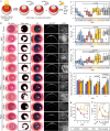
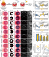
Fig. 2. Postoperative outcomes of cell injections in corneas with excimer laser-induced acute opacity.
a Experimental setup and timeline. The treatment outcomes were also compared to the non-treated corneas (see Supplementary Fig. 4 for the imaging results). b The corneal imaging showed that quiescent corneal stromal keratocytes (qCSKs) had the best therapeutic efficiency, followed by activated CSKs (aCSKs) and then stromal fibroblasts (SFs). SF injection also produced haze reduction albeit in a less efficient manner compared to the qCSKs, which was most likely caused by the increasing neovascularization (NV). The non-treated corneas exhibited persistently severe corneal haze and NV at any time point. The slit-lamp observation was supported by the haze (c), NV (d), and total grades (e). In the box plots, the center line shows the median score; box limits show the 1st and 3rd quartile; whiskers show minimum and maximum values; and points indicate outliers. Statistical significance was assessed with Kruskal–Wallis, followed by post hoc Dune-Bonferroni test. f The opacity was substantiated by the IVCM-based haze density measurements. The dash line indicates the mean of haze density preoperatively. g From a haze area point-of-view, the SFs and non-treated groups demonstrated almost no changes from PID0. The qCSKs showed the highest efficiency in haze area reduction, followed by aCSKs. The significant p values were relative to the qCSKs. h The % ΔCCT over time did not differ in the qCSKs, aCSKs, and SFs groups. On the other hand, the CCT reduction was also observed in the non-treated corneas, albeit at a lower rate. The significant p value was relative to the qCSKs. Data are presented as mean ± SEM. Statistical significance was analyzed with one-way ANOVA, followed by post hoc Tukey test. The qCSKs, aCSKs, SFs, and non-treated are represented by blue, red, yellow, and gray in the box plots, bar graphs, and line graphs. n = 6 rats in each group. Scale bars = 100 µm. Source data are provided as a Source Data file.
Fig. 3
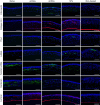
Fig. 3. Immunofluorescence staining of corneal fibrosis-associated proteins and keratan sulfate proteoglycans in laser-injured corneas undergoing cell therapies.
The corneas were harvested 21 days after cell injections. The quiescent corneal stromal keratocytes (qCSKs)-treated corneas exhibited little to no expression of Thy-1, α-smooth muscle actin (α-SMA), fibronectin, and collagen 3A1. The corneas that received activated CSKs (aCSKs) injections expressed Thy-1, α-SMA, fibronectin, and collagen 3A1 in the anterior stroma but at an attenuated level compared to the stromal fibroblasts (SFs)-injected corneas. The keratan sulfate (KS) and KS proteoglycans, keratocan and lumican, were largely absent in non-treated corneas but were re-expressed in the qCSK-treated corneal stroma close to the naïve corneal state. The aCSK- and SF-injected corneas also re-expressed the KS, keratocan, and lumican but their expression levels were lower than the naïve corneas and qCSK-treated corneas. Staining was repeated on 3 independent samples. Scale bars = 100 µm.
Fig. 4
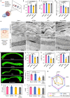
Fig. 4. Collagen fiber morphometry and organization in laser-ablated corneas undergoing cell therapies.
The corneas were harvested 21 days after cell injections. a Synchrotron small-angle X-ray scattering (SAXS) was employed to elucidate the interfibrillar distance (IFD), fibrillar diameter (FD), and degree of collagen fibril organization (matrix order) in the corneas centrally. The IFD (b) and FD (c) were only marginally different between all groups, namely the naïve, quiescent corneal stromal keratocytes (qCSKs)-treated, stromal fibroblasts (SFs)-treated, and non-treated corneas, with naïve corneas having the smallest IFD and FD. d The matrix order in the qCSK-injected corneas was akin to the naïve corneas and was significantly higher than the SFs-injected and non-treated samples. e Low-magnification transmission electron microscopy (TEM) images of the anterior stroma region revealed a more regular alternation of darker (collagen fibers that appeared in longitudinal orientation) and lighter (fibers that appeared in cross-sectional orientation) bands of lamellae in the naïve and qCSKs groups. At high magnification, especially within the darker band, the fibers were typically long and continuous in both naïve and qCSK-treated rats but appeared relatively shorter due to the interruption by the cross-sectionally appearing fibers in the lamellar layer. f Further collagen fiber profiles were characterized with a Histoindex Genesis 200 multiphoton microscope. The built-in analytical system then measured the tissue area ratio (TAR) (g), collagen area ratio in tissue (CART) (h), collagen fiber density (CFD) (i), collagen fiber count (CFC) (j), collagen fiber length (CFL) (k), and collagen area reticulation density (CARD) (l), revealing the restoration of collagen fiber profile by qCSKs to the naïve corneal state (m). In contrast, the collagen fiber profile of SF-injected corneas was closer to that of the non-treated corneas. Data are presented as mean ± SEM (n = 9 in each group for SAXS and n = 3 in each group for multiphoton microscopy). TEM was repeated on 3 independent samples. Statistical significance was analyzed with one-way ANOVA, followed by post hoc Tukey test. Naïve, qCSKs, SFs, and non-treated groups are represented by pink, blue, yellow, and gray, respectively, in the bar graphs and radar chart. Source data are provided as a Source Data file.
Fig. 5
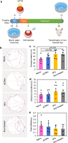
Fig. 5. Visual-dependent behavioral assessment of rats that underwent cell therapies with Morris water maze.
a Experimental timeline showing the swim training regime (TR), acute corneal haze induction with irregular phototherapeutic keratectomy (IrrPTK) and haze development, cell therapy, and closure of the eyelid of the non-lasered eye a day before the final test. b The swim path maps indicated that naïve and quiescent corneal stromal keratocytes (qCSKs)-treated rats performed better, e.g., traveled a shorter distance from the start point (black spots) to the escape platform (red spots) than the stromal fibroblasts (SFs) and non-treated groups. The qCSK-injected rats (blue bars) displayed similar escape latency (c), distance traveled (d), and path efficiency (e) as the naïve rats (pink bars). On the other hand, the SFs-treated (yellow bars) and non-treated (gray bars) rats had higher escape latency and distance traveled, and lower path efficiency than the naïve rats. Data are presented as mean ± SEM (n = 6 rats in each group, with each rat subjected to the trial thrice). Statistical significance was analyzed with one-way ANOVA, followed by post hoc Tukey test. Source data are provided as a Source Data file.
Fig. 6
Fig. 6. Postoperative outcomes of cell injections in corneas with excimer laser-induced chronic opacity.
a Experimental setup and timeline. The quiescent corneal stromal keratocytes (qCSKs) and stromal fibroblasts (SFs) injection outcomes were compared to the non-treated corneas. b The corneal imaging showed that qCSKs had the best therapeutic efficiency. SF injection exacerbated the haze and neovascularization (NV) severity in a similar manner to the non-treated corneas. The slit-lamp observation concurred with the haze (c), NV (d), and total grades (e), and was further substantiated by the in vivo confocal microscopy (IVCM)-based haze density measurements (f). The dash line indicates the mean of haze density preoperatively. In the box plots, the center line shows the median score; box limits show the 1st and 3rd quartile; whiskers show minimum and maximum values; and points indicate outliers. Statistical significance was analyzed with Kruskal–Wallis, followed by post hoc Dune-Bonferroni test. g The haze areas increased over time in the SF-injected and non-treated corneas. In contrast, treatment with qCSKs reduced the chronic haze area. The significant p values were relative to the qCSKs. h The central corneal thickness (CCT) returned to the preoperative state at a more rapid rate following qCSK injection compared to the non-treated group. The corneas remained thicker than the preoperated state even at 3 weeks after SF administration. Data are presented as mean ± SEM. Statistical significance was analyzed with one-way ANOVA, followed by post hoc Tukey test. The qCSKs, SFs, and non-treated are represented by blue, yellow, and gray in the box plots, bar graphs, and line graphs. n = 6 rats in each group. Scale bars = 100 µm. Source data are provided as a Source Data file.
Fig. 7
Fig. 7. Mechanism of haze clearance following quiescent corneal stromal keratocyte versus stromal fibroblast injections.
Following corneal injury, inflammatory and proliferative cytokines induced by the fibroblastic transformation of the native corneal stromal keratocytes (CSKs) result in abnormal extracellular matrix (ECM) or disorganized collagen fibers. Due to the relatively quiescent nature of injected quiescent CSKs (qCSKs), they are likely able to exert their intrinsic function to secrete the proper ECM and reorganize the collagen fibers. In contrast, the injected stromal fibroblasts (SFs) released cytokines/chemokines that compound the myofibroblastic transformation and inflammatory responses in the injured cornea. Cell injection of SFs likely results in the persistent presence of abnormal ECM, which in turn causes unabated corneal haze. Furthermore, the cytokines/chemokines naturally released by SFs likely contribute to increased neovascularization.

Similar articles

Cited by

References

    1. Jinabhai A, O’Donnell C, Radhakrishnan H, Nourrit V. Forward light scatter and contrast sensitivity in keratoconic patients. Cont. Lens Anterior Eye. 2012;35:22–27. doi: 10.1016/j.clae.2011.07.001. - DOI - PubMed
    1. Wang EY, et al. Global trends in blindness and vision impairment resulting from corneal opacity 1984-2020: a meta-analysis. Ophthalmology. 2023;130:863–871. doi: 10.1016/j.ophtha.2023.03.012. - DOI - PMC - PubMed
    1. Yam GHF, Riau AK, Funderburgh ML, Mehta JS, Jhanji V. Keratocyte biology. Exp. Eye Res. 2020;196:108062. doi: 10.1016/j.exer.2020.108062. - DOI - PubMed
    1. Jester JV, et al. The cellular basis of corneal transparency: evidence for ‘corneal crystallins’. J. Cell Sci. 1999;112:613–622. doi: 10.1242/jcs.112.5.613. - DOI - PubMed
    1. Kao WWY, Liu C-Y. Roles of lumican and keratocan on corneal transparency. Glycoconj. J. 2002;19:275–285. doi: 10.1023/A:1025396316169. - DOI - PubMed