. 2024 Jun 11;15(6):409.
doi: 10.1038/s41419-024-06808-1.

Glucose deprivation triggers DCAF1-mediated inactivation of Rheb-mTORC1 and promotes cancer cell survival

Affiliations

Glucose deprivation triggers DCAF1-mediated inactivation of Rheb-mTORC1 and promotes cancer cell survival

Miaomiao Li et al. Cell Death Dis. .

Abstract

Low glucose is a common microenvironment for rapidly growing solid tumors, which has developed multiple approaches to survive under glucose deprivation. However, the specific regulatory mechanism remains largely elusive. In this study, we demonstrate that glucose deprivation, while not amino acid or serum starvation, transactivates the expression of DCAF1. This enhances the K48-linked polyubiquitination and proteasome-dependent degradation of Rheb, inhibits mTORC1 activity, induces autophagy, and facilitates cancer cell survival under glucose deprivation conditions. This study identified DCAF1 as a new cellular glucose sensor and uncovered new insights into mechanism of DCAF1-mediated inactivation of Rheb-mTORC1 pathway for promoting cancer cell survival in response to glucose deprivation.


Conflict of interest statement

The authors declare no competing interests.

Figures

Fig. 1
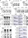
Fig. 1. Glucose deprivation induces ubiquitin-mediated degradation of Rheb.
AC Time-dependent downregulation of Rheb protein level in response to glucose deprivation, but not to amino acids or serum deprivation. Huh7, HCT116, and EC109 cells were grown in EBSS (A), glucose-free, amino acids-free, serum-free medium (B) or treated with 2-DG (20 mM) (C) for the indicated time and subjected to immunoblotting (IB) analysis. D Decreased stability of Rheb during glucose deprivation. Cells were grown in full medium (DMEM high-glucose medium) and glucose-free medium containing 100 µg/ml CHX, and the protein level of Rheb was analyzed at the indicated time points. E Slowed degradation of Rheb during glucose deprivation by proteasome inhibitor MG132. Cells were grown in glucose-free medium containing CHX (100 µg/ml) + DMSO (0.1%) or 10 µM MG132, and protein levels of Rheb were analyzed at the indicated time points for IB analysis. F Increased levels of ubiquitinated Rheb upon MG132 treatment. Cells were treated with 10 µM MG132, and immunoprecipitation (IP) was performed using IgG or Rheb antibody (Ab). The polyubiquitination of Rheb (Rheb-Ub) was detected with Ub Ab. G Promotion of polyubiquitination of Rheb under glucose deprivation. Cells were cultured with or without glucose-free medium with 10 µM MG132, and IP was performed using IgG or Rheb Ab. HJ Enhanced protein stability of Rheb by MG132. Cells were treated with 10 µM MG132 and collected at the indicated time (H) or treated with MG132 at different concentrations (I) for IB analysis. J Cells were treated with 100 µg/ml CHX + DMSO or 10 µM MG132 and harvested at the indicated time for IB analysis. WCL, whole cell lysate. All data were representative of at least three independent experiments (n = 3).
Fig. 2
Fig. 2. DCAF1 mediates K48-linked polyubiquitination of Rheb.
A Partial result of mass spectrometry analysis of the Rheb-associated immunoprecipitated complex. B, C Co-IP of Flag-Rheb and endogenous DCAF or Flag-DCAF1 and endogenous Rheb. 293 T cells transfected with an empty vector or Flag-Rheb (B) or Flag-DCAF1 (C), followed by IP analysis with anti-Flag and IB analysis as indicated. D, E IP analysis of endogenous DCAF1 and endogenous Rheb. IB analysis of co-immunoprecipitated endogenous DCAF1 and Rheb proteins using anti-Rheb (D) or anti-DCAF1 (E) antibody in Huh7, HCT116, and EC109 cells. An unrelated IgG was used as negative control. F Co-localization analysis of DCAF1 and Rheb. Huh7, HCT116, and EC109 cells were fixed and immunostained with antibodies against the indicated proteins. Representative images are shown. Scale bars: 25 μm. G DCAF1 promotes Rheb ubiquitination. 293T cells were transfected with recombinant plasmids as indicated and treated with 10 µM MG132, and subjected to IP with anti-Flag, followed by IB with anti-HA or anti-Flag. H, I Knockdown of DCAF1 attenuates Rheb polyubiquitination. H 293T cells were transfected with siDCAF1, Flag-Rheb, and HA-Ub as indicated, and subjected to IP with anti-Flag, followed by IB with anti-HA. I Huh7 cells were transfected with siDCAF1, and Rheb polyubiquitination was analyzed by IP with anti-Rheb, followed by IB with anti-Ub. J DCAF1 elevates the K48-linked, but not the K63-linked, polyubiquitination of Rheb. 293T cells were transfected with recombinant plasmids as indicated, treated with MG132, and subjected to IP with anti-Flag, followed by IB with anti-K48 Ub or anti- K63 Ub. All data were representative of at least three independent experiments (n = 3).
Fig. 3
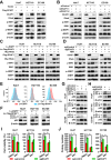
Fig. 3. DCAF1 regulates the degradation of Rheb.
A, B Overexpression of DCAF1 reduces the protein level of Rheb. 293T cells were transfected with different amounts of Flag-DCAF1 and same amount of HA-Rheb (A) or Huh7, HCT116, and EC109 cells were transfected with vector or Flag-DCAF1 (B). C, D Cells were collected and analyzed by IB as indicated. Knockdown of DCAF1 enhances the protein level of Rheb. 293T cells (C) and Huh7, HCT116, and EC109 cells (D) were transfected with siDCAF1 with (C) or without (D) Flag-Rheb, and analyzed by IB as indicated. E Treatment with MLN4924 restores the expression of Rheb in cell lines stably overexpressing DCAF1. Cells with stable overexpression of DCAF1 (Fv-Flag-DCAF1) or its counterpart (Lv-EGFP) were treated with MLN4924 or DMSO, and the protein levels of DCAF1 and Rheb were detected by IB using Flag Ab or Rheb Ab. F DCAF1 promotes Rheb ubiquitination. Cells with stable overexpression of DCAF1 or its counterpart were treated with 10 µM MG132. Rheb polyubiquitination was detected by IP using Rheb Ab and IB with Ub Ab. G Overexpression of DCAF1 decreases Rheb protein stability. Huh7, HCT116, and EC109 cells with stable overexpression of DCAF1 or its counterparts were treated with 100 µg/ml CHX. Cells were harvested at the indicated time points for IB analysis. H Silencing DCAF1 using CRISPR-Cas9 system (sgDCAF1) increases the protein level of Rheb. Cell proteins from sgDCAF1 and sgControl were collected and analyzed using IB. I DCAF1 depletion attenuates Rheb polyubiquitination. sgDCAF1 and sgControl cells were treated with 10 µM MG132, harvested, and subjected to IP with anti-Rheb, followed by IB with anti-Ub. J Silencing DCAF1 increases Rheb protein stability. Huh7, HCT116, and EC109 cells with stable silencing of DCAF1(sgDCAF1) and its counterpart (sgControl) were treated with 100 µg/ml CHX and harvested at the indicated time points for IB analysis. All data were representative of at least three independent experiments (n = 3).
Fig. 4
Fig. 4. DCAF1 regulates mTORC1 activity through Rheb.
A, B DCAF1 serves as a negative regulator of mTORC1 activity. Huh7, HCT116, and EC109 cells were transfected with Flag-DCAF1 (A) or siDCAF1 (B) and harvested for IB analysis as indicated. C, D DCAF1 regulates mTORC1 activity through Rheb. C Huh7, HCT116, and EC109 cells stable overexpression of DCAF1 or its counterparts were transfected with Myc-Rheb or left untransfected, and mTORC1 activity was evaluated by IB analysis. D Huh7, HCT116, and EC109 cells stable silencing of DCAF1 (sgDCAF1) and its counterparts were treated with NR1, an Rheb inhibitor, and mTORC1 activity was evaluated by IB analysis. E Overexpression of DCAF1 reduces cell size. FH The expression of DCAF1 affects autophagy. Cell proteins collected from Huh7, HCT116, and EC109 cells with stable overexpression of DCAF1 (F), transfected with siDCAF1 (G), or stably silencing DCAF1 (H) was analyzed by IB as indicated. I, J DCAF1 affects cell growth through Rheb. Huh7, HCT116, and EC109 cells with stable silencing of DCAF1 (sgDCAF1) and its counterpart (sgControl) were treated with DMSO or NR1 (Rheb inhibitor) for 24 h, and cell viability was detected (I). For long term growth, cells were treated with DMSO or NR1 for 10 days, and the number of colonies was counted (J). Data are represented as mean ± SEM. *p < 0.05 by Student’s t test (I and J). All data were representative of at least three independent experiments (n = 3).
Fig. 5
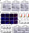
Fig. 5. Glucose deprivation transactivates DCAF1, promotes ubiquition-mediated degradation of Rheb, and inhibits mTORC1 activity.
A, B Silencing DCAF1 delays glucose deprivation-induced Rheb degradation and enhances the protein stability of Rheb. Stable sgDCAF1 and sgControl cells were cultured in glucose-free medium, treated without (A) or with (B) 100 µg/ml CHX, and collected at indicated times for IB analysis. C Stable sgDCAF1 cells were cultured with or without glucose-free medium, and the polyubiquitination of Rheb was analyzed by immunoprecipitation. D Huh7, HCT116, and EC109 cells were cultured in glucose-free medium and cell protein was collected at indicated times for IB analysis. E Huh7, HCT116, and EC109 cells were cultured in glucose-free medium and total RNA was collected at indicated times. The mRNA levels of DCAF1 were analyzed using quantitative-PCR (Q-PCR). F Cells were treated with 20 mM 2-DG, and total RNA was collected at indicated times. The DCAF1 mRNA levels were analyzed using Q-PCR. G, H Effect of 2-DG treatment on the protein level of DCAF1, Rheb, and mTORC1 activity in mouse model. The 6-week-old C57BL6 mice were randomized into two groups and treated with normal saline or 2-DG solution for 3 weeks. The liver (G) and brain (H) tissues were harvested, and protein expression was evaluated by IB analysis using specific antibodies as indicated. All data were representative of at least three independent experiments (n = 3).
Fig. 6
Fig. 6. DCAF1 enhances glucose deprivation-induced autophagy.
AD Stable Lv-Flag-DCAF1 promotes Rheb degradation, inhibits mTORC1 activity, and enhances glucose deprivation-induced autophagy. Huh7, HCT116, and EC109 cells with stably overexpressed DCAF1 were cultured in glucose-free medium for different durations and collected for IB analysis (A) or immunofluorescence detection of LC3 (B). Scale bars: 50 μm. The statistical analysis for panel B was shown in panel C. The cells treated as in panel B were collected and analyzed by IB (D). E, F Huh7, HCT116, and EC109 cells were cultured with and without glucose-free medium and treated with NR1 (E) or rapamycin (F). Cell proteins were collected and analyzed. G Stable sgDCAF1 or its counterparts were cultured with glucose-free medium. Cell proteins were collected at indicated times and analyzed using IB. Data are represented as mean ± SEM. *p < 0.05 by Student’s t test (C). All data were representative of at least three independent experiments (n = 3).
Fig. 7
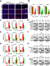
Fig. 7. DCAF1 promotes cancer cell survival and protects cancer cell from glucose deprivation-induced cell death.
A, B DCAF1 promotes cell survival under glucose deprivation. Huh7, HCT116, and EC109 cells with stable overexpression of DCAF1 (Lv-Flag-DCAF1) and its counterpart (Lv-EGFP) were cultured in glucose-free medium for 24 h. Cell proliferation was detected by EdU staining, and representative pictures are shown in panel A. Scale bars: 200 μm. Statistical analysis is shown in panel B. C, D DCAF1 protects cancer cells from cell death induced by glucose deprivation. Stable Lv-Flag-DCAF1 or control Lv-EGFP cells were cultured in glucose-free medium for 24 h. Cells were collected for apoptotic analysis using Annexin V-APC/7-AAD double staining FACS (C) and IB analysis (D). EH NR1 and rapamycin protect cancer cells from cell death induced by glucose deprivation. Huh7, HCT116, and EC109 cells were cultured in glucose-free medium and treated with NR1 (E, F) or rapamycin (G, H) for 24 h. Apoptosis was analyzed using Annexin V-FITC/PI double staining FACS (E, G) and IB analysis (F, H). I, J Silencing DCAF1 promotes glucose deprivation-induced cell death. Stable sgDCAF1 or control sgControl cells were cultured in normal glucose or glucose-free medium for 18 h. Apoptosis was analyzed using Annexin V-APC/7-AAD double staining FACS (I) and IB analysis (J). Data are represented as mean ± SEM. *p < 0.05 by Student’s t test (B, C, E, G, and I). All data were representative of at least three independent experiments (n = 3).

Similar articles

Cited by

References

    1. Boroughs LK, DeBerardinis RJ. Metabolic pathways promoting cancer cell survival and growth. Nat Cell Biol. 2015;17:351–9. doi: 10.1038/ncb3124. - DOI - PMC - PubMed
    1. Valvezan AJ, Manning BD. Molecular logic of mTORC1 signalling as a metabolic rheostat. Nat Metab. 2019;1:321–33. doi: 10.1038/s42255-019-0038-7. - DOI - PubMed
    1. Hung CM, Garcia-Haro L, Sparks CA, Guertin DA. mTOR-dependent cell survival mechanisms. Cold Spring Harb Perspect Biol. 2012;4:a008771. doi: 10.1101/cshperspect.a008771. - DOI - PMC - PubMed
    1. Zhao Y, Xiong X, Sun Y. DEPTOR, an mTOR inhibitor, is a physiological substrate of SCF(betaTrCP) E3 ubiquitin ligase and regulates survival and autophagy. Mol Cell. 2011;44:304–16. doi: 10.1016/j.molcel.2011.08.029. - DOI - PMC - PubMed
    1. Armijo ME, Campos T, Fuentes-Villalobos F, Palma ME, Pincheira R, Castro AF. Rheb signaling and tumorigenesis: mTORC1 and new horizons. Int J Cancer. 2016;138:1815–23. doi: 10.1002/ijc.29707. - DOI - PubMed

Publication types

MeSH terms

Substances