Review
. 2024 Jun 11;10(1):277.
doi: 10.1038/s41420-024-02014-2.

Regulated cell death in hypoxic-ischaemic encephalopathy: recent development and mechanistic overview

Affiliations
Review

Regulated cell death in hypoxic-ischaemic encephalopathy: recent development and mechanistic overview

Lingzhi Wu et al. Cell Death Discov. .

Abstract

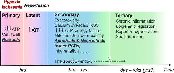
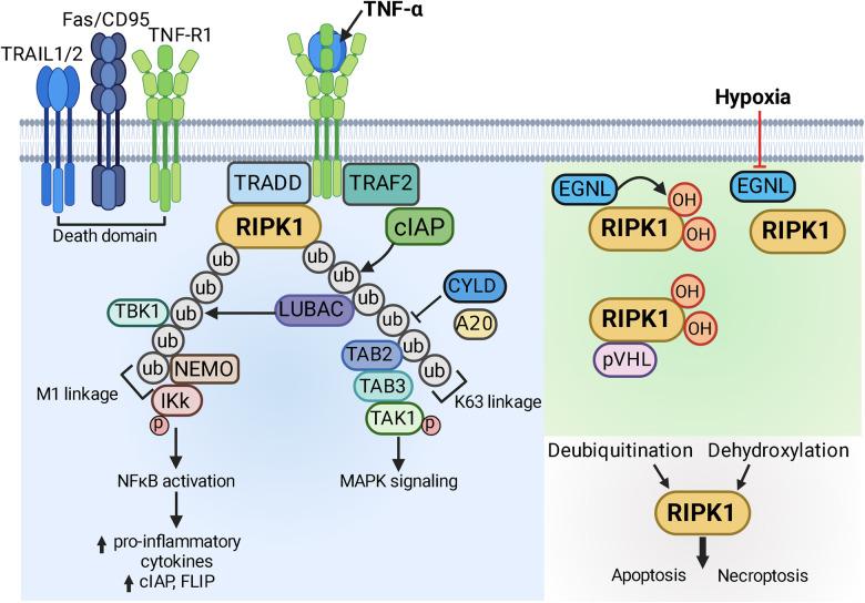
Hypoxic-ischaemic encephalopathy (HIE) in termed infants remains a significant cause of morbidity and mortality worldwide despite the introduction of therapeutic hypothermia. Depending on the cell type, cellular context, metabolic predisposition and insult severity, cell death in the injured immature brain can be highly heterogenous. A continuum of cell death exists in the H/I-injured immature brain. Aside from apoptosis, emerging evidence supports the pathological activation of necroptosis, pyroptosis and ferroptosis as alternative regulated cell death (RCD) in HIE to trigger neuroinflammation and metabolic disturbances in addition to cell loss. Upregulation of autophagy and mitophagy in HIE represents an intrinsic neuroprotective strategy. Molecular crosstalk between RCD pathways implies one RCD mechanism may compensate for the loss of function of another. Moreover, mitochondrion was identified as the signalling "hub" where different RCD pathways converge. The highly-orchestrated nature of RCD makes them promising therapeutic targets. Better understanding of RCD mechanisms and crosstalk between RCD subtypes likely shed light on novel therapy development for HIE. The identification of a potential RCD converging node may open up the opportunity for simultaneous and synergistic inhibition of cell death in the immature brain.


Conflict of interest statement

The authors declare no competing interests.

Figures

Fig. 1
Fig. 1. The distinct phases of hypoxic-ischaemic encephalopathy (HIE) injury in termed infants.
Brain hypoxia-ischaemia (I/R) triggers an immediate, early primary injury phase that is characterised by rapid ATP decline, cell swelling and necrotic cell death. A brief latent phase ensues from reperfusion with temporary ATP recovery but is only to be followed by further episodes of injury. The secondary injury phase encompasses a chain of pathophysiological events, including excitotoxicity, calcium overload, oxidative stress, secondary energy failure and increased mitochondrial permeability through mitochondrial permeability transition pore opening (mPTP, inner mitochondrial membrane) and mitochondrial outer membrane permeabilisation (MOMP). I/R insult also initiates a neuro-inflammatory response that may persist as chronic inflammation to alter brain development and neural plasticity for years. Evidences also point to a tertiary injury phase with long-term inflammatory/anti-inflammatory modulation and epigenetic regulation on neurogenesis, differentiation and myelination, which are also subjected to the later influences of sex hormones. A clinical therapeutic window of between 6 and 24 h after initial H/I insult has been identified, depending on the neuroprotective strategies, and attempts are undergoing to further extend the therapeutic window.
Fig. 2
Fig. 2. Post-translational modification of RIPK1.
Binding of the tumour necrosis factor superfamily (TNFSF) to the tumour necrosis factor receptor superfamily (TNFRSF) leads to TRADD (TNFR1-associated DD protein)-dependent recruitment of receptor-interacting protein kinase (RIPK1) through homotypic interaction. The E3 ubiquitin ligase cellular inhibitor of apoptosis 1 or 2 (cIAP1/2) is subsequently recruited by TNFR-associated factor 2 (TRAF2) to promote K63 ubiquitination (ub, grey circles) of RIPK1 to form the K63 polyubiquitin linkage. The K63 linkage facilitates the recruitment and activation of transforming growth factor β-activated kinase 1 (TAK1) via TAK-associated binding protein 2 and 3 (TAB2/3) to induce the pro-survival MAPK signalling pathway. The presence of K63 linkage further promotes the recruitment of linear ubiquitination assembly complex (LUBAC) to lead to M1 (linear) polyubiquitination of RIPK1 (ub, grey circlesO. The M1 ubiquitin linkage is recognised by NFκB essential modulator (NEMO) and inhibitor of κB kinases (IκK) to stimulate the NFκB signalling pathway and upregulate the transcription of pro-inflammatory cytokines; transcription of anti-apoptotic proteins such as cIAP and c-FLIP (cellular FLICE (FADD-like IL-1β-converting enzyme)-inhibitory peptide) is also activated by NFκB. Endogenous deubiquitinases such as CYLD and A20 can also be recruited to dissemble the M1 and K63 polyubiquitin linkages. Moreover, TAK1 and TANK-binding kinase 1 (TBK1) could mediate the inhibitory phosphorylation of RIPK1. The aggregation and stabilisation of RIPK1 with the collection of post-translational editing enzymes is termed as complex I. In addition to poly-ubiquitination, proline residues on RIPK1 are hydroxylated (OH, red circles) by the prolyl hydroxylase enzyme Egl nine homologues (EGLNs) and the hydroxylated RIPK1 interacts with von Hippel Lindau protein (pVHL) to prevent downstream RIPK1 signalling. Hypoxia prevents EGLNs-mediated RIPK1 prolyl hydroxylation to avoid the negative regulation by pVHL. Taken together, when RIPK1 becomes deubiquitinated (e.g. cIAP inhibition by SMAC/DIABLO or SMAC synthetics, NEMO knockout or TAK1 knockout, upregulation of CYLD and A20) and dehydroxylated during hypoxia, RIPK1 is released from an “inactive” state and resumes its kinase activity to activate apoptosis or necroptosis pathways. cIAP1/2 cellular inhibitor of apoptosis 1 or 2, c-FLIP cellular FLICE inhibitory peptide, CYLD cylindromatosis tumour suppressor protein, DIABLO direct inhibitor of apoptosis-binding protein with low pI, EGLN Egl nine homologues, FLICE FADD-like IL-1β-converting enzyme, IκK inhibitor of κB kinases, LUBAC linear ubiquitination assembly complex, MAPK mitogen activated protein kinase, NEMO NFκB essential modulator, NFκB nuclear factor kappa B, OH hydroxylation, RIPK1 receptor interacting protein kinase 1, SMAC second mitochondria-derived activator of caspase, TAB2/3 TAK-associated binding protein 2 and 3, TAK1 transforming growth factor β-activated kinase 1, TBK1 TANK-binding kinase 1, TNFSF tumour necrosis factor superfamily, TNFRSF tumour necrosis factor receptor superfamily, TRADD TNFR1-associated DD protein, TRAF2 TNFR-associated factor 2, ub ubiquitination, pVHL von Hippel Lindau protein. Created with BioRender.com.
Fig. 3
Fig. 3. Activation of RIPK1-dependent necroptosis in ischaemia-reperfusion brain injury.
In the hypoxic-ischaemic brain, neurons and endothelial cells are particularly susceptible to programmed cell death including necroptosis. Tumour necrosis factor-alpha (TNF-α) signalling through cell death receptor or direct activation of RIPK1 by cerebral ischaemia-reperfusion (I/R) initiates necroptosis, provided that RIPK1 is de-ubiquitinated and dehydroxylated (as shown in Fig. 2). When caspase-8 is absent or inhibited, for example, by endogenous X-linked inhibitor of apoptosis protein (XIAP), RIPK1 proceeds to recruit RIPK3 to facilitate RIPK3 oligomerisation (indicated by n) and trans-phosphorylation takes place between RIPK1 and RIPK3. Stabilised and phosphor-activated RIPK3 oligomer is necessary for the subsequent recruitment and activation of the pseudokinase mixed lineage kinase-domain like (MLKL), collectively forming the necrosome. RIPK3-mediated phosphorylation of MLKL leads to conformational change that is conducive for MLKL oligomerisation, whereby the MLKL oligomers can be comprised of three, four or six monomers (indicated by n). Oligomerised MLKL translocates to plasma membrane to perforate the lipid bilayer, the putative mechanism by which MLKL execute necroptosis. Necroptotic cell death is likely associated with release of immunogenic, cytosolic contents that could further stimulates the resident microglia to mount a pro-inflammatory response to propagate the neuro-inflammatory milieu (red double-way arrows). Moreover, membrane-localised MLKL may also interact with the endosomal sorting complex required for transport III (ESCRT-III) complex, to promote shedding of the damaged plasma membrane to preserve membrane integrity and delay necroptotic death. MLKL-containing necrosome may also translocate to the lipid membrane of endoplasmic reticulum and mitochondrion, the implication of which is currently being investigated. casp 8 caspase 8, ER endoplasmic reticulum, ESCRT-III endosomal sorting complex required for transport III, RIPK1 receptor-interacting protein kinase 1, RIPK3 receptor-interacting protein kinase 3, TNF-α tumour necrosis factor-alpha, XIAP X-linked inhibitor of apoptosis protein. Created with BioRender.com.
Fig. 4
Fig. 4. Activation of inflammasome and pyroptosis in ischaemia-reperfusion brain injury.
Following cerebral ischaemia and reperfusion, microglia and neurons have been shown to be prone to pyroptosis, and the concurrent release of mature interleukins make pyroptosis a highly pro-inflammatory type of programmed cell death. Hypoxia-ischaemia (H/I) challenge could trigger inflammasome assembly and pyroptosis through shared molecular triggers (see below) implicated in infectious and inflammatory conditions, and/or by activating of mechanism(s) specific for H/I and I/R that warrant future studies. Of the different canonical inflammasome configurations, NLRP3 inflammasome is the most well-characterised complex and comprises of NLR-family pyrin containing domain 3 (NLRP3), apoptosis-associated speck-like protein containing a caspase recruitment domain (ASC) and pro-caspase 1, the oligomerisation of which is facilitated by NIMA-related kinase 7 (NEK7). Assembly of the canonical inflammasome complex can be triggered by different stimuli converging at the level of K+ efflux (1). For example, extracellular ATP released during I/R could act as danger-associated molecular pattern (DAMP) to activate purinergic receptor P2X7 (P2X7R) and subsequently results in K+ efflux via tandem pore domain weak inward rectifying K+ channel (TWIK2). High-mobility group box 1 (HMGB1) was demonstrated as another DAMP that activates NLRP3 inflammasome during neonatal H/I brain injury, by signalling through receptor for advanced glycation end product (RAGE) (2). Moreover, cerebral I/R-induced mitochondrial stress and damage could lead to accumulation of ROS or mitochondrial DNA release (3), which had also been shown to drive NLRP3 inflammasome assembly. Subsequently, cleaved caspase-1 processes pro-interleukin 1beta (pro-IL1β) and pro-interleukin 18 (pro-IL18) to generate mature cytokines for extracellular release (a). Activated caspase-1 additionally cleaves gasdermin D (GSDMD) to promote N-terminal (NT)-GSDMD oligomersiation and pore formation on the plasma membrane (b), leading to pyroptosis and concurrent cytokine release. It remains unclear if the non-canonical inflammasome comprising of caspase-11 is activated during cerebral I/R and contributes to I/R-induced pyroptosis (b). ASC apoptosis-associated speck-like protein containing a caspase recruitment domain, GSDMD gasdermin D, H/I hypoxia-ischaemia, HMGB1 high mobility group box 1, IL-1β interleukin-1 beta, IL-18 interleukin-18, I/R ischaemia-reperfusion, LPS lipopolysaccharides, NEK7 NIMA-related kinase 7, NLRP3 NLR-family pyrin containing domain 3, ROS reactive oxygen species, P2X7R purinoceptor P2X7, RAGE receptor for advanced glycation end product, TWIK2 tandem pore domain weak inward rectifying K+ channel. Created with BioRender.com.
Fig. 5
Fig. 5. Activation of ferroptosis in ischaemia-reperfusion brain injury.
Compared to the adult brain, neonatal brain maybe more susceptible to hypoxia/ischaemia-induced ferroptosis owing to a higher poly-unsaturated fatty acids (PUFA) content prone to lipid peroxidation (lipids metabolism) and/or reduced antioxidation capacity from lower level of glutathione (amino acids metabolism). Common to both the adult and developing brains, increased brain iron deposition (iron metabolism) from ischaemia-reperfusion (I/R) will sensitise different cell types to ferroptosis. In the hypoxic-ischaemic brain, neuron and glia populations have been shown to be prone to ferroptosis. Ferroptosis is tightly regulated by balancing the metabolisms of amino acids, lipids and free iron. Glutathione (GSH) is required by the antioxidative enzyme glutathione peroxidase 4 (GPX4) to facilitate the conversion of toxic phospholipid hydroperoxides into non-toxic lipid alcohols therefore antagonising ferroptosis. The rate-limiting step in GSH biosynthesis is the extracellular import of cystine through Xc system (SLC3A2/SLC7A11). Excitotoxicity and high extracellular concentration of glutamate during cerebral I/R may hinder glutamate/cystine exchange to impair the biosynthesis of GSH. This in turn results in accumulation of phospholipid hydroperoxides at plasma membrane to cause membrane damage and drive ferroptosis. Endogenous antioxidants such as coenzyme Q10 (CoQ10) could also attenuate lipid peroxidation and ferroptosis. Cerebral I/R also leads to increased iron deposition in the brain, in part due to breaching of the blood–brain barrier, to sensitise susceptible cells to ferroptosis. Free iron ions are transported via transferrin receptor and intracellular iron concentration is regulated through binding to ferritin. Moreover, selective degradation of ferritin during autophagy (e.g. ferritinopathy) increases the cellular pool of labile iron ions to promote ferroptosis. In the brain, tau-mediated amyloid precursor protein (APP) transport to the plasma membrane to was shown to stabilise ferroportin (Fpt) and iron efflux. Labile iron ions enhance oxidation and peroxidation of poly-unsaturated fatty acids (PUFA) through Fenton reaction or lipooxygenases (LOX, e.g. LOX12, LOX15). Cellular pool of peroxidation-prone PUFAs, such phospholipid PUFA species containing the phosphotidylethanolamine moiety (PL-PUFA-(PE)) determines the cellular sensitivity to ferroptosis, and the readily oxidisable phospholipids are synthesised from PUFA in a two-step reaction involving acyl-coA synthetase long-chain family 4 (ACSL4) and lysophosphatidylcholine acyltransferase 3 (LPCAT3). ACSL4 acyl-coA synthetase long-chain family 4, APP amyloid precursor protein, CoQ10 coenzyme Q10, Fpt ferroportin, GPX4 glutathione peroxidase 4, GR glutathione reductase, GSH glutathione, H/I hypoxic-ischaemic, LOXs lipooxygenases, LPCAT3 lysophosphatidylcholine acyltransferase 3, NADP+ oxidised nicotinamide adenine dinucleotide phosphate, NADPH reduced nicotinamide adenine dinucleotide phosphate, PUFA poly-unsaturated fatty acids, PL-PUFA-PE phospholipid PUFA with phosphotidylethanolamine, PL-PUFA-PE-OH phospholipid PUFA alcohols with phosphotidylethanolamine, PL-PUFA-PE-OOH phospholipid PUFA hydroperoxides with phosphotidylethanolamine. Created with BioRender.com.
Fig. 6
Fig. 6. Activation of autophagy and mitophagy during ischaemia-reperfusion brain injury.
Hypoxia-ischaemia (H/I) potently activates key autophagy and mitophagy pathways, and studies on H/I brain injury have mostly focused on neuronal autophagy and mitophagy. Based on animal models, an important distinction between H/I-only (i.e. permanent MCAO) and ischaemia-reperfusion (I/R; i.e. transient MCAO or HIE) brain injury warrants attention, whereby activation of autophagy and/or mitophagy is mostly beneficial in the I/R paradigm and could be exploited as a neuroprotective strategy. Hypoxia-ischaemia activates Atg1/Unc-51 like autophagy activating kinase 1 (ULK1) and initiate isolation membrane formation through mechanistic target of rapamycin (mTOR) and AMP-activated protein kinase (AMPK) dual regulation. The plasma membrane sodium potassium ATPase (Na+/K+ ATPase) was identified as a novel regulator of AMPK in I/R brain injury, whereby hypoxia disrupts the interaction between Na+/K+ ATPase and AMPK to reinstate the kinase activity of AMPK and trigger autophagy. Cerebral ischaemia-reperfusion challenge also upregulates the core autophagy machineries, including microtubule-associated protein 1 light chain 3-II (LC3-II, lipidated with phosphatidylethanolamine, PE) and p62/sequestosome and the lysosome marker lysosomal membrane protein 2 (LAMP2). In addition, the lysosomal ion channel transmembrane protein 175 (TMEM175) is critical to (auto)lysosomal function and protects against neonatal I/R brain injury. In receptor-mediated mitophagy, hypoxia-ischaemia directly promotes the transcription and expression of BNIP3 and BNIP3L through hypoxia-inducible factor alpha (HIF-1α), to result in their increased mitochondrial localisation and BNIP3/BNIP3L-dependent mitophagy. Hypoxia also induces JNK1/2 signalling to phosphorylate BNIP3 at serine 60/threonine 66 residues to stabilise BNIP3 and promote mitophagy. Fun14-domain containing 1 (FUNDC1) is another hypoxia-sensitive mitophagy receptor and its phosphorylation status is finetuned by PGAM5, ULK1 and Src kinase, whereby dephosphorylation of serine 13 and tyrosine 18 and phosphorylation of serine 17 are necessary for driving FUNDC1-dependent mitophagy during hypoxia. Moreover, hypoxia/ischaemia could induce mitochondrial depolarisation that could indirectly trigger PINK1/Parkin-mediated ubiquitin-dependent mitophagy pathway, although direct evidence on hypoxia-induced PINK1/Parkin pathway is currently lacking in the context of H/I brain injury (dashed line). The intrinsic mitophagy receptors recruits LC3-II autophagosome to mitochondria destined for degradation via their LC3-interacting region (LIR), and similarly poly-ubiquitinated mitochondrial substrates could engage with LC3-II autophagosome via adaptor proteins (e.g. p62). AMPK AMP-activated protein kinase, BNIP3 B cell lymphoma 2 Interacting Protein 3, BNIP3L/NIX B cell lymphoma 2 Interacting Protein 3-like, FUNDC1 Fun14-domain containing 1, H/I hypoxia-ischaemia, HIE hypoxic-ischaemic encephalopathy, HIF-1α hypoxia inducible factor-1 alpha, I/R ischaemia-reperfusion, JNK1/2 c-Jun N-terminal kinase 1/2, LAMP2 lysosomal membrane protein 2, LC3 microtubule-associated protein 1 light chain 3, LIR LC3-interacting region, MCAO middle cerebral artery occlusion, mTOR mechanistic target of rapamycin, NDP52 nuclear dot protein 52, OPTN optineurin, TMEM175 transmembrane protein 175, PE phosphatidylethanolamine, PGAM5 phosphoglycerate mutase/protein phosphatase 5, PINK1 phosphatase and tensin/PTEN homologue-indced kinase 1, ULK1 Atg1/Unc-51 like autophagy activating kinase 1. Created with BioRender.com.
Fig. 7
Fig. 7. Biogenesis of autophagosome.
In mammalian cells, autophagosome biogenesis commences at the endoplasmic reticulum subdomains with the formation of an isolation membrane. The isolation membrane continues to expand and bend into a spherical shape, while various intracellular components are encapsulated into the expanding membrane, before the isolation membrane completely closes to form the mature autophagosome. The outer autophagosomal membrane fuses with lysosome to deliver the sequestered contents for degradation in the acidic environment of autolysosome. Autophagosome biogenesis is intricately coordinated by numerous autophagy-related gene (ATG) proteins, and can be triggered during cellular starvation, hypoxia, and endoplasmic reticulum stress. The dissociation of mTORC1 from the ULK/ATG13 complex initiates isolation membrane formation at the endoplasmic reticulum (ER) subdomain. ATG9-containing vesicles are recruited to the ULK/ATG13 complex, and the PI3K complex generates phosphatidylinositol 3-phosphate (PtdIns3P) to be inserted into the ATG9-vesicles for recruitment of downstream ATGs. ATG2/WD-repeat protein interacting with phosphoinositides (WIPI) complex is recruited to the ATG9 vesicles via PtdIns3P to facilitate membrane expansion. The ATG12/ATG5/ATG16L1 complex can be localised to the initiation membrane to catalyse the lipidation of ATG8-family proteins (LC3A/B, GABARAP), through the linkage WIPI2b that recognises PtdIns3P. In this regard, conjugation with the lipid phosphatidylethanolamine (PE) is required for insertion of the ATG8-family proteins into the expanding isolation membrane. ATG8-family protein is first cleaved by ATG4, before the lipidation reaction is sequentially catalysed by ATG7, ATG3 and ATG12/ATG5/ATG16L1 complex. Once anchored to the precursor autophagosome membrane, ATG8-family protein could trigger membrane tethering and expansion, recruit autophagy receptors on autophagic cargoes (e.g. mitophagy), as well as promote autophagosome-lysosome fusion. ATG autophagy-related protein, ER endoplasmic reticulum, GABARAP GABA type A receptor-associated protein, LC3 microtubule-associated protein 1 light chain 3, mTORC1 mammalian target of rapamycin complex 1, PE phosphatidylethanolamine, PtdIns3P phosphatidylinositol 3-phosphate, ULK unc-51 like autophagy activating kinase, WIPI WD-repeat protein interacting with phosphoinositides. Created with BioRender.com.
Fig. 8
Fig. 8. The molecular regulation of mitophagy.
Mitophagy can be broadly divided into (A) receptor-mediated mitophagy or (B) ubiquitination-dependent mitophagy. A BNIP3 and BNIP3L/NIX are cytosolic proteins that translocate to the mitochondrial outer membrane to form homodimers during hypoxia, with the LC3-interacting region (LIR) motif oriented towards the cytoplasm to recruit ATG8/LC3-containing autophagosome to induce mitophagy. The pro-mitophagy activity of BNIP3 and BNIP3L is positively regulated by phosphorylation at the indicated sites. BNIP3 may also heterodimerise with Bcl-XL to promote mitophagy. FUNDC1 can be positively and negatively (faded) regulated by phosphorylation at the indicated sites, through the concerted actions of different kinases and phosphatases. Bcl12L13 is another OMM-localised mitophagy receptor that requires phosphorylation to recruit LC3 and elicit mitophagy. B PINK1 undergoes basal degradation at mitochondria. When mitochondria are damaged/depolarised, PINK1 processing by PARL is interrupted and PINK1 becomes stabilised on to the OMM. PINK1 then phosphorylates the OMM-bound ubiquitin molecules to promote Parkin recruitment. PINK1 also phosphorylates Parkin to cause Parkin conformational change and to activate its E3 ubiquitin ligase activity. Parkin ubiquitinates many OMM proteins and stimulates the formation of polyubiquitin chains that are recognised by autophagy adaptor proteins, such as sequestosome 1 (SQSTM1/p62), nuclear dot protein 52 (NDP52/CALCOCO2) and optineurin (OPTN). The ubiquitin-bound adaptor proteins contain a LIR motif to recruit LC3-containing autophagosome for the induction of mitophagy. ATG8 autophagy-related protein 8, Bcl-XL B-cell lymphoma-extra large, Bcl12L13 B-cell lymphoma 2-like protein 13, BNIP3 B-cell lymphoma 2 Interacting Protein 3, BNIP3L/NIX B-cell lymphoma 2 Interacting Protein 3-like, FUNDC1 Fun14-domain containing 1, LC3 microtubule-associated protein 1 light chain 3, LIR LC3-interacting region, NDP52 nuclear dot protein 52, OMM outer mitochondrial membrane, OPTN optineurin, PINK1 phosphatase and tensin/PTEN homologue-indced kinase 1, SQSTM1/p62 sequestosome 1. Created with BioRender.com.

Similar articles

Cited by

References

    1. Kurinczuk JJ, White-Koning M, Badawi N. Epidemiology of neonatal encephalopathy and hypoxic-ischaemic encephalopathy. Early Hum Dev. 2010;86:329–38. doi: 10.1016/j.earlhumdev.2010.05.010. - DOI - PubMed
    1. Douglas-Escobar M, Weiss MD. Hypoxic-ischemic encephalopathy: a review for the clinician. JAMA Pediatr. 2015;169:397–403. doi: 10.1001/jamapediatrics.2014.3269. - DOI - PubMed
    1. Ferriero DM. Neonatal brain injury. N Engl J Med. 2004;351:1985–95. doi: 10.1056/NEJMra041996. - DOI - PubMed
    1. Lawn JE, Cousens S, Zupan J, Lancet Neonatal Survival Steering Team 4 million neonatal deaths: when? Where? Why? Lancet. 2005;365:891–900. doi: 10.1016/S0140-6736(05)71048-5. - DOI - PubMed
    1. Volpe JJ. Neonatal encephalopathy: an inadequate term for hypoxic-ischemic encephalopathy. Ann Neurol. 2012;72:156–66. doi: 10.1002/ana.23647. - DOI - PubMed