. 2024 Jun 11;15(1):4825.
doi: 10.1038/s41467-024-48920-y.

Space radiation damage rescued by inhibition of key spaceflight associated miRNAs

Affiliations

Space radiation damage rescued by inhibition of key spaceflight associated miRNAs

J Tyson McDonald et al. Nat Commun. .

Abstract

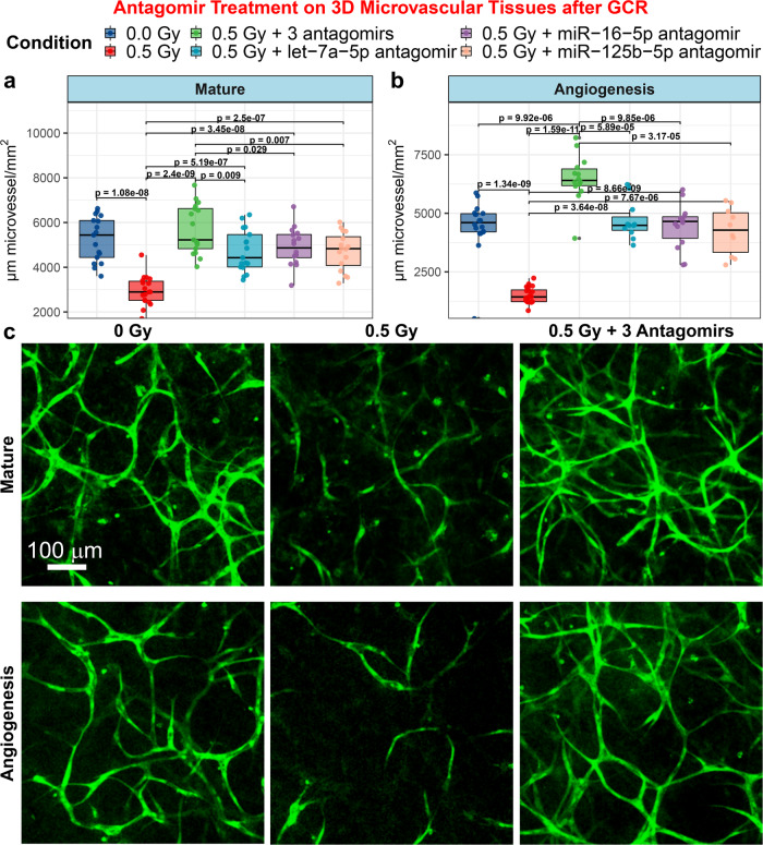
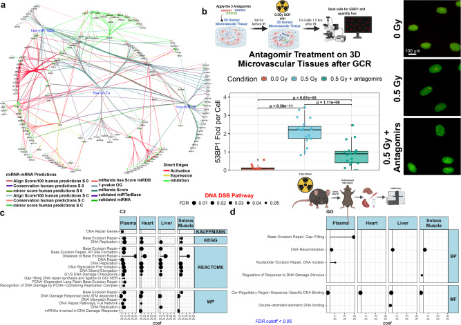
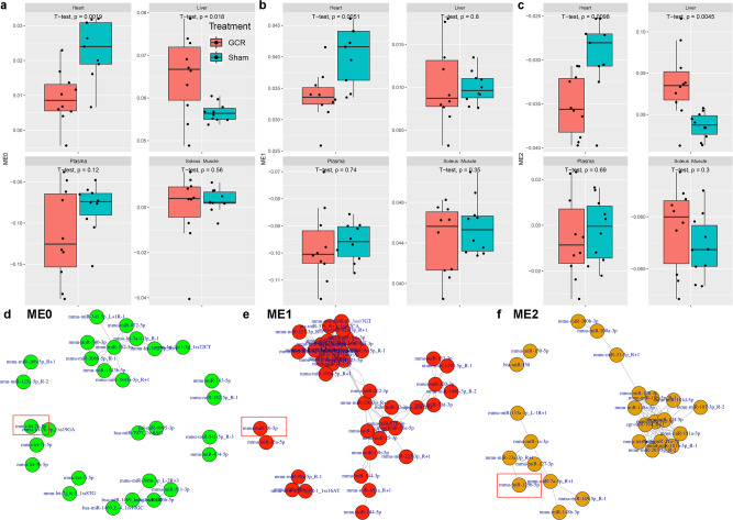
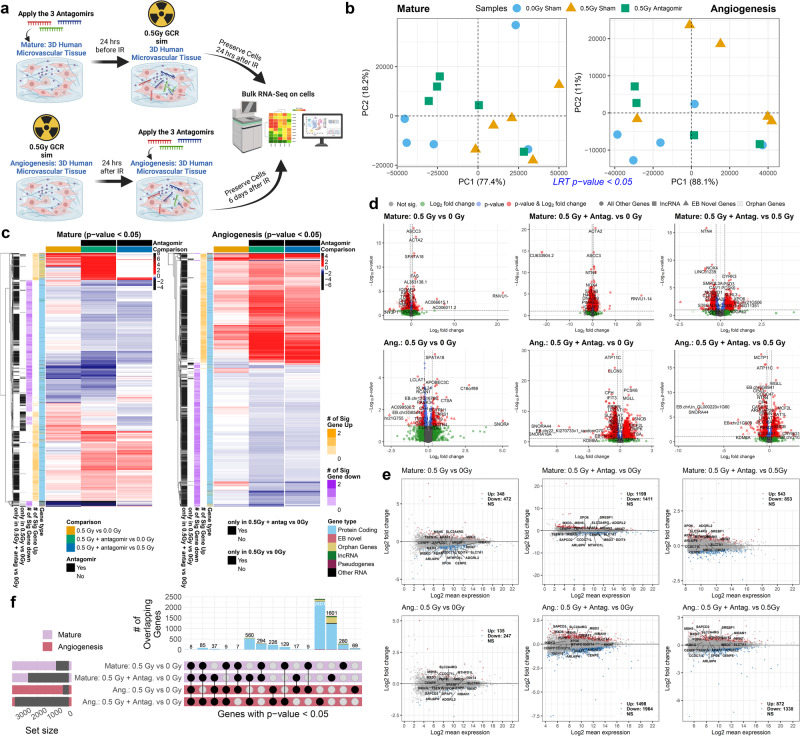
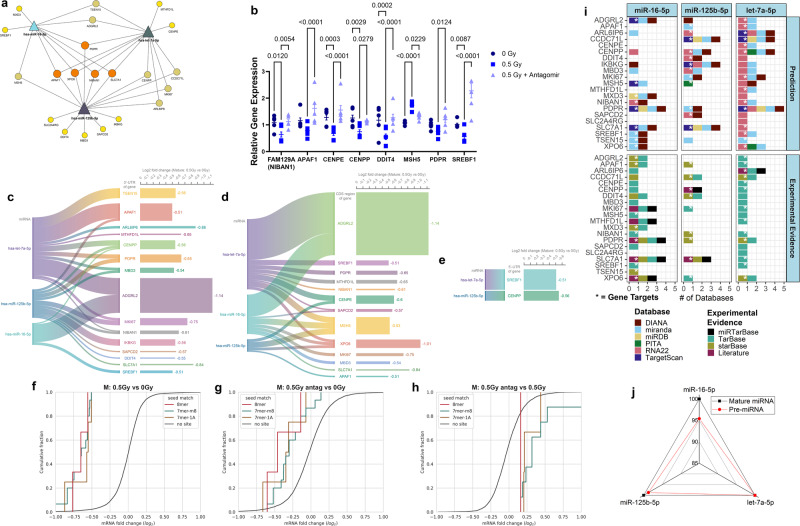
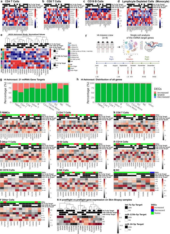
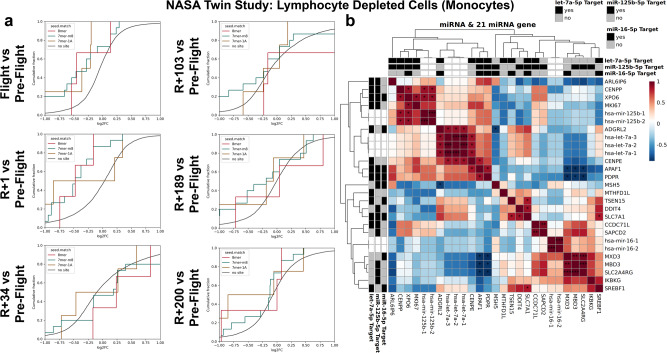
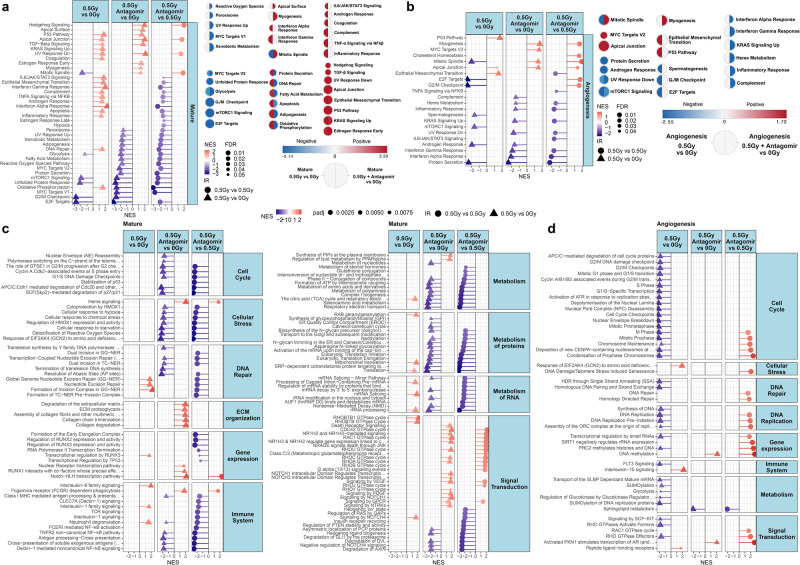
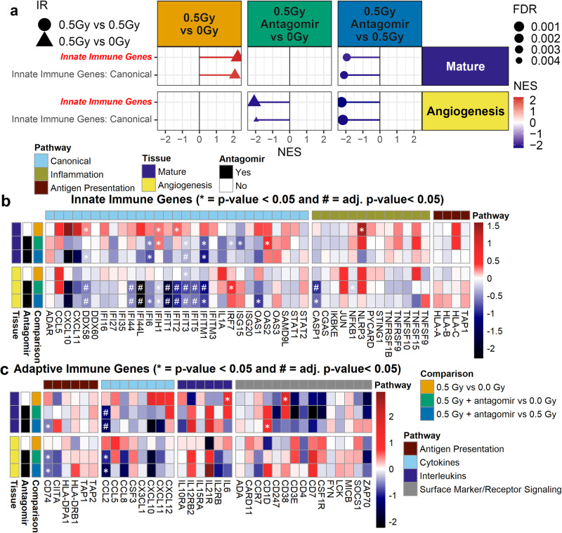
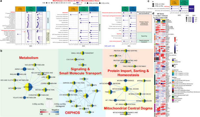
Our previous research revealed a key microRNA signature that is associated with spaceflight that can be used as a biomarker and to develop countermeasure treatments to mitigate the damage caused by space radiation. Here, we expand on this work to determine the biological factors rescued by the countermeasure treatment. We performed RNA-sequencing and transcriptomic analysis on 3D microvessel cell cultures exposed to simulated deep space radiation (0.5 Gy of Galactic Cosmic Radiation) with and without the antagonists to three microRNAs: miR-16-5p, miR-125b-5p, and let-7a-5p (i.e., antagomirs). Significant reduction of inflammation and DNA double strand breaks (DSBs) activity and rescue of mitochondria functions are observed after antagomir treatment. Using data from astronaut participants in the NASA Twin Study, Inspiration4, and JAXA missions, we reveal the genes and pathways implicated in the action of these antagomirs are altered in humans. Our findings indicate a countermeasure strategy that can potentially be utilized by astronauts in spaceflight missions to mitigate space radiation damage.


Conflict of interest statement

The authors declare no competing interests.

Figures

Fig. 1
Fig. 1. Antagomir treatment of 3D human umbilical vein endothelial cell (HUVEC) microvessel cell cultures.
a, b Quantification of formation of microvessels using 3D cultures of human microvessels irradiated with 0.5 Gy of simplified simulated galactic cosmic rays (GCR) compared to sham irradiated samples. Irradiated cultures were treated with or without antagomir-induced inhibition of miR-125b-5p, miR-16-5p, and/or let-7a-5p. For the boxplots the center line represents the median and the lines extending from both ends of the box indicates the quartile (Q) variability outside Q1 and Q3 to the minimum and maximum values. a mature and b angiogenesis microvessels. The p-values were determined by two-side multiple pairwise comparison. c Mature and angiogenesis microvessels fixed and fluorescently stained with 5-(4,6-dichlorotriazinyl) aminofluorescein (DTAF) after GCR irradiation, with or without antagomirs 24 h prior to irradiation. Scale bar = 100 µm. For mature the following biological independent samples were used: n = 17 for 0 Gy, n = 18 for 0.5 Gy, n = 17 for 0.5 Gy + 3 antagomirs, n = 17 for 0.5 Gy + let-7a-5p antagomir, n = 17 for 0.5 Gy + miR-16-5p antagomir, and n = 16 for 0.5 Gy + miR-125b-5p antagomir. For angiogenesis the following biological independent samples were performed: n = 18 for 0 Gy, n = 20 for 0.5 Gy, n = 14 for 0.5 Gy + 3 antagomirs, n = 11 for 0.5 Gy + let-7a-5p antagomir, n = 14 for 0.5 Gy + miR-16-5p antagomir, and n = 10 for 0.5 Gy + miR-125b-5p antagomir.
Fig. 2
Fig. 2. Quantification of DNA double strand break (DSB) repair foci with antagomir treatment and miRNA pathway analysis.
a Network representation for the DNA DSB repair gene targets for miR-125b-5p, miR-16-5p, and let-7a-5p generated by ClueGO in Cytoscape. As indicated in the figure legend, the color for the edges indicate either the predictions used for the miRNA-mRNA connection or the influence two different nodes will have on each other (i.e. Direct Edges). b Quantification of 53BP1 DNA repair foci in the mature 3D HUVEC microvessel cell culture 1.5 h after irradiation with 0.5 Gy of GCR. Representative images are shown on the right (scale bar = 20 µm). The p-values were determined by two-side multiple pairwise comparison. n = 3 biologically independent samples examined for each conditions and a total of the following random independent field of views for each condition: n = 12 field of views for 0 Gy and n = 19 field of views for both 0.5 Gy and 0.5 Gy + antagomirs. For the boxplot the center line represents the median and the lines extending from both ends of the box indicates the quartile (Q) variability outside Q1 and Q3 to the minimum and maximum values. The schematic of the experiment was created with BioRender.com. DNA DSB pathway-specific Gene Set Enrichment Analysis (GSEA) from c the curated chemical and genetic perturbations and canonical pathways collection (C2) and d the gene ontology (GO) collection using miRNA-sequencing data from different tissues (i.e. liver, heart, soleus muscle, and plasma) from C57BL/6 female mice irradiated with or without 0.5 Gy OF GCR exposure. Mice were euthanized (N = 10 irradiated and N = 10 sham controls) and tissues were harvested 24 h after irradiation. In Fig. 2 the schematics in panels b and d created with BioRender.com released under a Creative Commons Attribution-NonCommercial-NoDerivs 4.0 International license.
Fig. 3
Fig. 3. MiRNA based weighted gene co-expression network analysis (WGCNA) on different organs from GCR irradiated mice.
Boxplots displaying the expression of co-expressed miRNA network a Module 0 (ME0), b Module 1 (ME1), and c Module 2 (ME2) in heart, liver, plasma, and soleus muscle samples of mice that underwent Sham or GCR treatment. The y-axis represents the eigengene expression values of each network module. For the boxplots the center line represents the median and the lines extending from both ends of the box indicates the quartile (Q) variability outside Q1 and Q3 to the minimum and maximum values. The p-values were determined by two-side pairwise comparison. Network representation of highly correlated co-expressed miRNAs for d ME0, e ME1, and f ME2. The red box annotations highlight let-7a-5p, miR-16-5p, and miR-125b-5p. For all murine experiments n = 10 biologically independent animals examined with one beam time.
Fig. 4
Fig. 4. Global transcriptomic analysis reveals that antagomirs revert gene profiles closer to control samples.
a Schematic of the experimental profile for the mature and angiogenesis 3D cell culture models irradiated with 0.5 Gy of GCR and prepared for RNA-sequencing. There was n = 5 biological replicates for each condition. Created with BioRender.com released under a Creative Commons Attribution-NonCommercial-NoDerivs 4.0 International license. b Principal Component Analysis (PCA) of the significantly regulated genes (p-value < 0.05) for all conditions compared with likelihood ratio test (LTR) analysis for both mature and angiogenesis 3D microvessel cell culture models. c Heatmap of significantly regulated genes (p-value < 0.05) for mature and angiogenesis cell culture models. For each gene (i.e. row), the criteria for display was to have at least one comparison per gene to be significantly regulated. Then the trends for non-significant genes for that row were also displayed. The log2(fold-change) values are displayed. The side color bars indicate the number of significant genes that are either up- or down-regulated per row and also the type of gene. d Volcano and e MA plots for each comparison. f Upset plot displaying the overlapping significantly regulated genes (p-value < 0.05) for mature and angiogenesis models with and without antagomir treatment. For all RNA-seq data Wald test and the likelihood ratio test was used to generate the F statistic p-value. The adj. p-value plots are also provided in Supplementary Fig. 3.
Fig. 5
Fig. 5. 21 key gene targets for the three miRNAs restored back to control levels after antagomir treatment.
a Network display of the predicted miRNA gene targets by the mirDIP algorithm that were significantly regulated (p-value < 0.05) for 0.5 Gy versus 0 Gy, but were not statistically significant for 0.5 Gy with the antagomirs versus 0 Gy in the mature 3D cell culture model. For all RNA-seq data Wald test and the likelihood ratio test was used to generate the F statistic p-value. b Relative Gene Expression from PCR validation of 8 out of the 21 key genes. The p-values were determined by two-side multiple pairwise comparison. Data are presented as a dot plot of individual values with the mean values indicated with the +/−SEM. Binding probability of miRNAs to c 3’-UTR, d CDS, and e 5’-UTR of gene targets and their log2(fold change) in the presence or absence of radiation. Cumulative plots for the 21 key genes with comparing 0.5 Gy vs 0 Gy (f), 0.5 Gy + antagomir vs 0 Gy (g), and 0.5 Gy + antagomir vs 0.5 Gy (h) for the 3D mature microvessels. i Bar plot of the databases utilized by mirDIP (i.e. DIANA, miranda, miRDB, PITA, RNA22, and TargetScan) and experimental evidence for the 21 genes targeted by miR-16-5p, miR-125b-5p, and let-7a-5p. The experimental evidence for the targets have been compiled from TarBase, miRTarBase, starBase, and literature. We have also provided the details for the experimental evidence from these databases in Supplementary Table 1. The *mirDIP gene targets for each miRNA as shown in panel a. It is important to note that the databases provide literature references for all the experimental evidence and the literature indication in the figure is for additional literature evidence outside of these databases. j Radar plot illustrating homology of pre-miRNAs (red) and mature miRNAs (black) in humans and mice for miR-16-5p, miR-125b-5p, and let-7a-5p.
Fig. 6
Fig. 6. Expression of 21 genes rescued by the antagomir treatment in astronauts from the NASA Twin Study, JAXA CFE, and Inspiration4 (i4) missions.
ad Heatmaps displaying the 21 key gene profile for the different cell types (i.e. CD4 T cells, CD8 T cells, CD19 B cells, and lymphocyte deleted cell population) from RNA-seq on the NASA Twin Study. e Heatmap of the normalized plasma cell-free RNA expression values for the 21 key genes over time for the six astronauts over 120 days in space from JAXA study. The values shown are the averaged normalized expression values for all six astronauts for each time point during flight and post-flight. The three pre-flight time points were averaged together, since the changes for genes in the time leading up to flight are considered to be the same and part of the baseline values. For the time, L = Launch (i.e. meaning time after launch from Earth and length in space) and R = Return to Earth. f A schematic of the i4 experimental design created with BioRender.com released under a Creative Commons Attribution-NonCommercial-NoDerivs 4.0 International license. Global gene expression profile of the g 21 key genes and h all genes from scRNA-sequence data from the i4 astronauts. ir Heatmaps displaying the 21 key gene profile for the different cell types (i.e. PBMCs, CD4 T cells, CD8 T cells, Other T cells, B cells, CD14 cells, CD16 cells, NK cells, Dendritic cells (DC), and other cells) from scRNA-seq on the i4 astronauts. s Heatmap visualization of the 21 key genes on the i4 astronauts of relative expression changes in the postflight relative to preflight skin biopsy sample across four skin compartments including Outer Epidermis (OE), Inner Epidermis (IE), Outer Dermis (OD), and Vasculature (VA).
Fig. 7
Fig. 7. The interaction of the three miRNAs with the 21 gene targets from the NASA Twin Study in Monocytes.
a Cumulative plots for change over time with the 21 genes targeted by the three miRNAs using RNA-seq data from the NASA Twin Study data. Sequencing was performed on lymphocyte depleted cells (i.e. monocytes) from the Twin that was onboard the ISS for 340 days. There are comparisons over time starting with Flight versus Pre-Flight as well as comparing the different Return (R) to Earth timepoints from 1 to 200 days versus Pre-Flight. b Correlation plot of the three miRNAs (i.e. miR-16-5p, let-7a-5p, and miR-125b-5p) and the 21 gene targets for the overlapping time points from the miRNA-seq and mRNA-seq NASA Twin Study data. The gene targets for the miRNAs are indicated in the outer black (yes for gene target) and gray (no for gene target) rows and columns. Wald test and the likelihood ratio test was used to generate the F statistic p-value and significance for the correlation is shown by *p-value < 0.05.
Fig. 8
Fig. 8. Pathway analysis on the mature and angiogenesis 3D microvessel cell culture models revealing key functions being mitigated by the antagomirs after exposure to GCR irradiation.
Lollipop plots showing Gene Set Enrichment Analysis (GSEA) pathway analysis of the Hallmark pathways for a mature and b angiogenesis 3D cell culture models. The right of each plot displays circular nodes comparing 0.5 Gy versus 0 Gy (left side of node) with 0.5 Gy plus antagomir treatment versus 0 Gy (right side of node) analyzed with the Cytoscape plugin Enrichment Map. Only significantly regulated pathways with FDR < 0.25 are shown. GSEA Reactome pathway analysis of RNA-seq data on c mature and d angiogenesis 3D cell culture models displaying the pathways that are significantly altered by 0.5 Gy irradiation with or without antagomir treatment compared to 0 Gy. Gene sets that were significantly enriched in the same direction for the two treatment groups (both up or both down) are not shown leaving only opposing or single pathway enrichments. Only significantly regulated pathways with FDR < 0.05 are shown. The nominal enrichment score (NES) represents the relative degree a gene set is changed and is corrected for gene set size.
Fig. 9
Fig. 9. Antagomir countermeasure mitigates increases in innate immune activity.
a Lollipop plot of GSEA analysis on RNA-seq data using a custom curation of innate and adaptive immune pathways for mature and angiogenesis 3D microvessel cell culture models. The red italic bold font names indicate the primary pathway containing all genes from the sub-pathways. Only pathways with a FDR < 0.25 are shown. Heatmaps of the t-scores for the individual genes in the b innate and c adaptive immune custom pathways. (*p-value < 0.05). For all RNA-seq data Wald test and the likelihood ratio test was used to generate the F statistic p-value.
Fig. 10
Fig. 10. Mitigation of mitochondrial dysregulation and integrated stress response (ISR) with antagomir treatment on mature and angiogenesis 3D cell culture models irradiated with GCR.
a Lollipop plots for the GSEA analysis on mitochondrial pathways determined from MitoCarta/MitoPathway for 3D mature microvessel cell culture (FDR < 0.25). The red italic bold font names indicate the main general pathway containing all the genes from the sub-pathways. b Network representation analyzed with Cytoscape for the mitochondrial pathways from MitoCarta/MitoPathway. The node size indicates the number of significantly regulated genes associated with each pathway. Each node shows the pathway regulation with FDR < 0.25 for 0.5 Gy versus 0 Gy (left side) and 0.5 Gy with the antagomirs versus 0 Gy (right side). Genesets not significantly expressed by 0.5 Gy versus 0 Gy are highlighted in yellow. c GSEA analysis on the custom integrated stress response (ISR) pathways for mature and angiogenesis RNA-seq data. The red italic bold font names indicate the main general pathway containing all the genes from the sub-pathways. Only significantly regulated with pathways with FDR < 0.25 are shown. d Heatmaps of the t-scores for the individual genes in the ISR custom pathways. (*p-value < 0.05). For all RNA-seq data Wald test and the likelihood ratio test was used to generate the F statistic p-value.

Similar articles

Cited by

  • An Update on Neuroaging on Earth and in Spaceflight.
    Kuznetsov NV, Statsenko Y, Ljubisavljevic M. Kuznetsov NV, et al. Int J Mol Sci. 2025 Feb 18;26(4):1738. doi: 10.3390/ijms26041738. Int J Mol Sci. 2025. PMID: 40004201 Free PMC article. Review.
  • Presymptomatic microRNA-based biomarker signatures for the prognosis of localized radiation injury in mice.
    Ancel L, Gueguen J, Thoër G, Marçais J, Chemloul A, Le Guen B, Benderitter M, Tamarat R, Souidi M, Benadjaoud MA, Flamant S. Ancel L, et al. PLoS One. 2025 Aug 8;20(8):e0329737. doi: 10.1371/journal.pone.0329737. eCollection 2025. PLoS One. 2025. PMID: 40779592 Free PMC article.
  • miRTarBase 2025: updates to the collection of experimentally validated microRNA-target interactions.
    Cui S, Yu S, Huang HY, Lin YC, Huang Y, Zhang B, Xiao J, Zuo H, Wang J, Li Z, Li G, Ma J, Chen B, Zhang H, Fu J, Wang L, Huang HD. Cui S, et al. Nucleic Acids Res. 2025 Jan 6;53(D1):D147-D156. doi: 10.1093/nar/gkae1072. Nucleic Acids Res. 2025. PMID: 39578692 Free PMC article.
  • A second space age spanning omics, platforms and medicine across orbits.
    Mason CE, Green J, Adamopoulos KI, Afshin EE, Baechle JJ, Basner M, Bailey SM, Bielski L, Borg J, Borg J, Broddrick JT, Burke M, Caicedo A, Castañeda V, Chatterjee S, Chin CR, Church G, Costes SV, De Vlaminck I, Desai RI, Dhir R, Diaz JE, Etlin SM, Feinstein Z, Furman D, Garcia-Medina JS, Garrett-Bakelman F, Giacomello S, Gupta A, Hassanin A, Houerbi N, Irby I, Javorsky E, Jirak P, Jones CW, Kamal KY, Kangas BD, Karouia F, Kim J, Kim JH, Kleinman AS, Lam T, Lawler JM, Lee JA, Limoli CL, Lucaci A, MacKay M, McDonald JT, Melnick AM, Meydan C, Mieczkowski J, Muratani M, Najjar D, Othman MA, Overbey EG, Paar V, Park J, Paul AM, Perdyan A, Proszynski J, Reynolds RJ, Ronca AE, Rubins K, Ryon KA, Sanders LM, Glowe PS, Shevde Y, Schmidt MA, Scott RT, Shirah B, Sienkiewicz K, Sierra MA, Siew K, Theriot CA, Tierney BT, Venkateswaran K, Hirschberg JW, Walsh SB, Walter C, Winer DA, Yu M, Zea L, Mateus J, Beheshti A. Mason CE, et al. Nature. 2024 Aug;632(8027):995-1008. doi: 10.1038/s41586-024-07586-8. Epub 2024 Jun 11. Nature. 2024. PMID: 38862027 Free PMC article. Review.
  • Taking the 3Rs to a higher level: replacement and reduction of animal testing in life sciences in space research.
    Vinken M, Grimm D, Baatout S, Baselet B, Beheshti A, Braun M, Carstens AC, Casaletto JA, Cools B, Costes SV, De Meulemeester P, Doruk B, Eyal S, Ferreira MJS, Miranda S, Hahn C, Helvacıoğlu Akyüz S, Herbert S, Krepkiy D, Lichterfeld Y, Liemersdorf C, Krüger M, Marchal S, Ritz J, Schmakeit T, Stenuit H, Tabury K, Trittel T, Wehland M, Zhang YS, Putt KS, Zhang ZY, Tagle DA. Vinken M, et al. Biotechnol Adv. 2025 Jul-Aug;81:108574. doi: 10.1016/j.biotechadv.2025.108574. Epub 2025 Apr 1. Biotechnol Adv. 2025. PMID: 40180136 Review.

References

    1. Patel ZS, et al. Red risks for a journey to the red planet: The highest priority human health risks for a mission to Mars. npj Microgravity. 2020;6:1–13. doi: 10.1038/s41526-020-00124-6. - DOI - PMC - PubMed
    1. Roy-O’Reilly M, Mulavara A, Williams T. A review of alterations to the brain during spaceflight and the potential relevance to crew in long-duration space exploration. npj Microgravity. 2021;7:1–9. doi: 10.1038/s41526-021-00133-z. - DOI - PMC - PubMed
    1. Strollo F, Gentile S, Strollo G, Mambro A, Vernikos J. Recent Progress in Space Physiology and Aging. Front Physiol. 2018;9:1551. doi: 10.3389/fphys.2018.01551. - DOI - PMC - PubMed
    1. Vernikos J, Schneider VS. Space, gravity and the physiology of aging: parallel or convergent disciplines? A mini-review. Gerontology. 2010;56:157–166. doi: 10.1159/000252852. - DOI - PubMed
    1. Afshinnekoo E, et al. Fundamental Biological Features of Spaceflight: Advancing the Field to Enable Deep-Space Exploration. Cell. 2020;183:1162–1184. doi: 10.1016/j.cell.2020.10.050. - DOI - PMC - PubMed