Review
. 2024 Aug;38(12):2392-2405.
doi: 10.1038/s41433-024-03154-6. Epub 2024 Jun 11.

Electrodiagnostic tests of the visual pathway and applications in neuro-ophthalmology

Affiliations
Review

Electrodiagnostic tests of the visual pathway and applications in neuro-ophthalmology

Antonio Calcagni et al. Eye (Lond). 2024 Aug.

Abstract

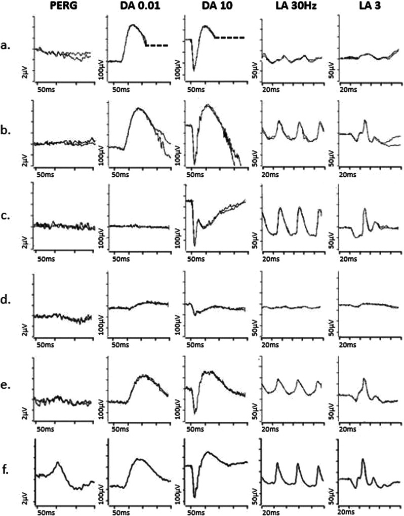
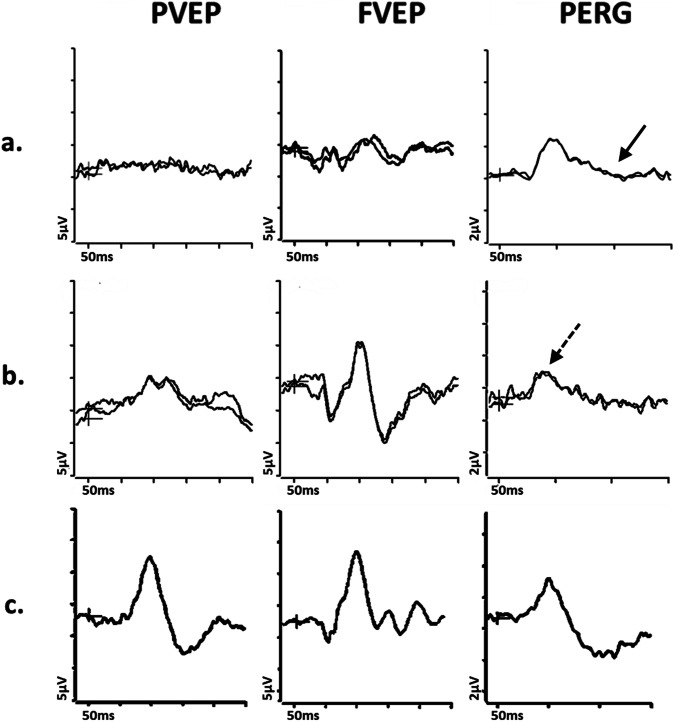
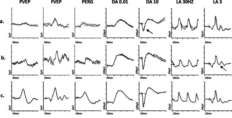
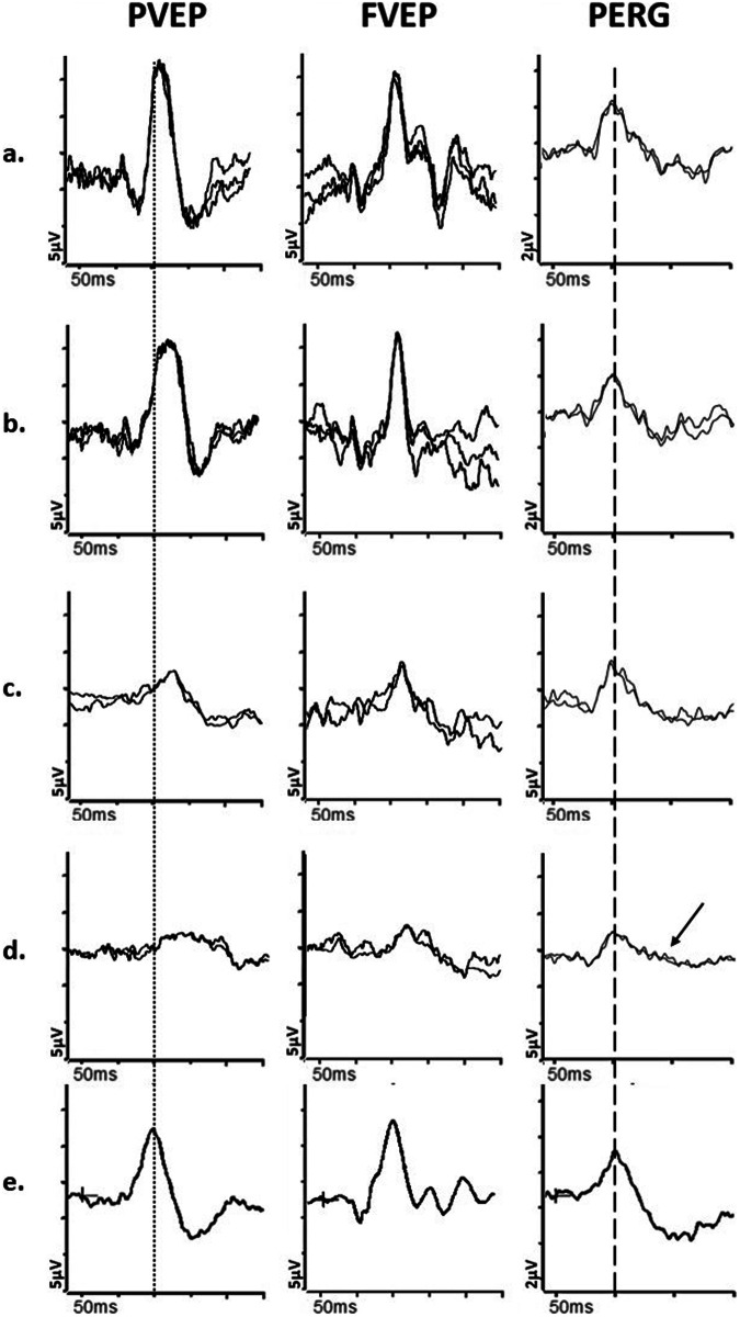
This article describes the main visual electrodiagnostic tests relevant to neuro-ophthalmology practice, including the visual evoked potential (VEP), and the full-field, pattern and multifocal electroretinograms (ffERG; PERG; mfERG). The principles of electrophysiological interpretation are illustrated with reference to acquired and inherited optic neuropathies, and retinal disorders that may masquerade as optic neuropathy, including ffERG and PERG findings in cone and macular dystrophies, paraneoplastic and vascular retinopathies. Complementary VEP and PERG recordings are illustrated in demyelinating, ischaemic, nutritional (B12), and toxic (mercury, cobalt, and ethambutol-related) optic neuropathies and inherited disorders affecting mitochondrial function such as Leber hereditary optic neuropathy and dominant optic atrophy. The value of comprehensive electrophysiological phenotyping in syndromic diseases is highlighted in cases of SSBP1-related disease and ROSAH (Retinal dystrophy, Optic nerve oedema, Splenomegaly, Anhidrosis and Headache). The review highlights the value of different electrophysiological techniques, for the purposes of differential diagnosis and objective functional phenotyping.

摘要: 本文介绍了与神经眼科临床相关的主要视觉电生理诊断试验, 包括视觉诱发电位(VEP), 以及全视野ERG, 图形ERG以及多焦ERG(ffERG;PERG;mfERG)。电生理学检查的基本原则以及对获得性和遗传性视神经病变, 以及与视神经病变需要鉴别的其它视网膜病变, 如视锥细胞与黄斑营养不良、副肿瘤和血管视网膜病变等。VEP和PERG在脱髓鞘、缺血、营养性(B12)和毒性(汞、钴和乙胺丁醇相关)视神经病变以及影响线粒体功能的遗传性疾病的诊断中起到了重要作用, 如Leber遗传性视神经病变和显性视神经萎缩。在SSBP1相关疾病和ROSAH(视网膜营养不良、视神经水肿、脾肿大、多汗和头痛)的病例中, 电生理改变在全身性疾病中的诊断性价值尤为突出。本综述强调了不同的电生理技术在鉴别诊断和客观的视网膜功能以及疾病表型方面的价值。.


Conflict of interest statement

NJ: Consultancy fees received from Chiesi, AC, MMN, AGR: None

Figures

Fig. 1
Fig. 1. Electrophysiological findings in cases of retinal pathology.
Examples of pattern ERGs and full-field ERGs are shown from one eye of a patient with cone dystrophy (a), macular dystrophy (b), melanoma associated retinopathy (c), from right and left eyes of a patient with a right central retinal artery occlusion (d, e) and from a representative normal subject for comparison (f). Recordings showed a high degree of inter-ocular symmetry in all but the patient with a central retinal artery occlusion. All patient traces are superimposed to demonstrate reproducibility. The DA 0.01, DA 10 and LA3 ERGs include a 20 ms pre-stimulus delay. The patient with cone dystrophy has abnormal LA ERGs consistent with generalised (peripheral) cone system dysfunction, with an undetectable PERG P50 component in keeping with severe macular involvement (a). In macular dystrophy PERG P50 is undetectable, and ffERGs are normal (b), indicating dysfunction confined to the macula. In melanoma associated retinopathy the ffERGs are pathognomonic for generalised On bipolar cell dysfunction, including an electronegative DA10 ERG and specific distortions of the LA 3 ERG waveform (c). In CRAO there is unilateral severe generalised inner retinal dysfunction of rod and cone systems, manifest as an electronegative DA10 ERG, with severe LA ERG abnormalities, reflecting severe inner retinal dysfunction (including On and Off bipolar cell dysfunction); of note, patients with long-standing CRAO frequently have additional reduction of the dark adapted (DA3 and DA10) ERG a-wave amplitudes (although relatively mild compared with b-wave reductions). A significant loss of the normal dark-adapted cone system contribution to the DA3 and DA10 ERGs (normally mixed rod and cone system responses) is likely contributory, but the exact mechanism is unknown.
Fig. 2
Fig. 2. Electrophysiological findings in cases of Leber hereditary optic neuropathy and Dominant optic atrophy.
Examples of pattern reversal VEPs (PVEP), flash VEPs (FVEP) and pattern ERGs (PERG) are shown from one eye of a patient with (a) Leber Hereditary Optic Neuropathy (LHON); b Dominant Optic Atrophy; and c from a representative normal subject for comparison. In these cases recordings showed a high degree of inter-ocular symmetry and are shown for one eye only. All patient traces are superimposed to demonstrate reproducibility. The patient with LHON (a) has an undetectable PVEP and the FVEP severely reduced with additional waveform distortion, consistent with severe optic nerve dysfunction. The PERG shows P50 preservation of amplitude but mild shortening of peak time and the N95 component is selectively reduced (solid arrow) in keeping with severe macular retinal ganglion cell dysfunction. The patient with DOA (b) shows a subnormal and distorted PVEP, preserved FVEP and reduction in the PERG N95:P50 ratio, with additional P50 peak time shortening (arrow with broken lines), consistent with early retinal ganglion cell involvement.
Fig. 3
Fig. 3. Electrophysiological findings in SSBP1- and ALPK1- related pathologies.
Examples of pattern reversal VEPs (PVEP), flash VEPs (FVEP), pattern ERGs (PERG) and full-field ERGs are shown from one eye of a patient with (a) SSBP1-related pathology; b ALPK1-related pathology (ROSAH syndrome); and (c) from a representative normal subject for comparison. Recordings showed a high degree of inter-ocular symmetry and are shown for one eye only. All patient traces are superimposed to demonstrate reproducibility. The DA 0.01, DA 10 and LA3 ERGs include a 20 ms pre-stimulus delay. The patient with SSBP1-related disease (a) shows a subnormal PVEP, a borderline FVEP and reduction in the PERG N95:P50 ratio, with additional P50 peak time shortening, in keeping with macular retinal ganglion cell dysfunction; the DA ERGs are subnormal, with reduction (solid arrow) in the DA10 a-wave, which localises dysfunction at the level of the photoreceptors; LA ERGs are borderline normal. The patient with ALPK1-related pathology (b) shows a subnormal and delayed PVEP, a normal FVEP and reduction in the PERG N95:P50 ratio, in keeping with macular retinal ganglion cell dysfunction; the DA ERGs are borderline normal; LA ERGs are subnormal, indicative of generalised cone system dysfunction, with additional PhNR attenuation (arrow with broken lines), suggestive of global retinal ganglion cell loss.
Fig. 4
Fig. 4. Electrophysiological findings in cases of optic nerve demyelination.
Examples of pattern reversal VEPs (PVEP), flash VEPs (FVEP) and pattern ERGs (PERG) from a and b, a patient with demyelination in the one eye; (c and d), a patient with demyelination in the left eye and sub-clinical involvement of the right eye; e from a representative normal subject for comparison. All patient traces are superimposed to demonstrate reproducibility. Peak time differences are highlighted by the vertical dotted line (PVEP P100) and vertical dashed line (PERG P50). The first patient, with demyelination in the left eye (b) has a PVEP P100 of mildly delayed peak time and normal amplitude, consistent with mild optic nerve conduction delay. The flash VEP is normal. The PERG N95:P50 ratio, P50 peak time and amplitude are normal. The asymptomatic right eye (a) shows normal PVEP, FVEP and PERG. The second patient’s left eye (d) shows a PVEP P100 of markedly delayed peak time and subnormal amplitude, consistent with severe optic nerve conduction delay. Additionally, the FVEP is moderately subnormal and mildly delayed. There is severe PERG N95:P50 ratio reduction (arrow), consistent with severe retinal ganglion cell dysfunction; P50 peak time is normal. The asymptomatic right eye (c) shows a PVEP P100 of moderately delayed peak time and subnormal amplitude, consistent with mild to moderate optic nerve conduction delay and sub-clinical involvement. The right eye PERG is normal.
Fig. 5
Fig. 5. Electrophysiological findings in a patient with non-arteritic ischaemic optic neuropathy.
Example of pattern reversal VEP (PVEP), flash VEP (FVEP) and pattern ERG (PERG) from the right (a) and left (b) eye of a patient with non-arteritic anterior ischaemic optic neuropathy (AION); and from a representative normal subject (c) for comparison. All patient traces are superimposed to demonstrate reproducibility. Peak time differences are highlighted by the vertical dotted line (PVEP P100) and vertical dashed line (PERG P50). The left eye with AION (b) has a PVEP P100 component of reduced amplitude, poorly formed N135 component, and normal peak time and the FVEP has an altered waveform compared with the contralateral healthy eye, consistent with moderately severe optic nerve dysfunction. The PERG shows P50 preservation but the N95:P50 ratio is markedly reduced (arrow) consistent with marked RGC involvement. The right eye (a) is unaffected, with a normal PVEP, FVEP and PERG.
Fig. 6
Fig. 6. Electrophysiological findings in cases of nutritional and toxic neuropathies.
Examples of pattern reversal VEPs (PVEP), flash VEPs (FVEP) and pattern ERGs (PERG) from a patient with chronic alcohol withdrawal syndrome and Vitamin B12 deficiency (a); from a patient initially suspected of LHON that had developed cobalt toxicity due to cobalt leaching out from the femoral heads following multiple total hip replacements (b); from a patient initially suspected of LHON and subsequently diagnosed with mercury toxicity, demonstrated by high levels of mercury in urine, resulting from a prolonged and restricted diet of fish high in methyl mercury (c); from a case of ethambutol toxicity following treatment for lymph node TB (d); from a representative normal subject for comparison (e). All recordings showed a high degree of inter-ocular symmetry and are shown for one eye only. All patient traces are superimposed to demonstrate reproducibility. Peak time differences are highlighted by the vertical dotted line (PVEP P100) and vertical dashed line (PERG P50). The patient with Vit B12 deficiency (a) has an undetectable PVEP and the FVEP is severely reduced, consistent with severe optic nerve dysfunction. The PERG N95:P50 ratio is severely reduced (arrow); P50 peak time is markedly shortened with marginal reduction in amplitude, consistent with severe RGC involvement. The patient with Cobalt toxicity (b) shows an undetectable PVEP and FVEP of reduced amplitude, consistent with moderately severe optic neuropathy; in addition, there is severe reduction of the PERG N95:P50 ratio (arrow), with mild shortening of P50 peak time, consistent with retinal ganglion cell dysfunction. The PERG P50 amplitude is preserved. The patient with Mercury toxicity (c) shows moderate PERG N95:P50 ratio reduction (arrow) and shortened P50 peak time, consistent with moderate retinal ganglion cell dysfunction; PERG P50 amplitude is preserved. In addition, the PVEP is undetectable and the FVEP amplitude is of marginally reduced amplitude, consistent with optic nerve involvement. The patient with Ethambutol toxicity (d) shows a marginally delayed and subnormal PVEP P100, and normal FVEP, consistent with mild optic nerve dysfunction. The PERG P50 peak time and amplitude are normal but the N95:P50 ratio is reduced (arrow), consistent with retinal ganglion cell involvement.
Fig. 7
Fig. 7. Multichannel pattern onset-offset VEPs in a case of albinism.
Recordings a and b: from a patient with ocular albinism and (c): from a representative normal subject for comparison, using five scalp electrodes positioned over the occiput; 2 electrodes over the right hemisphere; two electrodes over the left hemisphere; 1 electrode over the midline. a Pattern onset-offset VEPs from stimulation of the right eye of the patient with albinism are present and of significantly larger amplitude from the left hemisphere (bold arrows) compared with the right hemisphere (dashed arrows), consistent with contralateral predominance; VEP responses from stimulation of the left eye (b) are present and of significantly larger amplitude from the right hemisphere (bold arrows) compared with the left hemisphere (dashed arrows), consistent with contralateral predominance. c VEP responses from the normal subject are of similar amplitude and peak time from the left and right hemisphere.

References

    1. Odom JV, Bach M, Brigell M, Holder GE, McCulloch DL, Mizota A, et al. ISCEV standard for clinical visual evoked potentials: (2016 update). Doc Ophthalmol. 2016;133:1–9. 10.1007/s10633-016-9553-y - DOI - PubMed
    1. Robson AG, Frishman LJ, Grigg J, Hamilton R, Jeffrey BG, Kondo M, et al. ISCEV Standard for full-field clinical electroretinography (2022 update). Doc Ophthalmol. 2022;144:165–77. 10.1007/s10633-022-09872-0 - DOI - PMC - PubMed
    1. Hoffmann MB, Bach M, Kondo M, Li S, Walker S, Holopigian K, et al. ISCEV standard for clinical multifocal electroretinography (mfERG) (2021 update). Doc Ophthalmol. 2021;142:5–16. 10.1007/s10633-020-09812-w - DOI - PMC - PubMed
    1. Constable PA, Bach M, Frishman LJ, Jeffrey BG, Robson AG. International Society for Clinical Electrophysiology of V. ISCEV Standard for clinical electro-oculography (2017 update). Doc Ophthalmol. 2017;134:1–9. 10.1007/s10633-017-9573-2 - DOI - PMC - PubMed
    1. Thompson DA, Bach M, Sustar Habjan M, Viswanathan S, Robson AG. ISCEV Standard for clinical pattern electroretinography (2024 update). Doc Ophthalmol. 2024;148:75–85. 10.1007/s10633-024-09970-1 - DOI - PMC - PubMed

MeSH terms