

Iodine-125 seed inhibits proliferation and promotes apoptosis of cholangiocarcinoma cells by inducing the ROS/p53 axis
- PMID: 38862667
- PMCID: PMC11166828
- DOI: 10.1007/s10142-024-01392-1
Iodine-125 seed inhibits proliferation and promotes apoptosis of cholangiocarcinoma cells by inducing the ROS/p53 axis
Abstract
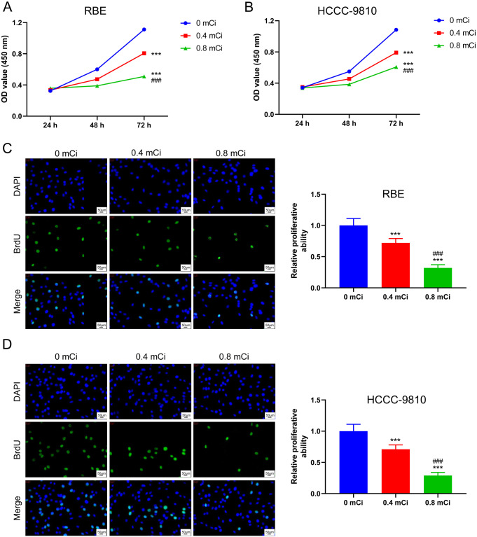
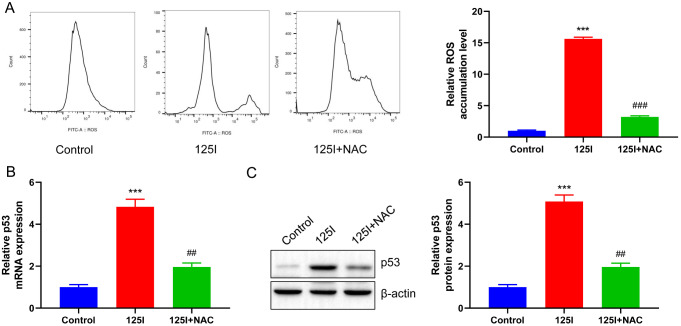
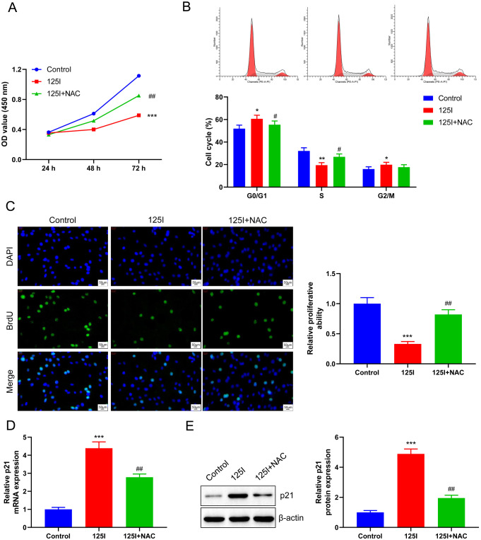
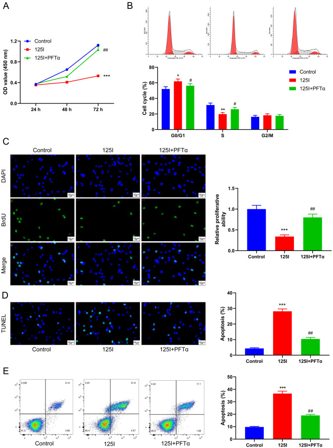
With advances in radioactive particle implantation in clinical practice, Iodine-125 (125I) seed brachytherapy has emerged as a promising treatment for cholangiocarcinoma (CCA), showing good prognosis; however, the underlying molecular mechanism of the therapeutic effect of 125I seed is unclear. To study the effects of 125I seed on the proliferation and apoptosis of CCA cells. CCA cell lines, RBE and HCCC-9810, were treated with reactive oxygen species (ROS) scavenger acetylcysteine (NAC) or the p53 functional inhibitor, pifithrin-α hydrobromide (PFTα). Cell counting kit-8 (CCK-8) assay, 5-bromo-2-deoxy-uridine (BrdU) staining, and terminal deoxynucleotidyl transferase (TdT)-mediated dUTP nick end labeling (TUNEL) assay and flow cytometry assay were performed to test the radiation-sensitivity of 125I seed toward CCA cells at different radiation doses (0.4 mCi and 0.8 mCi). 2,7-dichlorofluorescein diacetate (DCF-DA) assay, real-time quantitative polymerase chain reaction (RT-qPCR), and western blot analysis were performed to assess the effect of 125I seed on the ROS/p53 axis. A dose-dependent inhibitory effect of 125I seeds on the proliferation of CCA cells was observed. The 125I seed promoted apoptosis of CCA cells and induced the activation of the ROS/p53 pathway in a dose-dependent manner. NAC or PFTα treatment effectively reversed the stimulatory effect of 125I seed on the proliferation of CCA cells. NAC or PFTα suppressed apoptosis and p53 protein expression induced by the 125I seed. 125I seed can inhibit cell growth mainly through the apoptotic pathway. The mechanism may involve the activation of p53 and its downstream apoptotic pathway by up-regulating the level of ROS in cells.
Keywords: Apoptosis; Cholangiocarcinoma; Iodine-125 seed; Proliferation; ROS/p53 pathway.
© 2024. The Author(s).
Conflict of interest statement
The authors declare no competing interests.
Figures


Similar articles
-
Mechanistic insights into 125I seed implantation therapy for Cholangiocarcinoma: focus on ROS-Mediated apoptosis and the role of GPX2.J Cancer Res Clin Oncol. 2024 Jun 25;150(6):324. doi: 10.1007/s00432-024-05840-0. J Cancer Res Clin Oncol. 2024. PMID: 38914724 Free PMC article.
-
MCM2 promotes the proliferation, migration and invasion of cholangiocarcinoma cells by reducing the p53 signaling pathway.Yi Chuan. 2022 Mar 20;44(3):230-244. doi: 10.16288/j.yczz.21-426. Yi Chuan. 2022. PMID: 35307646
-
Photodynamic therapy inhibits cancer progression and induces ferroptosis and apoptosis by targeting P53/GPX4/SLC7A11 signaling pathways in cholangiocarcinoma.Photodiagnosis Photodyn Ther. 2024 Jun;47:104104. doi: 10.1016/j.pdpdt.2024.104104. Epub 2024 Apr 26. Photodiagnosis Photodyn Ther. 2024. PMID: 38679154
-
TRAIL Enhances Shikonin Induced Apoptosis through ROS/JNK Signaling in Cholangiocarcinoma Cells.Cell Physiol Biochem. 2017;42(3):1073-1086. doi: 10.1159/000478758. Epub 2017 Jun 29. Cell Physiol Biochem. 2017. PMID: 28662515
-
Knockdown of tripartite motif 59 (TRIM59) inhibits proliferation in cholangiocarcinoma via the PI3K/AKT/mTOR signalling pathway.Gene. 2019 May 25;698:50-60. doi: 10.1016/j.gene.2019.02.044. Epub 2019 Feb 27. Gene. 2019. PMID: 30822475
Cited by
-
Evaluating the efficacy of 8Spheres microsphere embolization combined with iodine‑125 seed implantation in advanced refractory lung cancer: A retrospective study.Oncol Lett. 2025 Apr 9;29(6):285. doi: 10.3892/ol.2025.15031. eCollection 2025 Jun. Oncol Lett. 2025. PMID: 40247985 Free PMC article.
-
Radioactive stent versus normal stent insertion for inoperable malignant biliary obstruction: a systematic review and meta-analysis.Surg Endosc. 2025 Apr;39(4):2692-2700. doi: 10.1007/s00464-025-11571-1. Epub 2025 Mar 6. Surg Endosc. 2025. PMID: 40047865
References
MeSH terms
Substances
LinkOut - more resources
Full Text Sources
Research Materials
Miscellaneous
