. 2024 Jul;8(7):854-871.
doi: 10.1038/s41551-024-01201-x. Epub 2024 Jun 11.

Deep-learning-enabled antibiotic discovery through molecular de-extinction

Affiliations

Deep-learning-enabled antibiotic discovery through molecular de-extinction

Fangping Wan et al. Nat Biomed Eng. 2024 Jul.

Abstract

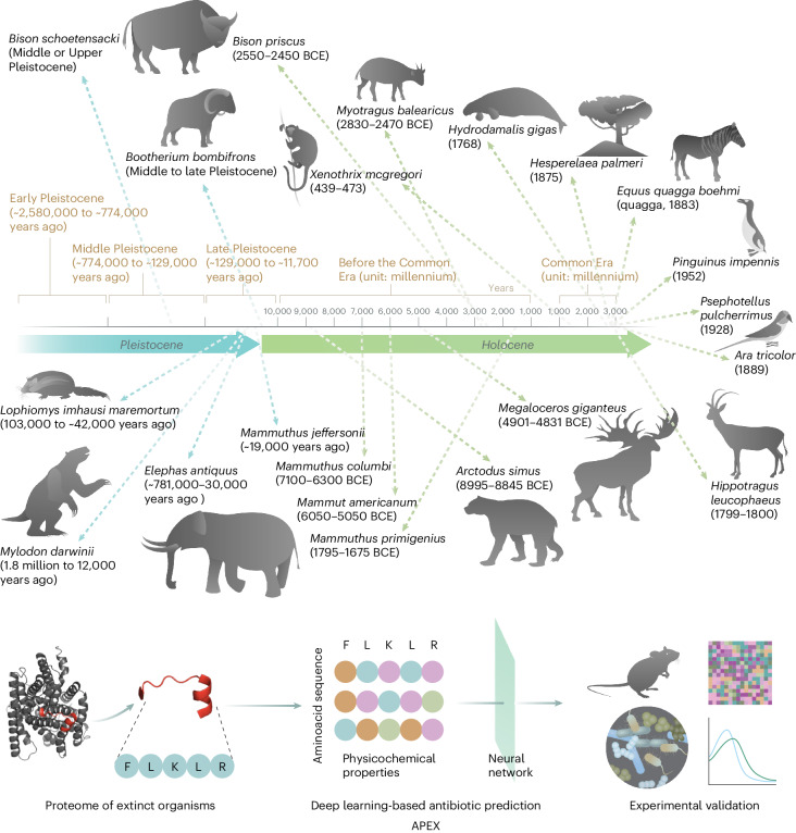
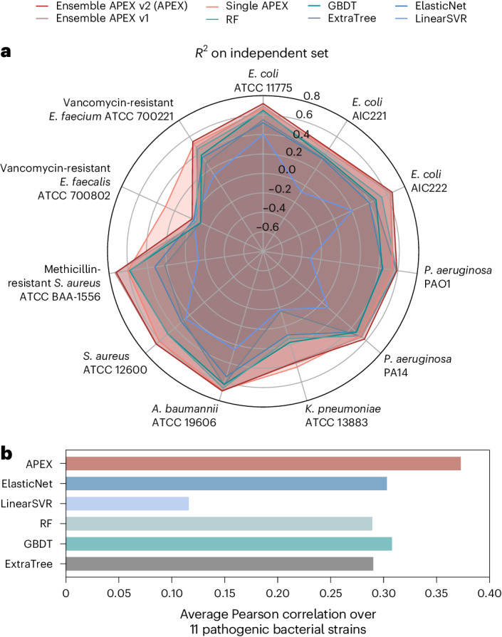
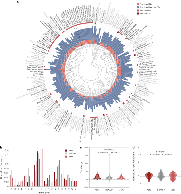
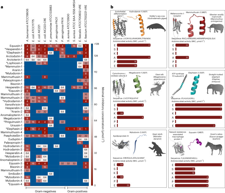
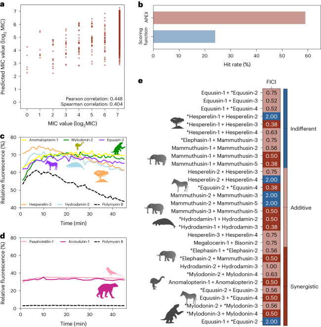
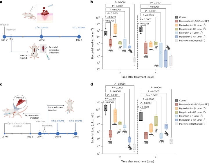
Molecular de-extinction aims at resurrecting molecules to solve antibiotic resistance and other present-day biological and biomedical problems. Here we show that deep learning can be used to mine the proteomes of all available extinct organisms for the discovery of antibiotic peptides. We trained ensembles of deep-learning models consisting of a peptide-sequence encoder coupled with neural networks for the prediction of antimicrobial activity and used it to mine 10,311,899 peptides. The models predicted 37,176 sequences with broad-spectrum antimicrobial activity, 11,035 of which were not found in extant organisms. We synthesized 69 peptides and experimentally confirmed their activity against bacterial pathogens. Most peptides killed bacteria by depolarizing their cytoplasmic membrane, contrary to known antimicrobial peptides, which tend to target the outer membrane. Notably, lead compounds (including mammuthusin-2 from the woolly mammoth, elephasin-2 from the straight-tusked elephant, hydrodamin-1 from the ancient sea cow, mylodonin-2 from the giant sloth and megalocerin-1 from the extinct giant elk) showed anti-infective activity in mice with skin abscess or thigh infections. Molecular de-extinction aided by deep learning may accelerate the discovery of therapeutic molecules.


Conflict of interest statement

C.F.-N. provides consulting services to Invaio Sciences and is a member of the Scientific Advisory Boards of Nowture S.L., Peptidus and Phare Bio. The de la Fuente Lab has received research funding or in-kind donations from United Therapeutics, Strata Manufacturing PJSC and Procter & Gamble, none of which were used in support of this work.

Figures

Fig. 1
Fig. 1. Molecular de-extinction of antibiotics from ancient proteomes using deep learning.
All available proteomes of extinct organisms were mined by APEX, our deep learning algorithm. Amino acid sequences ranging from 8 to 50 amino acid residues within proteins from extinct organisms were inputted into multitask deep learning models that trained on both public and in-house peptide data to evaluate the potential antimicrobial activity. The highest ranked peptides based on predicted antimicrobial activities were then selected and thoroughly characterized against clinically relevant pathogens both in vitro and in animal models. The mechanism of action, physicochemical features and synergistic interactions of these peptides were also assayed. The dates report the approximate extinction date or period for the organisms studied. The protein and peptide structures shown in the figure were created with PyMOL Molecular Graphics System, version 2.1 Schrödinger, LLC.
Fig. 2
Fig. 2. APEX prediction performance and comparison with other models.
a, Radar chart showing R2 correlation in terms of species-specific antimicrobial activity prediction on an independent dataset (a held-out subset from our in-house peptide dataset) for various ML models. The radius reflects the R2 value for each of the models. APEX variants outperformed the baseline ML methods for most of the pathogens analysed. RF, random forest; GBDT, gradient boosting decision tree; ExtraTree, extra-tree regressor; ElasticNet, elastic net; LinearSVR, linear support vector regression. b, Mean of species-wise Pearson correlation of log2-transformed MICs between values obtained experimentally and predicted by various ML models. Evaluated dataset: 69 peptides were synthesized and tested.
Fig. 3
Fig. 3. Antimicrobials identified by APEX in extinct organisms and their composition and physicochemical properties.
a, Phylogenetic tree showing the extinct organisms scanned by APEX. Circular bars denote the log10-transformed average active (red) and inactive (blue) EPs discovered by APEX. A peptide was considered active when its predicted median MIC against the bacterial strains tested was ≤80 μmol l−1. The values were normalized by the number of proteins per organism scanned. The organisms whose EPs were selected for validation are highlighted in bold type. Extinct organisms that presented active EPs validated experimentally are indicated by a light red square and, within that group, those organisms encoding extinct sequences absent in extant organisms are highlighted with a dark red square. b, Amino acid frequency in AEPs and MEPs compared with known AMPs from the DBAASP database. AEPs present a higher frequency of the basic residue K, the aliphatic residue V, and uncharged polar residues (M, Q and T) than MEPs. c,d, Distribution of two physicochemical properties for peptides with predicted antimicrobial activity (AEPs and MEPs) and AMPs from DBAASP: net charge (c) and normalized hydrophobicity (d). Net charge directly influences the initial electrostatic interactions between the peptide and negatively charged bacterial membranes, and hydrophobicity directly influences the interactions of the peptide with lipids in the membrane bilayers. EPs from extinct organisms are slightly less hydrophobic and similarly have a net positive charge, compared with EPs from the modern human proteome or peptides from DBAASP. Statistical significance in c and d was determined using two-tailed t-tests followed by Mann–Whitney test; P values are shown in the graph. The solid line inside each box represents the mean value obtained for each group.
Fig. 4
Fig. 4. Antimicrobial activity profiles of sequences from the proteomes of extinct organisms.
a, Heat map of the antimicrobial activities (μmol l−1) of the active antimicrobial agents from extinct organisms against 11 clinically relevant pathogens, including strains resistant to conventional antibiotics. Briefly, 106 bacterial cells and serially diluted EPs (0–128 μmol l−1) were incubated at 37 °C. One day post treatment, the optical density at 600 nm was measured in a microplate reader to evaluate bacterial growth in the presence of the EPs from extinct organisms. MIC values in the heat map are the arithmetic mean of the replicates in each condition. b, Examples of active AEPs and MEPs from various extinct organisms, their parent protein and their activity profile against ESKAPE pathogens (Enterococcus spp., S. aureus, K. pneumoniae, A. baumannii, P. aeruginosa, E. coli). Antimicrobial activity is expressed as the MIC (μmol l−1), and activity bars are presented as −log2 MIC. The data for the assays in a are the mean, and the experiments were performed in three independent replicates. AEPs in a are indicated by an asterisk (*). The protein and peptide structures shown in the figure were created with PyMOL Molecular Graphics System, version 2.1 Schrödinger, LLC.
Fig. 5
Fig. 5. Antimicrobial activity, mechanism of action and synergy of antimicrobials from the proteomes of extinct organisms.
a, Pan-bacterial Pearson and Spearman correlations of log2-transformed MICs between experimentally validated values and values predicted by APEX. b, Comparison between the hit rates of APEX and the scoring function previously described by Torres et al. to detect antibiotics in the modern human proteome. c, Cytoplasmic membrane depolarization by five antimicrobials from extinct organisms. The A. baumannii membrane was more strongly depolarized by the peptides than by the antibiotic polymyxin B. d, NPN permeabilization assays showing the effect of two antimicrobials from extinct organisms on the outer membrane of A. baumannii. Higher permeability was observed with the peptides than with the antibiotic polymyxin B. e, Heat map showing interactions between antimicrobials identified by APEX, expressed as the FICI. Most of the tested EP pairs from extinct organisms either synergized or had an additive effect against A. baumannii and P. aeruginosa; the latter was only tested against the peptide pair composed of equusin-1 and equusin-2 shown in the last row of the heat map. The data for the assays in ce are the mean, and the experiments were performed in three independent replicates. AEPs in e are indicated by an asterisk (*).
Fig. 6
Fig. 6. Anti-infective activity of antibiotic molecules in animal models.
a, Schematic of the skin abscess mouse model used to assess the anti-infective activity of selected antimicrobials from extinct organisms (n = 6) against A. baumannii ATCC 19606. b, The molecules mammuthusin-2 (M. primigenius), hydrodamin-1 (H. gigas), megalocerin-1 (Megalocerus sp.), elephasin-2 (E. antiquus) and mylodonin-2 (M. darwinii), administered at their MIC in a single dose, inhibited the proliferation of the infection for up to 4 days after treatment compared to the untreated control group. Elephasin-2 and mylodonin-2 cleared the infection in some of the mice, with activity comparable to that of the antibiotic used as control, polymyxin B. c, Schematic of the neutropenic thigh infection mouse model in which EPs from extinct organisms were injected intraperitoneally. Anti-infective activity against A. baumannii ATCC 19606 was assessed 2 and 4 days after intraperitoneal peptide administration (n = 6). d, Two days after intraperitoneal injection, mylodonin-2 at its MIC reduced A. baumannii ATCC19606 infection as much as polymyxin B, compared to the untreated control group. Four days post treatment, mammuthusin-2 and elephasin-2 showed the same level of activity as polymyxin B. Statistical significance in b and d was determined using one-way ANOVA followed by Dunnett’s test; P values are shown in the graph. For the boxplots, the centre line represents the mean, the box limits the first and third quartiles and the whiskers (minima and maxima) 1.5 × the interquartile range. The solid line inside each box represents the mean value obtained for each group. Panels a and c were created with BioRender.com. Source data

Similar articles

Cited by

References

    1. New Report Calls for Urgent Actionto Avert Antimicrobial Resistance Crisis (World Health Organization, 2019).
    1. Zuckerkandl, E. & Pauling, L. Molecules as documents of evolutionary history. J. Theor. Biol.8, 357–366 (1965). - PubMed
    1. Maasch, J. R. M. A., Torres, M. D. T., Melo, M. C. R. & de la Fuente-Nunez, C. Molecular de-extinction of ancient antimicrobial peptides enabled by machine learning. Cell Host Microbe31, 1260–1274 (2023). - PMC - PubMed
    1. Wong, F., de la Fuente-Nunez, C. & Collins, J. J. Leveraging artificial intelligence in the fight against infectious diseases. Science381, 164–170 (2023). - PMC - PubMed
    1. Porto, W. F. et al. In silico optimization of a guava antimicrobial peptide enables combinatorial exploration for peptide design. Nat. Commun.9, 1490 (2018). - PMC - PubMed

LinkOut - more resources