. 2024 Jun 11;4(1):111.
doi: 10.1038/s43856-024-00544-5.

Impact of gallbladder hypoplasia on hilar hepatic ducts in biliary atresia

Affiliations

Impact of gallbladder hypoplasia on hilar hepatic ducts in biliary atresia

Nanae Miyazaki et al. Commun Med (Lond). .

Abstract

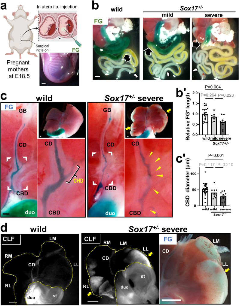
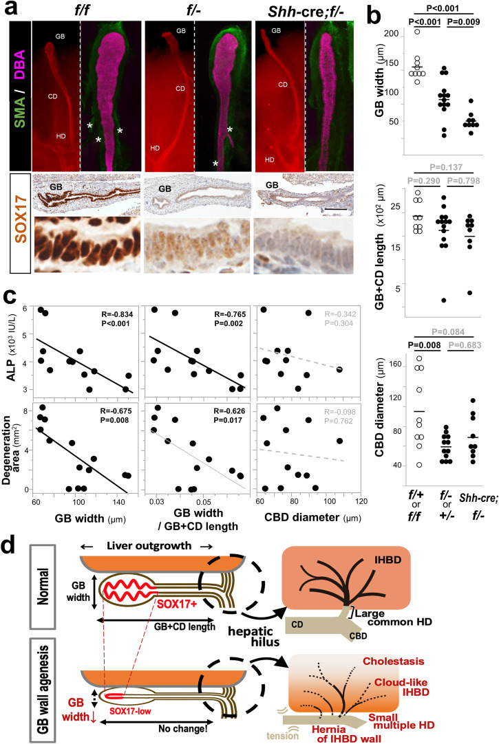
Background: Biliary atresia (BA) is an intractable disease of unknown cause that develops in the neonatal period. It causes jaundice and liver damage due to the destruction of extrahepatic biliary tracts,. We have found that heterozygous knockout mice of the SRY related HMG-box 17 (Sox17) gene, a master regulator of stem/progenitor cells in the gallbladder wall, exhibit a condition like BA. However, the precise contribution of hypoplastic gallbladder wall to the pathogenesis of hepatobiliary disease in Sox17 heterozygous embryos and human BA remains unclear.

Methods: We employed cholangiography and histological analyses in the mouse BA model. Furthermore, we conducted a retrospective analysis of human BA.

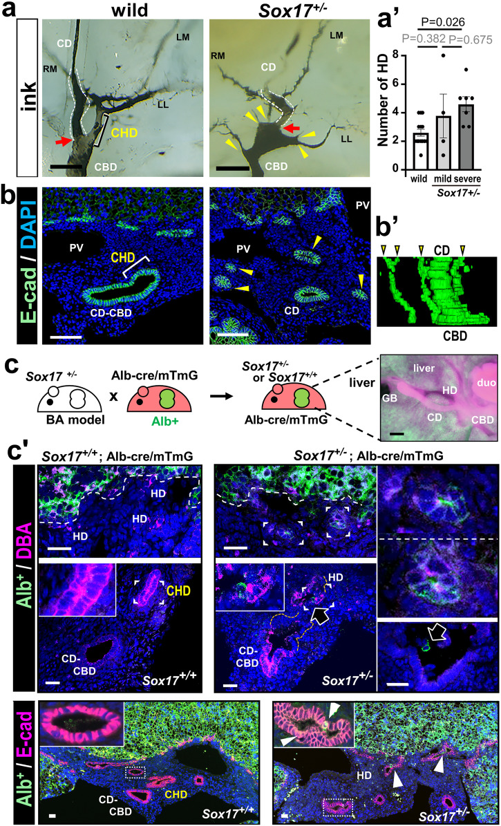
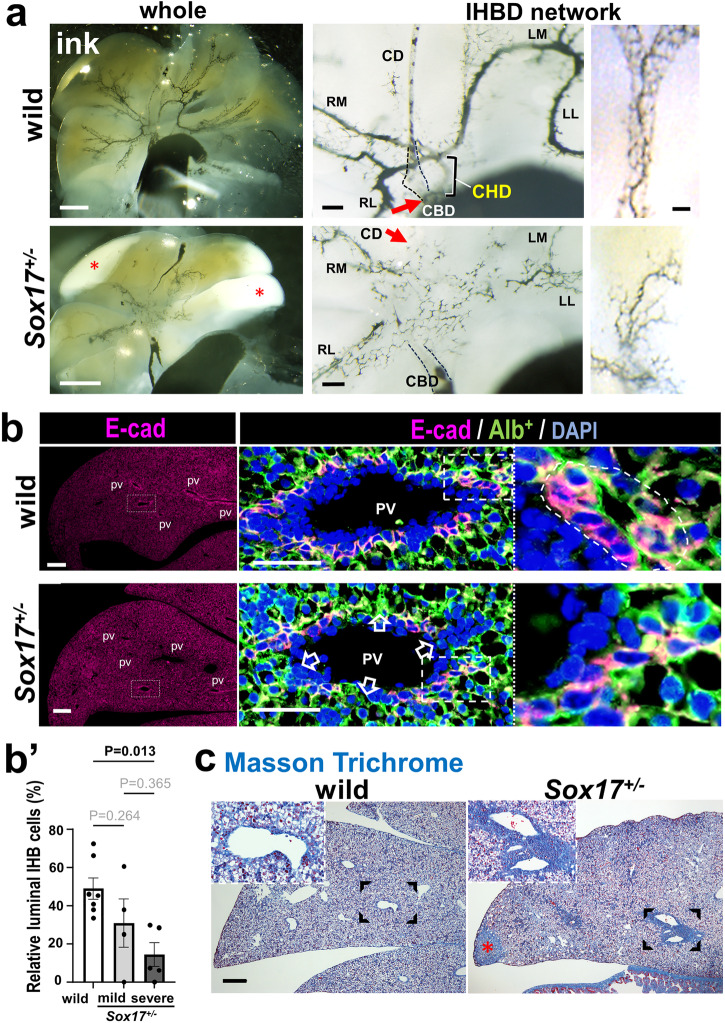
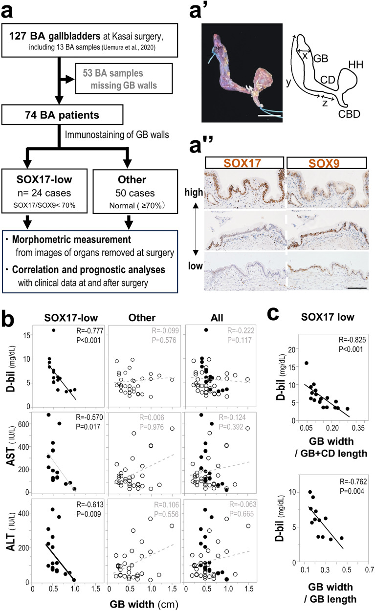
Results: We show that gallbladder wall hypoplasia causes abnormal multiple connections between the hilar hepatic bile ducts and the gallbladder-cystic duct in Sox17 heterozygous embryos. These multiple hilar extrahepatic ducts fuse with the developing intrahepatic duct walls and pull them out of the liver parenchyma, resulting in abnormal intrahepatic duct network and severe cholestasis. In human BA with gallbladder wall hypoplasia (i.e., abnormally reduced expression of SOX17), we also identify a strong association between reduced gallbladder width (a morphometric parameter indicating gallbladder wall hypoplasia) and severe liver injury at the time of the Kasai surgery, like the Sox17-mutant mouse model.

Conclusions: Together with the close correlation between gallbladder wall hypoplasia and liver damage in both mouse and human cases, these findings provide an insight into the critical role of SOX17-positive gallbladder walls in establishing functional bile duct networks in the hepatic hilus of neonates.

Plain language summary

Biliary atresia (BA) is a disease in newborns that causes a serious liver condition due to damage to the bile ducts (the pathways that carry bile juice). Although reduced function of a key gene called Sox17, which is essential for forming the gallbladder wall, has been observed in some BA cases, the link between gallbladder issues and liver damage is unknown. This study has shown how damage spreads through the bile ducts in the liver around the time of birth when there are problems in the gallbladder wall due to reduced SOX17 function. The findings indicate that proper growth of the gallbladder wall during this critical period is essential for forming a normal network of bile ducts in the developing liver. This discovery is promising for early diagnosis and better treatment of BA in newborns.


Conflict of interest statement

The authors declare no competing interests.

Figures

Fig. 1
Fig. 1. Imaging of bile flow in intrauterine Sox17+/− embryos by a cholangiography.
a Schematic illustrations of Intrauterine embryos injected intraperitoneally with Fast green FCF (FG) to visualize the bile flow in vivo. The lower right inset shows the image of the isolated embryo with intraperitoneal FG injection. b, c FG outflows into the fetal intestine in the Sox17+/− embryos with (severe) or without (mild) liver damages, together with wild-type littermate, at embryonic days (E) 18.5 at 90 min after FG injection. In b, large black arrow and small white arrow indicate the reachable edge of FG (blue) and endogenous bile (yellow), respectively. In c, the FG-positive (FG+) extrahepatic biliary structure in the enlarged view of the hepatic hilar area represented by the bracket, showing the multiple small hepatic ducts (yellow arrowheads) in the severe Sox17+/− embryo, instead of the large common hepatic duct (CHD) in the wild-type littermate. Bar graph in (b’) shows relative FG+ gut length (wild-type: n = 20, mild Sox17+/−: n = 10, severe Sox17+/−: n = 9; maternal: n = 6). The bar graph in c’ shows FG+ minimum luminal diameter of the common bile duct (CBD) (wild-type: n = 30, mild Sox17+/−: n = 9, severe Sox17+/−: n = 12; maternal: n = 8). Data are presented as mean ± s.e.m (by one-way ANOVA followed by Tukey’s test). d Whole liver images of wild-type and severe Sox17+/− embryos injected at E18.5 with a mixture of FG and Cholyl-lysyl-fluorescein (CLF; fluorescein-labeled bile acid), showing ectopic accumulation of both two bile tracers in the peripheral hepatic lobules (yellow arrows). CBD common bile duct, CD cystic duct, CHD common hepatic duct, duo duodenum, GB gallbladder, LL left lateral lobe, LM left medial lobe, RM right medial lobe, RL right lateral lobe, st stomach. Scale bars, 1 mm (a, b, d), 250 μm (c).
Fig. 2
Fig. 2. Ectopic formation of multiple hilar hepatic ducts and extrahepatic herniation of intrahepatic duct walls in severe Sox17+/− embryos prior to birth.
a, b Multiple small hepatic ducts (yellow arrowheads) visualized by retrograde cholangiography using black ink injection from the fetal duodenum (a, a’), anti-E-cadherin (E-cad) immunostaining (b; green fluorescence) and 3D construction of the DBA (Dolichos biflorus agglutinin; bile duct marker) lectin stained serial sections (b’) in Sox17+/− embryos at embryonic days (E) 18.5. In the wild-type embryo, one large common hepatic duct (CHD) connects to the cystic duct (red arrow). The bar graph in a’ shows the numbers of hilar hepatic ducts per whole liver (mean ± s.e.m; by one-way ANOVA followed by Tukey’s test [wild-type: n = 14, mild Sox17+/−: n = 4, severe Sox17+/−: n = 7, maternal: n = 7]). c Schematic representation of Sox17+/+ and Sox17+/− mice (biliary atresia [BA] model) in an Alb-cre; ROSAmTmG (Albumin [Alb] – Green Fluorescent Protein [GFP]+) background (left in c). Whole mount Alb- GFP+ liver (right in c) and immunostaining (c’) of intrahepatic (green; anti-GFP staining) and extrahepatic (magenta; DBA or anti-E-cad staining) bile duct walls at the hepatic hilus of E18.5 embryos. The ectopic Alb-GFP+ cells in hepatic ducts are protruding (white arrowhead) from the hepatic parenchyma (dashed lines). Note the contribution of several GFP+ cells in the damaged duct walls in Sox17+/− embryos (black arrows). The insets show the enlarged view of the area represented by the bracket. CD cystic duct, CBD common bile duct, CHD common hepatic duct, HD hepatic duct, LL left lateral lobe, LM left medial lobe, RM right medial lobe, PV portal vein, GB gallbladder, duo duodenum, Scale bars, 250 μm (a), 100 μm (b), 25 μm (c).
Fig. 3
Fig. 3. Cloud-like fine networks of intrahepatic bile ducts, together with early signs of hepatic fibrosis in the severe Sox17+/− embryos prior to birth.
a Retrograde cholangiography (black ink), showing a cloud-like intrahepatic bile duct (IHBD)network in the severe Sox17+/− embryo at embryonic days (E) 18.5. Note the poor development of networks near the damaged peripheral edge (red asterisk). Red arrows show that common hepatic duct (CHD) connects to the cystic duct (red arrow). b, b’ Anti-E-cadherin immunostaining and the morphometric analyses of the stained sections, showing the reduced cell population of intrahepatic ducts with apical lumen per total E-cad+ cells in Sox17+/− embryos, as compared with wild-type littermates. In b Alb (Albumin)-GFP (Green fluorescent Protein)+ (green)/E-cad (E-cadherin)+ (red) intrahepatic ducts (yellow for double positive) around portal vein in the peripheral lobular regions of the Sox17+/+ and Sox17+/− embryos at E18.5 (Alb-cre; ROSAmTmG background). The construction of IHBDs in the Sox17+/− embryos is defective (open arrows). In b’, the bar graph shows the relative E-cad+ cell population of luminal duct per total cells (mean ± s.e.m.; by one-way ANOVA followed by Tukey’s test [wild-type: n = 7, mild Sox17+/−: n = 4, severe Sox17+/−: n = 5; maternal: n = 8]). c Masson’s trichrome in liver lobules of severe Sox17+/− embryos. In c, Masson trichrome staining sections, showing the fibrosis (blue) in the peripheral margins (red asterisk). CBD common bile duct, CD cystic duct, CHD common hepatic duct, IHBD intrahepatic bile duct, PV portal vein, RL right lateral lobe, RM right medial lobe, LM left medial lobe, LL left lateral lobe. Scale bars, 1 mm (left in a), 200 μm (middle in a, left in b and c), 100 μm (right in a), 50 μm (middle in b).
Fig. 4
Fig. 4. Impact of gallbladder hypoplasia on the liver injury in human BA cases with aberrant SOX17-low gallbladder walls at the time of Kasai surgery.
a-a”, Study design to analyze the characteristics and prognosis of the Biliary atresia (BA) cases with aberrant SOX17-low gallbladder walls. Of 127 gallbladder specimens from BA patients, 74 showed preserved gallbladder walls at the time of Kasai surgery. Among the 74 samples, 24 gallbladders were classified as having aberrant gallbladder walls with reduced SOX17 expression level (less than 70% of the SOX17/SOX9 index) by Immunohistochemical analysis of SOX17 and SOX9 (brown color). In a’, gross anatomical image and scheme of an extrahepatic duct resected by Kasai surgery in a BA patient. CBD: common bile duct, CD: cystic duct, GB: gallbladder, HH: hepatic hilum, x = GB width, y = GB length, z = CD length. In a”, human BA gallbladder walls of two serial sections of gallbladder body that were stained with anti-SOX17 and anti-SOX9 antibody (SOX17/SOX9 indices = 115.9%, 56.3% and 6.5% from top [high] to bottom [low]). b, c, Spearman correlation coefficients between gallbladder width (b, GB width [cm]; c, relative GB width per gallbladder-cystic duct [GB + CD] length or per GB length) and serum markers (direct bilirubin [D-bil, mg/dl], aspartate aminotransferase [AST, IU/l] or alanine aminotransferase [ALT, IU/l]) in the SOX17-low BA cases (solid circle), other cases (open circle), and both of them (All) (SOX17-low: n = 17, Other: n = 34, All: n = 51). Each point shows the value (black solid line, p < 0.01; gray solid line, 0.01 ≤ p < 0.05; gray dashed line, p ≥ 0.05). Scale bar, 1 cm (a’), 100 µm (a”).
Fig. 5
Fig. 5. Effects of excessive gallbladder wall hypoplasia on gallbladder shape and liver injury in Sox17-mutant embryos just prior to birth.
a Whole-mount dolichos biflorus agglutinin (DBA; magenta) and anti-alpha-smooth muscle actin (SMA; green)-double staining (upper panels) and anti-SOX17 immunostaining (brown) of sagittal sections of distal sac-like structures (i.e., presumptive gallbladder; lower panels) of Sox17flox/flox (f/f), Sox17flox/− (f/−) and Shh-cre; Sox17flox/− (Shh-cre; f/−) embryos at embryonic days (E) 18.5. The border between gallbladder and cystic duct walls in the Shh-cre; f/− embryos cannot be defined even by anti-SMA staining for gallbladder-specific smooth muscle layers in the presumptive gallbladder region. asterisks, vascular smooth muscle. b Dot plots of the gallbladder (GB) width (i.e., maximum diameter of DBA-positive distal sac-like structure), gallbladder-cystic duct (GB + CD) length and minimum common bile duct (CBD) diameter (y-axis) in three genotypes (i.e., control [f/+ and f/f], heterozygous [f/− and +/−] and homozygous [Shh-cre; f/−] deletion of two Sox17 alleles (x-axis). Note significant reduction in GB width and CBD diameter by Tukey’s honestly significant difference test), in contrast to no change in GB + CD length among three genotypes (Shh-cre; f/−: n = 9, f/− or +/−: n = 14, f/+ or f/f: n = 9). c Spearman rank correlation tests between GB width, relative GB width per GB + CD length and minimum CBD diameter in x axis and liver injury level (i.e., serum Alkaline phosphatase [ALP, IU/l] level and degeneration area) on the y-axis (black solid line, p < 0.01; gray solid line, 0.01 ≤ p < 0.05; gray broken line, p ≥ 0.05) (n = 13). d Schematic illustration of the ripple effects of GB wall hypoplasia on the intrahepatic duct (IHBD) network. Hypoplastic GB wall with reduced SOX17 expression causes reduced GB width, albeit of no change in GB + CD length (left in d), and it simultaneously causes abnormal formation of a hilar bile duct network (right in d) as follows: i) deformation of a large common hepatic duct (i.e.; multiple small hepatic ducts); ii) extrahepatic herniation of the IHBD wall; iii) a cloud-like immature IHBD network near the hepatic hilus; and iv) peripheral cholestasis. CBD common bile duct, CD cystic duct, GB gallbladder, IHBD intrahepatic bile duct, HD hepatic duct. Scale bar in a, 100 µm.

Similar articles

Cited by

References

    1. Lemaigre FP. Development of the Intrahepatic and Extrahepatic Biliary Tract: A Framework for Understanding Congenital Diseases. Annu. Rev. Pathol. 2020;15:1–22. doi: 10.1146/annurev-pathmechdis-012418-013013. - DOI - PubMed
    1. Spence JR, et al. Sox17 regulates organ lineage segregation of ventral foregut progenitor cells. Dev. Cell. 2009;17:62–74. doi: 10.1016/j.devcel.2009.05.012. - DOI - PMC - PubMed
    1. Uemura M, et al. Expression and function of mouse Sox17 gene in the specification of gallbladder/bile-duct progenitors during early foregut morphogenesis. Biochem. Biophys. Res. Commun. 2010;391:357–363. doi: 10.1016/j.bbrc.2009.11.063. - DOI - PubMed
    1. Higashiyama H, et al. Embryonic cholecystitis and defective gallbladder contraction in the Sox17-haploinsufficient mouse model of biliary atresia. Development. 2017;144:1906–1917. doi: 10.1242/dev.147512. - DOI - PMC - PubMed
    1. Higashiyama H, et al. Anatomy and development of the extrahepatic biliary system in mouse and rat: a perspective on the evolutionary loss of the gallbladder. J. Anat. 2018;232:134–145. doi: 10.1111/joa.12707. - DOI - PMC - PubMed

LinkOut - more resources