. 2024 Oct;45(10):2077-2091.
doi: 10.1038/s41401-024-01318-4. Epub 2024 Jun 11.

ABBV-744 alleviates LPS-induced neuroinflammation via regulation of BATF2-IRF4-STAT1/3/5 axis

Affiliations

ABBV-744 alleviates LPS-induced neuroinflammation via regulation of BATF2-IRF4-STAT1/3/5 axis

Le-le Wang et al. Acta Pharmacol Sin. 2024 Oct.

Abstract

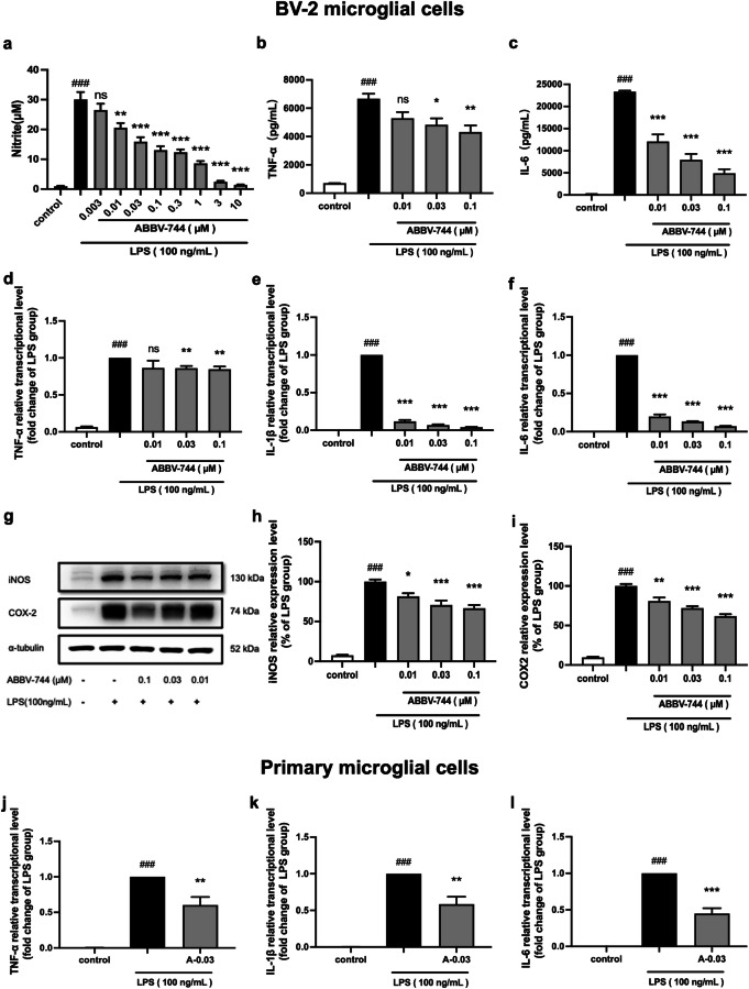
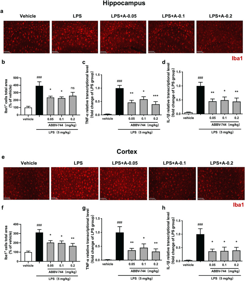
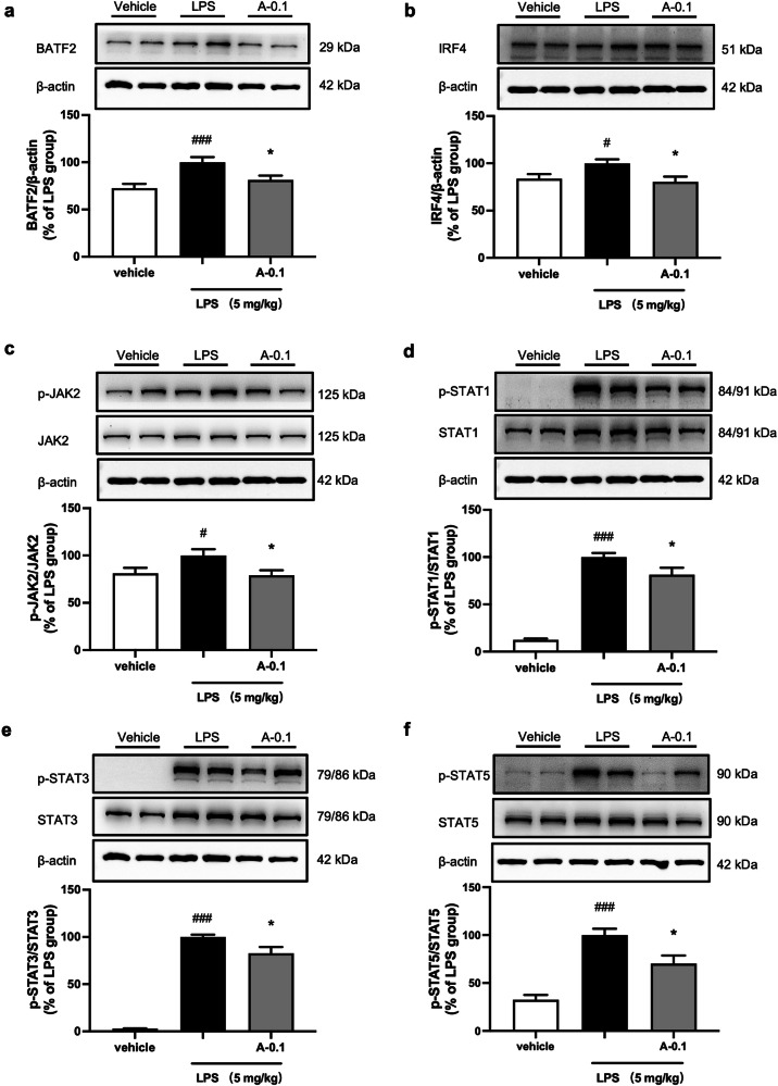
Suppression of neuroinflammation using small molecule compounds targeting the key pathways in microglial inflammation has attracted great interest. Recently, increasing attention has been gained to the role of the second bromodomain (BD2) of the bromodomain and extra-terminal (BET) proteins, while its effect and molecular mechanism on microglial inflammation has not yet been explored. In this study, we evaluated the therapeutic effects of ABBV-744, a BD2 high selective BET inhibitor, on lipopolysaccharide (LPS)-induced microglial inflammation in vitro and in vivo, and explored the key pathways by which ABBV-744 regulated microglia-mediated neuroinflammation. We found that pretreatment of ABBV-744 concentration-dependently inhibited the expression of LPS-induced inflammatory mediators/enzymes including NO, TNF-α, IL-1β, IL-6, iNOS, and COX-2 in BV-2 microglial cells. These effects were validated in LPS-treated primary microglial cells. Furthermore, we observed that administration of ABBV-744 significantly alleviated LPS-induced activation of microglia and transcriptional levels of pro-inflammatory factors TNF-α and IL-1β in mouse hippocampus and cortex. RNA-Sequencing (RNA-seq) analysis revealed that ABBV-744 induced 508 differentially expressed genes (DEGs) in LPS-stimulated BV-2 cells, and gene enrichment and gene expression network analysis verified its regulation on activated microglial genes and inflammatory pathways. We demonstrated that pretreatment of ABBV-744 significantly reduced the expression levels of basic leucine zipper ATF-like transcription factor 2 (BATF2) and interferon regulatory factor 4 (IRF4), and suppressed JAK-STAT signaling pathway in LPS-stimulated BV-2 cells and mice, suggesting that the anti-neuroinflammatory effect of ABBV-744 might be associated with regulation of BATF2-IRF4-STAT1/3/5 pathway, which was confirmed by gene knockdown experiments. This study demonstrates the effect of a BD2 high selective BET inhibitor, ABBV-744, against microglial inflammation, and reveals a BATF2-IRF4-STAT1/3/5 pathway in regulation of microglial inflammation, which might provide new clues for discovery of effective therapeutic strategy against neuroinflammation.

Keywords: ABBV-744; BATF2; BD2; IRF4; JAK/STAT; microglial inflammation.


Conflict of interest statement

The authors declare no competing interests.

Figures

Fig. 1
Fig. 1. Effect of ABBV-744 on LPS-induced inflammatory responses in microglial cells.
ac BV-2 cells were pretreated with ABBV-744 (0.003, 0.01, 0.03, 0.1, 0.3, 1, 3, 10 μM) for 2 h and then exposed to LPS (100 ng/mL) for 24 h (n = 5). The production of NO was measured using nitric oxide detection kit (a). The levels of pro-inflammatory factors TNF-α (b) and IL-6 (c) were measured using ELISA. d-f BV-2 cells were pretreated with ABBV-744 (0.01, 0.03, 0.1 μM) for 2 h and then exposed to LPS (100 ng/mL) for 3 h (n = 5). The mRNA levels of TNF-α (d), IL-1β (e) and IL-6 (f) were measured by quantitative real-time PCR. gi BV-2 cells were pretreated with ABBV-744 (0.01, 0.03, 0.1 μM) for 2 h and then exposed to LPS (100 ng/mL) for 24 h (n = 5). The protein levels of iNOS, COX2 and α-tubulin were measured using immunoblot analysis (g). The quantification of the intensities of iNOS (h) and COX2 (i) relative to α-tubulin are shown. jl Primary microglial cells were pretreated with ABBV-744 (0.03 μM) for 2 h and then exposed to LPS (100 ng/mL) for 3 h (n = 5). The mRNA levels of TNF-α (j), IL-1β (k), and IL-6 (l) were measured by quantitative real-time PCR. Comparisons between control group and LPS group were generated with unpaired two-tailed Student’s t-test. Comparisons between LPS group and ABBV-744-treated groups were generated with One-way ANOVA followed by Dunnett’s multiple comparison test. Data are shown as the mean ± SEM, ###P < 0.001 vs. the control group, *P < 0.05, **P < 0.01, ***P < 0.001, ns (no significant difference) vs. the LPS group. A-0.03: 0.03 μM ABBV-744.
Fig. 2
Fig. 2. Effect of ABBV-744 on LPS-induced activation of microglia and transcription of pro-inflammatory genes in the hippocampus and cortex of mice.
ah C57BL/6J mice were treated with vehicle (5% DMSO + 40% PEG300 + 55% saline; p.o.) or ABBV-744 (0.05, 0.1, 0.2 mg/kg; p.o.) daily for 2 days, followed by injection with saline or LPS (5 mg/kg; i.p.) for 1 day. Mice were sacrificed 24 h after LPS treatment (n = 7–8). a Activation of microglia in the CA1 region of mice hippocampus was detected by immunofluorescence staining using an anti-Iba1 antibody. Scale bar = 100 μm. b Quantification of Iba1 positive cells in the hippocampus of mice (n = 7–8). The mRNA levels of TNF-α (c) and IL-1β (d) in the hippocampus were measured by quantitative real-time PCR (n = 7–8). e Activation of microglia in the prefrontal cortex of mice was detected by immunofluorescence staining using an anti-Iba1 antibody. Scale bar = 100 μm. f Quantification of Iba1 positive cells in the cortex of mice (n = 7–8). The mRNA levels of TNF-α (g) and IL-1β (h) in the cortex were measured by quantitative real-time PCR (n = 7-8). Comparisons between control group and LPS group were generated with unpaired two-tailed Student’s t-test. Comparisons between the LPS group and ABBV-744-treated groups were generated with One-way ANOVA followed by Dunnett’s multiple comparison test. Data are shown as the mean ± SEM, ###P < 0.001 vs. the vehicle group, *P < 0.05, **P < 0.01, ***P < 0.001, ns (no significant difference) vs. the LPS group. For all immunofluorescence and qPCR experiments: vehicle group, LPS group and 0.1 mg/kg ABBV-744 treatment group, n = 8; 0.05 mg/kg and 0.2 mg/kg ABBV-744 treatment groups, n = 7.
Fig. 3
Fig. 3. Analysis of RNA-seq experiment. a-d BV-2 cells were stimulated with LPS (100 ng/mL) for 3 h following pretreatment with ABBV-744 (0.03 μM) for 2 h.
RNA sequencing and analysis were performed (P < 0.05 and two-fold difference, n = 4). a Volcano plot of DEGs between LPS and LPS + ABBV-744 groups. The blue dot and red dot represent that the expression level of genes has difference, the gray dot represents no difference. b The enrichment chord diagram displays top 5 GO terms and corresponding genes among 508 DEGs between the LPS group versus the LPS + ABBV-744 group. c The KEGG functional annotation bar chart shows four major categories of KEGG metabolic pathways. d The KEGG functional enrichment analysis line chart shows top 20 pathways enriched by DEGs.
Fig. 4
Fig. 4. Effect of ABBV-744 on LPS-induced expression levels of BATF2, IRF4, and JAK/STAT signaling pathway in BV-2 cells.
a, b BV-2 cells were pretreated with ABBV-744 (0.03 μM) for 2 h and then exposed to LPS (100 ng/mL) for 3 h (n = 4–5). The mRNA levels of BATF2 (a) and IRF4 (b) were measured by quantitative real-time PCR. c BV-2 cells were pretreated with ABBV-744 (0.03 μM) for 2 h and then exposed to LPS (100 ng/mL) for 15 min (n = 5). The protein level of BATF2 was measured by Western blot. The quantification of the intensity of BATF2 relative to β-actin is shown. dh BV-2 cells were pretreated with ABBV-744 (0.03 μM) for 2 h and then exposed to LPS (100 ng/mL) for 3 h (n = 6–7). Protein levels of IRF4 (d), p-JAK2 and JAK2 (e), p-STAT1 and STAT1 (f), p-STAT3 and STAT3 (g), p-STAT5 and STAT5 (h) were measured by Western blot and β-actin or GAPDH were used as a loading control. Comparisons of groups were generated with unpaired two-tailed Student’s t-test. Data are shown as the mean ± SEM, ##P < 0.01, ###P < 0.001 vs. the control group, **P < 0.01, ***P < 0.001 vs. the LPS group. A-0.03: 0.03 μM ABBV-744.
Fig. 5
Fig. 5. Effect of knocking down of BATF2 or IRF4 and suppressing JAK-STAT signaling pathway on LPS-induced expression levels of pro-inflammatory genes in BV-2 cells.
af BV-2 cells were transfected with small interfering RNAs for 24 h and then exposed to LPS (100 ng/mL) for 3 h (n = 7). The mRNA levels of TNF-α (a), IL-1β (b) and IL-6 (c) were measured by quantitative real-time PCR after knocking down of BATF2. The mRNA levels of TNF-α (d), IL-1β (e) and IL-6 (f) were measured by quantitative real-time PCR after knocking down of IRF4. gi BV-2 cells were pretreated with AG490 (10 μM) for 2 h and then exposed to LPS (100 ng/mL) for 3 h (n = 7). The mRNA levels of TNF-α (g), IL-1β (h) and IL-6 (i) were measured by quantitative real-time PCR. Comparisons of groups were generated with unpaired two-tailed Student’s t-test. Data are shown as the mean ± SEM, ###P < 0.001 vs. the control group, *P < 0.05, ***P < 0.001 vs. the LPS group. AG-10: 10 μM AG490.
Fig. 6
Fig. 6. Effect of knocking down of BATF2 on IRF4 and JAK-STAT signaling pathway in LPS-stimulated BV-2 cells.
ae BV-2 cells were transfected with BATF2 small interfering RNA for 24 h and then exposed to LPS (100 ng/mL) for 3 h (n = 6). Protein levels of IRF4 (a), p-JAK2 and JAK2 (b), p-STAT1 and STAT1 (c), p-STAT3 and STAT3 (d), p-STAT5 and STAT5 (e) were measured by Western blot and β-actin was used as a loading control. fi BV-2 cells were transfected with IRF4 small interfering RNA for 24 h and then exposed to LPS (100 ng/mL) for 3 h (n = 5–7). Protein levels of p-JAK2 and JAK2 (f), p-STAT1 and STAT1 (g), p-STAT3 and STAT3 (h), p-STAT5 and STAT5 (i) were measured by Western blot and β-actin was used as a loading control. Comparisons of groups were generated with unpaired two-tailed Student’s t-test. Data are shown as the mean ± SEM, ###P < 0.001 vs. the NC (negative control) group, *P < 0.05, **P < 0.01, ***P < 0.001, ns (no significant difference) vs. the NC + LPS group.
Fig. 7
Fig. 7. Effect of ABBV-744 on LPS-induced expression levels of BATF2, IRF4, and JAK-STAT signaling pathway in LPS-injected mice.
af C57BL/6J mice were treated with vehicle (5% DMSO + 40% PEG300 + 55% saline; p.o.) or ABBV-744 (0.1 mg/kg; p.o.) daily for 2 days, followed by injection with saline or LPS (5 mg/kg; i.p.) for 1 day. Mice were sacrificed 3 h after LPS treatment (n = 12). Protein levels of BATF2 (a), IRF4 (b), p-JAK2 and JAK2 (c), p-STAT1 and STAT1 (d), p-STAT3 and STAT3 (e), p-STAT5 and STAT5 (f) were measured by Western blot and β-actin was used as a loading control. Comparisons of groups were generated with unpaired two-tailed Student’s t-test. Data are shown as the mean ± SEM, #P < 0.05, ###P < 0.001 vs. the vehicle group, *P < 0.05 vs. the LPS group. A-0.1: 0.1 mg/kg ABBV-744.

References

    1. Prinz M, Jung S, Priller J. Microglia biology: one century of evolving concepts. Cell. 2019;179:292–311. - PubMed
    1. Colonna M, Butovsky O. Microglia function in the central nervous system during health and neurodegeneration. Annu Rev Immunol. 2017;35:441–68. - PMC - PubMed
    1. Borst K, Dumas AA, Prinz M. Microglia: Immune and non-immune functions. Immunity. 2021;54:2194–208. - PubMed
    1. Wright-Jin EC, Gutmann DH. Microglia as dynamic cellular mediators of brain function. Trends Mol Med. 2019;25:967–79. - PMC - PubMed
    1. Lecca D, Jung YJ, Scerba MT, Hwang I, Kim YK, Kim S, et al. Role of chronic neuroinflammation in neuroplasticity and cognitive function: a hypothesis. Alzheimers Dement. 2022;18:2327–40. - PMC - PubMed

MeSH terms

Substances

LinkOut - more resources