. 2024 Jun 11;30(1):82.
doi: 10.1186/s10020-024-00837-4.

ERRFI1 exacerbates hepatic ischemia reperfusion injury by promoting hepatocyte apoptosis and ferroptosis in a GRB2-dependent manner

Affiliations

ERRFI1 exacerbates hepatic ischemia reperfusion injury by promoting hepatocyte apoptosis and ferroptosis in a GRB2-dependent manner

Hang Zhao et al. Mol Med. .

Abstract

Background: Programmed cell death is an important mechanism for the development of hepatic ischemia and reperfusion (IR) injury, and multiple novel forms of programmed cell death are involved in the pathological process of hepatic IR. ERRFI1 is involved in the regulation of cell apoptosis in myocardial IR. However, the function of ERRFI1 in hepatic IR injury and its modulation of programmed cell death remain largely unknown.

Methods: Here, we performed functional and molecular mechanism studies in hepatocyte-specific knockout mice and ERRFI1-silenced hepatocytes to investigate the significance of ERRFI1 in hepatic IR injury. The histological severity of livers, enzyme activities, hepatocyte apoptosis and ferroptosis were determined.

Results: ERRFI1 expression increased in liver tissues from mice with IR injury and hepatocytes under oxygen-glucose deprivation/reoxygenation (OGD/R) conditions. Hepatocyte-specific ERRFI1 knockout alleviated IR-induced liver injury in mice by reducing cell apoptosis and ferroptosis. ERRFI1 knockdown reduced apoptotic and ferroptotic hepatocytes induced by OGD/R. Mechanistically, ERRFI1 interacted with GRB2 to maintain its stability by hindering its proteasomal degradation. Overexpression of GRB2 abrogated the effects of ERRFI1 silencing on hepatocyte apoptosis and ferroptosis.

Conclusions: Our results revealed that the ERRFI1-GRB2 interaction and GRB2 stability are essential for ERRFI1-regulated hepatic IR injury, indicating that inhibition of ERRFI1 or blockade of the ERRFI1-GRB2 interaction may be potential therapeutic strategies in response to hepatic IR injury.

Keywords: Apoptosis; ERRFI1; Ferroptosis; GRB2; Hepatic ischemia and reperfusion injury.


Conflict of interest statement

The authors declare that they have no competing interests.

Figures
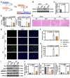
Fig. 1
Fig. 1
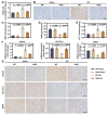
ERRFI1 deficiency ameliorated IR-induced hepatic injury and hepatocellular apoptosis. (A) Schematic diagram showed the hepatocyte-specific ERRFI1 knockout strategy and ischemia reperfusion model. (B, C) Western blot analysis of ERRFI1 protein level in liver tissues from wild type (WT) mice and hepatocyte-specific ERRFI1-knockout (ERRFI1-HKO) mice with sham treatment or ischemia for 90 min followed by reperfusion for 6 h, and quantitative analysis is shown. (D) Liver function assessed by ALT and AST of mice with different treatment. (E) Liver pathology was determined by H&E staining (scale bar: 100 μm). (F) Suzike’s injury score was used to assess the degree of injury based on H&E staining. (G, H) TUNEL staining of apoptotic cells in liver tissues from WT mice and ERRFI1-HKO mice under different conditions (scale bar: 50 μm), and quantification showing the percentage of apoptotic cells. (I, J) Western blot analysis of Bax, Bcl-2, and cleaved caspase-3 in liver tissues from WT mice and ERRFI1-HKO mice after IR injury. For statistical analysis, one-way ANOVA was used (n = 6)
Fig. 2
Fig. 2
ERRFI1 deficiency protected against hepatic IR-induced ferroptosis. (A) Intracellular ROS level was determined by DCFH-DA staining after hepatic IR. (B) Immunohistochemical staining of 8-OHdG in liver tissues (scale bar: 50 μm). (C, D) The content of MDA and the level of GSH in the livers of mice subjected to sham treatment or to an induction of IR. (E) Hepatic Fe2+ content in each group. (F) The mRNA levels of ACSL4, SLC7A11, and GPX4 in liver tissues of mice with different treatments. (G) Representative immunohistochemical images of ACSL4, SLC7A11, and GPX4 in liver tissues (scale bar: 50 μm). For statistical analysis, one-way ANOVA was used (n = 6)
Fig. 3
Fig. 3
Knockdown of ERRFI1 inhibited apoptosis of hepatocytes induced by hypoxic-reoxygenation. (A) Expression of ERRFI1 in L-02 cells following hypoxia/reoxygenation (H/R) and ERRFI1 knockdown at the mRNA level was determined by real-time PCR. (B) Cell viability of ERRFI1-silenced L-02 cells after OGD/R exposure. (C, D) Apoptosis-positive cells were detected by TUNEL staining (scale bar: 50 μm). (E, F) Western blot analysis of Bax, Bcl-2, and cleaved caspase-3 in L-02 cells under indicated conditions. For statistical analysis, one-way ANOVA was used (n = 3)
Fig. 4
Fig. 4
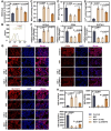
Knockdown of ERRFI1 suppressed OGD/R-induced ferroptosis in hepatocytes. (A) ROS level in ERRFI1-silenced L-02 cells exposed to hypoxia/reoxygenation. (B) Flow cytometry analysis of lipid peroxidation using C11-BODIPY 581/591 in L-02 cells under indicated conditions. (C, D) MDA content and GSH level in L-02 cells cultured under indicated conditions. (E) Fe2+ content in ERRFI1-silenced L-02 cells after OGD/R exposure was determined. (F) Real-time PCR showed the mRNA levels of ACSL4, SLC7A11, and GPX4 in response to ERRFI1 knockdown under H/R conditions. (G, H) Fluorescence immunostaining of ACSL4, SLC7A11, and GPX4 in L-02 cells transfected with sh-ERRFI1 during H/R injury (scale bar: 20 μm). For statistical analysis, one-way ANOVA was used (n = 3)
Fig. 5
Fig. 5
ERRFI1 directly interacted with GRB2 and maintained its stability by hindering its proteasomal degradation. (A-C) Transcript and protein levels of GRB2 in ERRFI1-silenced L-02 cells were detected. (D) Co-immunoprecipitation of ERRFI1 and GRB2. L-02 cells were subjected to GRB2 immunoprecipitation and subsequent immunoblotting of ERRFI1 and GRB2. (E, F) L-02 cells were transfected with sh-ERRFI1 and treated with CHX for the indicated times. Western blot analysis showed the expression of GRB2. (G, H) The expression of GRB2 in sh-ERRFI1-transfected L-02 cells with or without MG132 treatment. For statistical analysis, Student’s t test was used (n = 3)
Fig. 6
Fig. 6
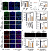
ERRFI1 facilitated OGD/R-induced injury of hepatocytes in a GRB2-dependent manner. (A-D) L-02 cells were transfected with GRB2 overexpression plasmid or vector plasmid, followed by 4 h of hypoxia and 12 h of reoxygenation. Cell apoptosis was detected by TUNEL staining. Scale bar: 20 μm (A, B). Lipid peroxidation was measured using C11-BODIPY 581/591 (C). Fe2+ content was determined (D). (E, F) L-02 cells cotransfected with sh-ERRFI1 and GRB2 overexpression plasmid were subjected to OGD/R stimulation in the presence or absence of 20 µM Z-VAD-FMK (ZVF, an apoptosis inhibitor). L-02 cells were treated with 5 µM camptothecin (CPT, an apoptosis inducer) as positive control at the same time as OGD/R stimulation. TUNEL staining of L-02 cells under indicated conditions was performed (scale bar: 20 μm). (G-L) L-02 cells cotransfected with sh-ERRFI1 and GRB2 overexpression plasmid were subjected to OGD/R stimulation in the presence or absence of 5 µM ferrostatin-1 (Fer-1, a ferroptotic inhibitor). L-02 cells were treated with 10 µM erastin (a ferroptotic inducer) as positive control at the same time as OGD/R stimulation. DCFH-DA staining was used to detect ROS production in cells (G). Fe2+ content in cells was measured by a commercial kit (H). The protein expression of GPX4 in cells was analyzed by immunofluorescence. Scale bar: 20 μm (I, K). Cell death was detected by propidium iodide (PI) staining. Scale bar: 50 μm (J, L). For statistical analysis, student’s t test and one-way ANOVA were used (n = 3)

Similar articles

Cited by

References

    1. Amatschek S, Koenig U, Auer H, Steinlein P, Pacher M, Gruenfelder A, et al. Tissue-wide expression profiling using cDNA subtraction and microarrays to identify tumor-specific genes. Cancer Res. 2004;64(3):844–56. doi: 10.1158/0008-5472.CAN-03-2361. - DOI - PubMed
    1. Bellini M, Pest MA, Miranda-Rodrigues M, Qin L, Jeong JW, Beier F. Overexpression of MIG-6 in the cartilage induces an osteoarthritis-like phenotype in mice. Arthritis Res Therapy. 2020;22(1):119. doi: 10.1186/s13075-020-02213-z. - DOI - PMC - PubMed
    1. Cai J, Yi FF, Yang L, Shen DF, Yang Q, Li A et al. Targeted expression of receptor-associated late transducer inhibits maladaptive hypertrophy via blocking epidermal growth factor receptor signaling. Hypertension (Dallas, Tex: 1979). 2009;53(3):539 – 48. - PMC - PubMed
    1. Chen YC, Colvin ES, Maier BF, Mirmira RG, Fueger PT. Mitogen-inducible gene 6 triggers apoptosis and exacerbates ER stress-induced β-cell death. Mol Endocrinol (Baltimore Md) 2013;27(1):162–71. doi: 10.1210/me.2012-1174. - DOI - PMC - PubMed
    1. Chen W, Zhong H, Wang X, Pang Q, Zhuang J, Hu J, et al. Mig6 reduces inflammatory mediators production by regulating the activation of EGFR in LPS-induced endotoxemia. J Cell Physiol. 2018;233(9):6975–83. doi: 10.1002/jcp.26488. - DOI - PubMed