. 2024 Jun 11;19(1):82.
doi: 10.1186/s13020-024-00959-1.

Xinbao Pill ameliorates heart failure via regulating the SGLT1/AMPK/PPARα axis to improve myocardial fatty acid energy metabolism

Affiliations

Xinbao Pill ameliorates heart failure via regulating the SGLT1/AMPK/PPARα axis to improve myocardial fatty acid energy metabolism

Linjie Pan et al. Chin Med. .

Abstract

Background: Heart failure (HF) is characterized by a disorder of cardiomyocyte energy metabolism. Xinbao Pill (XBW), a traditional Chinese medicine formulation integrating "Liushen Pill" and "Shenfu Decoction," has been approved by China Food and Drug Administration for the treatment of HF for many years. The present study reveals a novel mechanism of XBW in HF through modulation of cardiac energy metabolism.

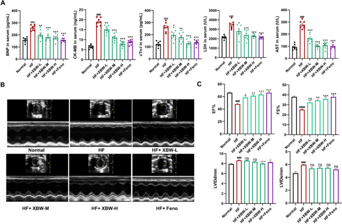
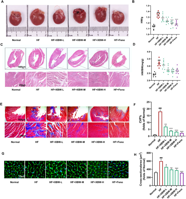
Methods: In vivo, XBW (60, 90, 120 mg/kg/d) and fenofibrate (100 mg/kg/d) were treated for six weeks in Sprague-Dawley rats that were stimulated by isoproterenol to induce HF. Cardiac function parameters were measured by echocardiography, and cardiac pathological changes were assessed using H&E, Masson, and WGA staining. In vitro, primary cultured neonatal rat cardiomyocytes (NRCMs) were induced by isoproterenol to investigate the effects of XBW on myocardial cell damage, mitochondrial function and fatty acid energy metabolism. The involvement of the SGLT1/AMPK/PPARα signalling axis was investigated.

Results: In both in vitro and in vivo models of ISO-induced HF, XBW significantly ameliorated cardiac hypertrophy cardiac fibrosis, and improved cardiac function. Significantly, XBW improved cardiac fatty acid metabolism and mitigated mitochondrial damage. Mechanistically, XBW effectively suppressed the expression of SGLT1 protein while upregulating the phosphorylation level of AMPK, ultimately facilitating the nuclear translocation of PPARα and enhancing its transcriptional activity. Knockdown of SGLT1 further enhanced cardiac energy metabolism by XBW, while overexpression of SGLT1 reversed the cardio-protective effect of XBW, highlighting that SGLT1 is probably a critical target of XBW in the regulation of cardiac fatty acid metabolism.

Conclusions: XBW improves cardiac fatty acid energy metabolism to alleviate HF via SGLT1/AMPK/PPARα signalling axis.

Keywords: AMPK/PPARα axis; Heart failure; Myocardial energy metabolism; SGLT1; Xinbao Pill.


Conflict of interest statement

The authors declare that they have no competing interests.

Figures

Fig. 1
Fig. 1
XBW effectively mitigates ISO-induced myocardial hypertrophy in NRCMs and modulates the expression of enzymes involved in myocardial fatty acid metabolism. AB In NRCMS, Western Blot detected alterations in β-MHC and ANP protein levels, indicators of ISO-induced myocardial hypertrophy, as well as CD36, CPT-1B, and ACADM, markers of cardiac FA energy metabolism. CD ISO-induced alterations in cell surface area of NRCMs within 48 h. The cell surface area was meticulously evaluated via Rhodamine-Phalloidin staining. Scale bar: 60 μm. E The viability of NRCMs was assessed using the MTT assay after incubation with various concentrations of XBW for 24 h. FG After ISO stimulation and drug treatment, the WB analysis assessed the protein contents of β-MHC, ANP, ACADM, CD36, and CPT-1B. HI The alterations in cell surface area following XBW administration were quantified. Scale bar: 60 μm. Compared with Ctrl group, ###P < 0.001, ##P < 0.01, #P < 0.05, ns: no significance; compared with ISO-induced group, ***P < 0.001, **P < 0.01, *P < 0.05, ns: no significance
Fig. 2
Fig. 2
XBW improves cardiac function in rats with heart failure induced by ISO. A Myocardial injury markers, including BNP, CK-MB, cTn-I, LDH, and AST, were analyzed in the serum of rats from different groups. B Echocardiography M-mode images from the left ventricles of various experimental groups are presented as representative examples. C Cardiac function was evaluated by quantifying echocardiographic parameters such as EF%, FS%, LVIDd, and LVIDs. Compared with Normal group, ###P < 0.001, ##P < 0.01, #P < 0.05; compared with HF group, ***P < 0.001, **P < 0.01, *P < 0.05, ns: no significance, n = 7. HF: ISO subcutaneous injection induced-HF group; HF + XBW-L: XBW treatment group (low dose: 60 mg/kg); HF + XBW-M: XBW treatment group (medium dose: 80 mg/kg); HF + XBW-H: XBW treatment group (high dose: 120 mg/kg); HF + Feno: fenofibrate treatment group (100 mg/kg)
Fig. 3
Fig. 3
XBW ameliorates structural abnormalities in myocardial tissue of heart failure rats induced by ISO. A Intact cardiac morphological images of rats in various experimental groups. B Heart weights of rats in each group. C Representative HE-stained heart sections. Scale bar: 1000 μm. Scale bar: 250 μm. D Heart weight-to-body weight ratio of rats. E Masson-stained heart sections in each group. Scale bar: 100 μm. F Quantitative analysis of collagen volume in the left ventricles. G WGA-stained sections of representative rat hearts from each group. Scale bar: 25 μm. H Quantitative analysis of the cross-sectional area. Compared with Normal group, ###P < 0.001, ##P < 0.01, #P < 0.05; compared with HF group, ***P < 0.001, **P < 0.01, *P < 0.05
Fig. 4
Fig. 4
XBW exhibits cardioprotective effects by mitigating the pathological alterations associated with HF and modulating fatty acid metabolism in ISO-induced HF rats. AB Immunohistochemical staining was performed to assess the expression of ANP protein. Scale bar: 30 μm. C–D WB was employed to determine the protein levels of markers associated with hypertrophy and fibrosis in cardiac tissue. EF Immunohistochemistry was utilized to examine the expression of CPT-1B protein. Scale bar: 30 μm. GH The ACADM, CD36, and CPT-1B protein levels were assessed using WB analysis. Compared with Normal group, ###P < 0.001, ##P < 0.01, #P < 0.05; compared with HF group, ***P < 0.001, **P < 0.01, *P < 0.05, ns: no significance
Fig. 5
Fig. 5
XBW significantly enhances cardiac energy levels and alleviates mitochondrial functional damage in ISO-induced NRCMs. A The XF Cell Mito Stress Test was employed to assess alterations in OCR levels in ISO-induced NRCMs. B The ATP content levels in NRCMs following drug intervention. C The mitochondrial membrane potential of NRCMs was investigated using the JC-1 Fluorescent probe. Scale bar: 60 μm. D The mitochondrial morphology of NRCMs was visualized via the Mito-Tracker Red CMXRos probe. Scale bar: 60 μm or 20 μm. E WB analysis was employed to assess the alterations in the levels of PGC-1α and NRF1, which are associated with mitochondrial biogenesis. Independent experiments were performed at least three times with similar results. Compared with Ctrl group, ###P < 0.001, ##P < 0.01, #P < 0.05; compared with ISO-induced group, ***P < 0.001, **P < 0.01, *P < 0.05, ns: no significance
Fig. 6
Fig. 6
XBW may modulate the AMPK/PPARα signalling axis through SGLT1 inhibition, thereby enhancing myocardial energy supply. A Venn diagram of the intersection between XBW and HF. B PPI network interaction diagram. C GO and KEGG analyses of dominant targets. DF, H WB was employed to assess the changes of the SGLT1/AMPK/PPARα axis following XBW treatment. G Immunofluorescence was utilized to detect the protein expression of PPARα in NRCMs. Scale bar: 25 μm. I PPARα luciferase activity was quantified using a dual luciferase reporter gene assay. Independent experiments were performed at least three times with similar results. Compared with Ctrl group, ###P < 0.001, ##P < 0.01, #P < 0.05; compared with ISO-induced group, *** P < 0.001, **P < 0.01, *P < 0.05, ns: no significance
Fig.7
Fig.7
XBW may modulate the AMPK/PPARα signalling axis through SGLT1 inhibition, thereby enhancing myocardial energy supply. AB Immunohistochemical staining was employed to assess the protein expression of SGLT1 in myocardial tissue from each experimental group. Scale bar: 30 μm. C–D PPARα protein levels were evaluated using immunohistochemistry. Scale bar: 30 μm. EH WB analysis was utilized to determine the expression levels of SGLT1 and PPARα proteins, as well as the ratio of p-AMPK to AMPK protein expression. I ATP content in myocardial tissue from each experimental group was analyzed. Compared with Normal group, ###P < 0.001, ##P < 0.01, #P < 0.05; compared with HF group, ***P < 0.001, **P < 0.01, *P < 0.05, ns: no significance
Fig. 8
Fig. 8
SGLT1 silencing enhances the potential of XBW in attenuating myocardial pathologic changes and enhancing myocardial fatty acid energy metabolism. A The effects of XBW on alterations in the protein levels of myocardial hypertrophy markers β-MHC and ANP after SGLT1 silence. B The effect of XBW on intracellular glucose transport after silencing SGLT1 was measured by flow cytometry. C The protein levels of FN and α-SMA, which serve as biomarkers for myocardial fibrosis, were assessed via WB analysis. DE The effect of XBW on changes in cell surface area visualized through Rhodamine-Phalloidin staining following SGLT1 interference. Scale bar: 100 μm. F NRCMs’ ATP content level. G Immunofluorescence was employed to examine PPARα expression after silencing SGLT1. Scale bar: 10 μm. HI WB analysis was conducted to evaluate the protein expression of SGLT1, PPARα, and myocardial fatty acid energy metabolism indicators CD36, CPT-1B, and ACADM. Independent experiments were performed at least three times with similar results. Compared with Ctrl group, ###P < 0.001, ##P < 0.01, #P < 0.05; compared with ISO-induced group, ***P < 0.001, **P < 0.01, *P < 0.05
Fig. 9
Fig. 9
SGLT1 overexpression blocked the effectiveness of XBW in treating myocardial pathologic changes and improving cardiac fatty acid energy metabolism A The protein levels of β-MHC and ANP under SGLT1 overexpression. B The effect of XBW on intracellular glucose transport subjected to SGLT1 overexpression. C The protein levels of FN and α-SMA were assessed via WB analysis. DE Cell surface area was determined via Rhodamine-Phalloidin staining. Scale bar: 100 μm F Cellular ATP content levels were assessed. G IF analysis disclosed alterations in PPARα protein expression. HI The protein contents of SGLT1, PPARα, CD36, CPT-1B, and ACADM were assessed using WB analysis to investigate the impact of SGLT1 overexpression across different experimental groups. Independent experiments were performed at least three times with similar results. Compared with Ctrl group, ###P < 0.001, ##P < 0.01, #P < 0.05; compared with ISO-induced group, ***P < 0.001, **P < 0.01, *P < 0.05, ns: no significance

Similar articles

Cited by

References

    1. Truby LK, Rogers JG. advanced heart failure: epidemiology, diagnosis, and therapeutic approaches. JACC Heart failure. 2020;8(7):523–536. doi: 10.1016/j.jchf.2020.01.014. - DOI - PubMed
    1. Ziaeian B, Fonarow GC. Epidemiology and aetiology of heart failure. Nat Rev Cardiol. 2016;13(6):368–378. doi: 10.1038/nrcardio.2016.25. - DOI - PMC - PubMed
    1. Wasyluk W, Nowicka-Stążka P, Zwolak A. Heart metabolism in sepsis-induced cardiomyopathy-unusual metabolic dysfunction of the heart. Int J Environ Res Public Health. 2021;18(14):7598. doi: 10.3390/ijerph18147598. - DOI - PMC - PubMed
    1. Lopaschuk GD, Collins-Nakai RL, Itoi T. Developmental changes in energy substrate use by the heart. Cardiovasc Res. 1992;26(12):1172–1180. doi: 10.1093/cvr/26.12.1172. - DOI - PubMed
    1. Bertero E, Maack C. Metabolic remodelling in heart failure. Nat Rev Cardiol. 2018;15(8):457–470. doi: 10.1038/s41569-018-0044-6. - DOI - PubMed