Review
. 2024 Jun 11;17(1):44.
doi: 10.1186/s13045-024-01559-0.

Deciphering the performance of macrophages in tumour microenvironment: a call for precision immunotherapy

Affiliations
Review

Deciphering the performance of macrophages in tumour microenvironment: a call for precision immunotherapy

Belén Toledo et al. J Hematol Oncol. .

Abstract

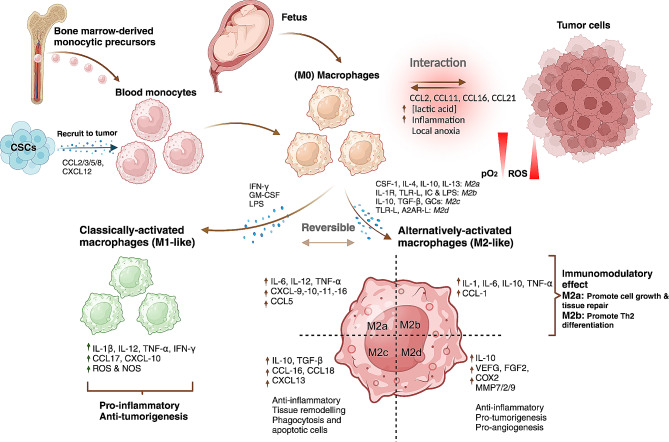
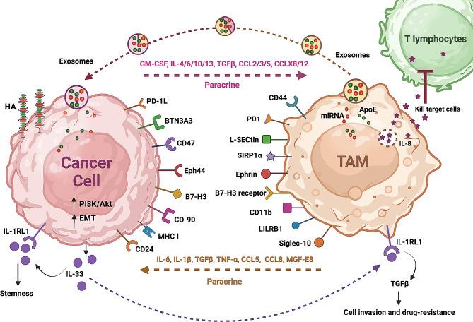
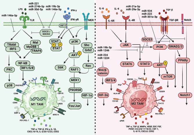
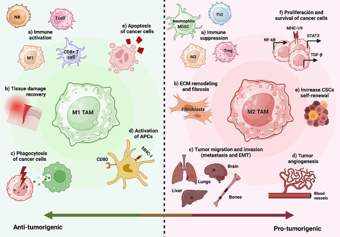
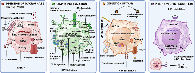
Macrophages infiltrating tumour tissues or residing in the microenvironment of solid tumours are known as tumour-associated macrophages (TAMs). These specialized immune cells play crucial roles in tumour growth, angiogenesis, immune regulation, metastasis, and chemoresistance. TAMs encompass various subpopulations, primarily classified into M1 and M2 subtypes based on their differentiation and activities. M1 macrophages, characterized by a pro-inflammatory phenotype, exert anti-tumoural effects, while M2 macrophages, with an anti-inflammatory phenotype, function as protumoural regulators. These highly versatile cells respond to stimuli from tumour cells and other constituents within the tumour microenvironment (TME), such as growth factors, cytokines, chemokines, and enzymes. These stimuli induce their polarization towards one phenotype or another, leading to complex interactions with TME components and influencing both pro-tumour and anti-tumour processes.This review comprehensively and deeply covers the literature on macrophages, their origin and function as well as the intricate interplay between macrophages and the TME, influencing the dual nature of TAMs in promoting both pro- and anti-tumour processes. Moreover, the review delves into the primary pathways implicated in macrophage polarization, examining the diverse stimuli that regulate this process. These stimuli play a crucial role in shaping the phenotype and functions of macrophages. In addition, the advantages and limitations of current macrophage based clinical interventions are reviewed, including enhancing TAM phagocytosis, inducing TAM exhaustion, inhibiting TAM recruitment, and polarizing TAMs towards an M1-like phenotype. In conclusion, while the treatment strategies targeting macrophages in precision medicine show promise, overcoming several obstacles is still necessary to achieve an accessible and efficient immunotherapy.

Keywords: Cancer cell; Immunity; Immunotherapy; Polarization; Tumour microenvironment; Tumour-associated macrophages.


Conflict of interest statement

The authors declare no conflict of interest.

Figures

Fig. 1
Fig. 1
The diverse cellular origins of TAMs, give rise to two primary subpopulations with distinct roles in shaping the immune response. Tissue macrophages typically arise from circulating monocytes originating in the bone marrow, undergoing differentiation into M0 macrophages, which subsequently polarize into M1 or M2 states in response to microenvironmental signals. Notably, macrophages can also originate from embryonic precursors during early fetal development, bypassing monocytic intermediates. Interactions between macrophages and tumour cells begin as early as the M0 stage, influencing subsequent polarization and recruiting additional macrophages to the tumour site via chemoattractants released by both CSCs and tumour cells. The M1 phenotype, primarily induced by factors like IFN-γ, LPS, and GM-CSF, leads to the secretion of pro-inflammatory cytokines such as IL-6, IL-12, IL-23, and TNF-α, contributing to enhanced inflammatory responses and cytotoxic effects on pathogens and tumour cells. In contrast, the M2 phenotype encompasses subtypes like M2a, M2b, M2c, and M2d, each influenced by specific stimuli such as CSF-1, IL-4, IL-13, and IL-10, which are associated with parasite infection, tissue remodelling, allergic diseases, and angiogenesis. Under typical conditions of the TME, characterized by low oxygen levels, high lactic acid levels, inflammation, and oxidative stress, macrophages tend to adopt the M2 phenotype, marked by elevated levels of IL-10, TGF-β, pro-angiogenic factors, and tissue-remodelling enzymes like MMPs. Consequently, this M2 phenotype promotes angiogenesis, immunosuppression, and tumour progression. Abbreviations: C-C motif chemokine ligand, (CCL) Cancer stem cells (CSCs), colony stimulating factor 1 (CSF-1), chemokine C-X-C motif ligand (CXCL), glucocorticoid (GC), granulocyte macrophage colony-stimulating factor (GM-CSF), immune complex (IC), interferon-gamma (IFN-γ), interleukin (IL), IL-1 receptor antagonist (IL-1ra), lipopolysaccharide (LPS), major histocompatibility complex (MHC), matrix metalloproteinases (MMPs), nitric oxide (NO), reactive oxygen species (ROS), tumour-associated macrophages (TAMs), transforming growth factor beta (TGF-β), tumour microenvironment (TME), tumour necrosis factor alpha (TNF⍺), toll-like receptor (TLR)
Fig. 2
Fig. 2
Crosstalk between tumour cells and tumour-associated macrophages (TAMs). TAMs secrete chemokines and cytokines such as IL-6, IL-8, and IL-10, which actively contribute to cancer advancement. Notably, IL-8 released by TAMs exerts cytotoxic effects on T lymphocytes. Additionally, various juxtacrine interactions between tumour cells and TAMs play a pivotal role in inducing immunosuppression. The PD-1/L1 signalling pathway further exacerbates tumour immune evasion by impeding the normal functioning of macrophages and other immune effector cells. Furthermore, the interaction of B7-H3 with its receptor has been implicated in the inhibition of T lymphocytes, thus facilitating tumour immune evasion. The SIRPα/CD47 and CD24/Siglec-10 pathways serve as the “do-not-eat-me” signal, wherein tumour cells over-expressing CD47 and CD24 are recognized as self-normal cells, thereby evading phagocytosis by macrophages. Another significant mechanism of tumour evasion involves LILRB1/MHC class I component β2-microglobulin, which inhibits the phagocytosis of tumour cells by macrophages. Exosomes facilitate intercellular communication by transporting various molecules, including exosomal mRNA, circRNA, lncRNA, miRNA, lipids, and proteins. Interestingly, exosomes exhibit dynamic alterations in their cargo during transit from the origin to the destination cell. ApoE is highly expressed in TAMs and is transferred, along with other molecules, via exosomes to cancer cells, activating the PI3K-Akt signalling pathway and promoting cytoskeletal remodeling, EMT, and cancer cell migration. Other juxtacrine mechanisms, such as Eph44-ephrin interaction, regulate immune cell processes, including proliferation, survival, apoptosis, activation, and migration. CD44, a transmembrane adhesion molecule, plays a crucial role in binding and metabolizing hyaluronic acid (HA) and serves as an effective phagocytic receptor, influencing inflammation, phagocytosis, and multi-drug resistance. Interactions such as CD44-HA, BTN3N3–L-SECtin, CD90-CD11b, and Eph44-ephrin also also trigger signals that support the maintenance of cancer stem cells. Furthermore, IL-33 released by tumour cells sustains stemness via autocrine interaction with IL-1RL1, while also promoting tumour cell invasion and drug resistance through TAM-mediated TGF-β release and TAM proliferation and differentiation in a paracrine manner. This intricate interplay results in the amplification of the aforementioned crosstalk. Abbreviations: Apolipoprotein E (ApoE), C-C motif chemokine Ligand, (CCL), circular RNA (circRNA), epithelial-mesenchymal transition (EMT), granulocyte-macrophage colony-stimulating factor (GM-CSF), hyaluronic acid (HA), interleukin (IL), Janus kinase (JAK), long non-coding RNA (lncRNA), macrophage colony-stimulating factor (M-CSF), milk fat globule-EGF factor 8 (MGF-E8), major histocompatibility complex (MHC), microRNA (miRNA), messenger RNA (mRNA), programmed death-ligand 1 (PD-L1), sialic acid binding Ig-like lectin (Siglec), signal transducer and activator of transcription 3 (STAT3), tumour-associated macrophages (TAMs), transforming growth factor beta (TGF-β), tumour necrosis factor alpha (TNF⍺)
Fig. 3
Fig. 3
Principal pathways implicated in the activation of tumour-associated macrophages (TAMs). Macrophages of the M1 phenotype express innate immune receptors like Toll-like receptors (TLRs) during their development and maturation, enabling recognition of pathogen-associated molecular patterns such as LPS from microbial surfaces. Ligand binding to TLR4 by LPS activates downstream signalling pathways, including the MyD88-dependent pathway or the IRF5-dependent pathway, leading to further signalling via the NF-κB pathway or the p38-MAPK pathway, respectively. These pathways collectively promote the expression of inflammatory factors and polarization of M1 macrophages. Additionally, polarization towards M1 can be induced by IFN-y binding to its receptor, activating the JAK/STAT1 and PI3K/AKT/Fos/Jun pathways. The latter is also activated upon ligand binding of receptor tyrosine kinases like MER. Inhibitors such as SOCS1/3 can inhibit both the TLR4/MyD88/NF-κB and JAK/STAT1 pathways, thus hindering M1 polarization activity by blocking upstream signalling of these pathways. Conversely, M2 polarization primarily occurs through the interaction of IL-4/6 with their receptors, activating the JAK/STAT3/6 signalling pathway. Moreover, activation of the TGF-βR results in downstream signalling via the PI3K/Akt/mTOR and TGF-βR/Smad/PPARy pathways. Additionally, activation of the Wnt/β-catenin signalling pathway by tumour-derived Wnt ligands stimulates M2 polarization. Notch signalling also contributes to M2 polarization through a positive feed-forward loop, promoting production of IL-6, IL-10, and IL-12. Inhibition of these pathways by SOCS3 prevents M2 polarization. Certain exosomal miRNAs regulate macrophage polarization by affecting the mentioned signal pathways or transcription factors. Furthermore, detection of oxidative stress leads to upregulation of HIF-1α and HIF-2α, with HIF-1α favoring M1 polarization and HIF-2α promoting M2 polarization. Abbreviations: protein kinase B (AKT), hypoxia-inducible factors (HIF), interferon regulatory factors (IRF), mitogen-activated protein kinase kinase (MEK), mammalian target of rapamycin (mTOR), bone marrow differentiation factor 88 (MyD88), nuclear factor kappa B (NF-κB), LPS Phosphatidylinositide 3-kinases (PI3K), protein kinase C (PKC), peroxisome proliferator-activated receptor (PPAR), rapidly accelerated fibrosarcoma (RAF), suppressors of cytokine signalling (SOCS), signal transducer and activator of transcription (STAT), Toll-like receptor (TLR), TNF receptor associated factors (TRAF)
Fig. 4
Fig. 4
Tumour-associated macrophages (TAMs) are involved in various anti- and pro-oncogenic processes. A key characteristic of macrophages is their intrinsic plasticity, the two extremes of which have been identified as M1-type and M2-type polarization. Depending on the paracrine signals they receive as well as the type of tissue, microenvironment and stage of the tumour, they may lead to one phenotype or another. On the left side of the figure, anti-tumoural M1-like macrophages are depicted. These macrophages contribute to T cell recruitment and immune activation, particularly by stimulating NK cells. They exhibit direct cytotoxic and phagocytic effects on tumour cells. Additionally, M1-like macrophages aid in tissue repair, promote the maturation of antigen-presenting cells (APCs) necessary for efficient antigen presentation, and actively induce apoptosis in cancer cells. On the right side of the figure, pro-tumoural M2-like macrophages are illustrated. These macrophages, conditioned by the hypoxic TME, release immunosuppressive mediators, support tumour proliferation, angiogenesis, invasion, and metastasis. They induce epithelial-mesenchymal-transition, facilitate tissue remodeling and inflammation, and enhance the self-renewal rate of CSCs
Fig. 5
Fig. 5
TAM-targeting treatment approaches. An overview of the most promising strategies to combat tumour progression by targeting TAMs in cancer therapy. The approaches are categorized into inhibition of macrophage recruitment (red), repolarization of TAMs (green), depletion of TAMs (yellow), or promotion of phagocytosis (purple)

Similar articles

Cited by

References

    1. Kvansakul M, Hinds MG. The molecular basis of chemotherapy resistance in cancer. Oncogene. 2021;40(45):6199–215.
    1. Toledo B et al. Dual role of fibroblasts educated by Tumour in Cancer Behavior and therapeutic perspectives. Int J Mol Sci. 2022;23(24). - PMC - PubMed
    1. De Palma M, Lewis CE. Cancer: macrophages limit chemotherapy. Nature. 2011;472(7343):303–4. doi: 10.1038/472303a. - DOI - PubMed
    1. Hughes R, et al. Perivascular M2 macrophages stimulate Tumor Relapse after Chemotherapy. Cancer Res. 2015;75(17):3479–91. doi: 10.1158/0008-5472.CAN-14-3587. - DOI - PMC - PubMed
    1. DeNardo DG, Ruffell B. Macrophages as regulators of tumour immunity and immunotherapy. Nat Rev Immunol. 2019;19(6):369–82. doi: 10.1038/s41577-019-0127-6. - DOI - PMC - PubMed

Publication types