. 2024 Jun;19(6):e2300659.
doi: 10.1002/biot.202300659.

Development and validation of CYP26A1 inhibition assay for high-throughput screening

Affiliations

Development and validation of CYP26A1 inhibition assay for high-throughput screening

Srilatha Sakamuru et al. Biotechnol J. 2024 Jun.

Abstract

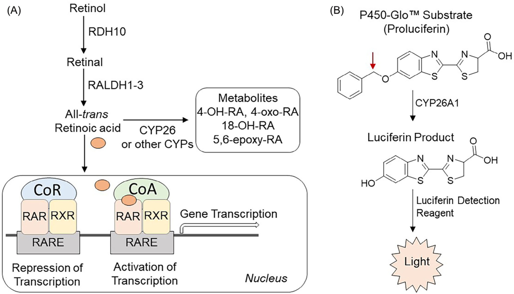
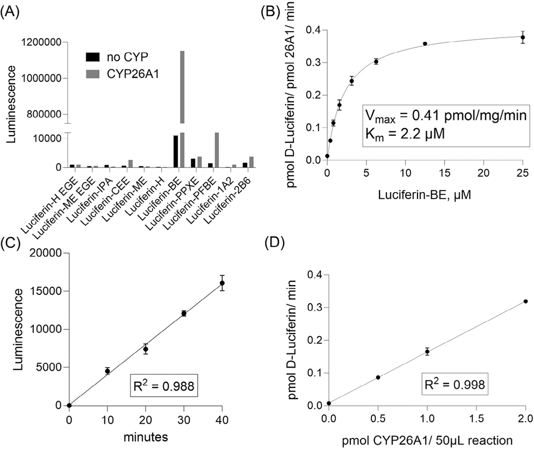
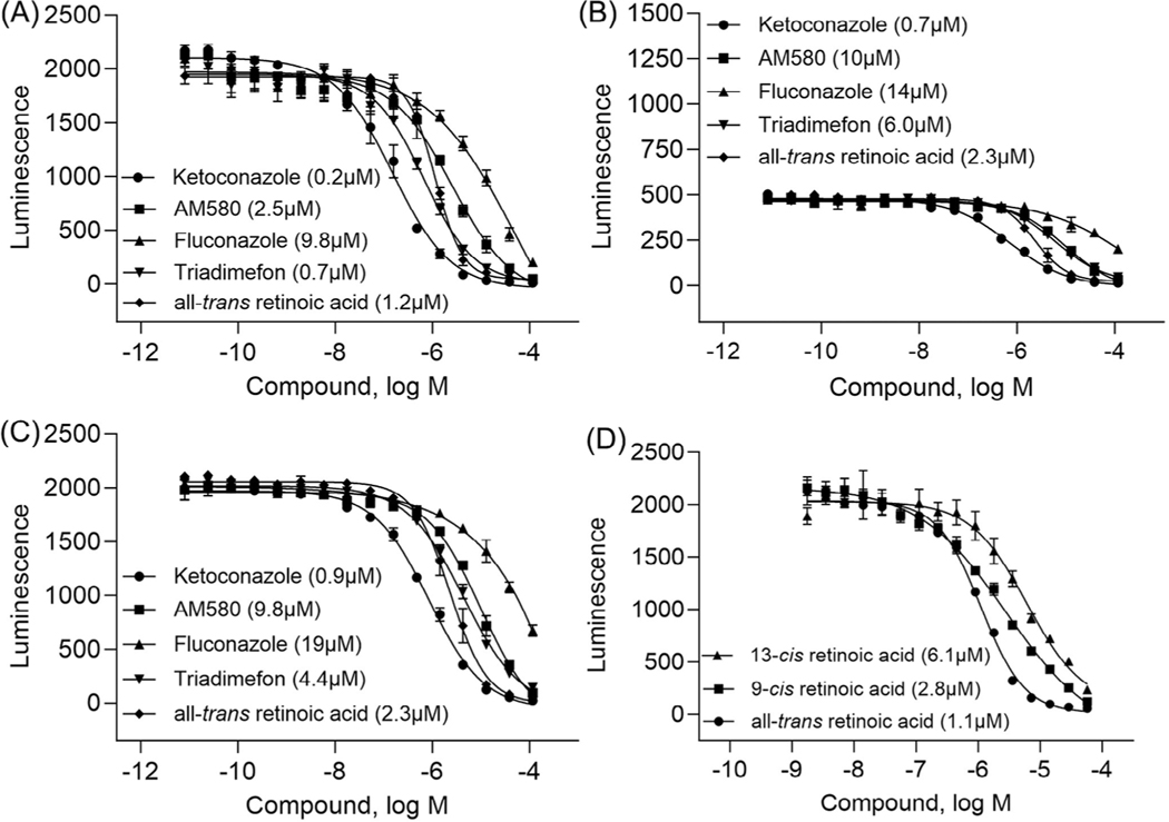
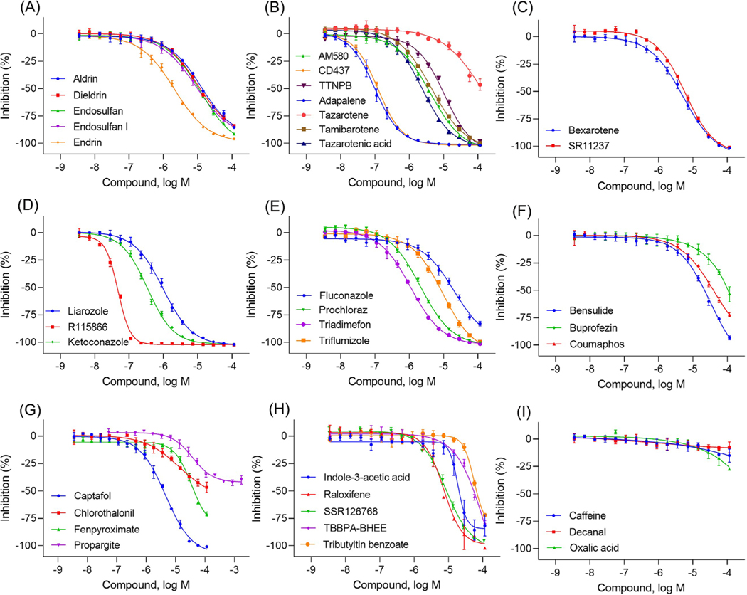
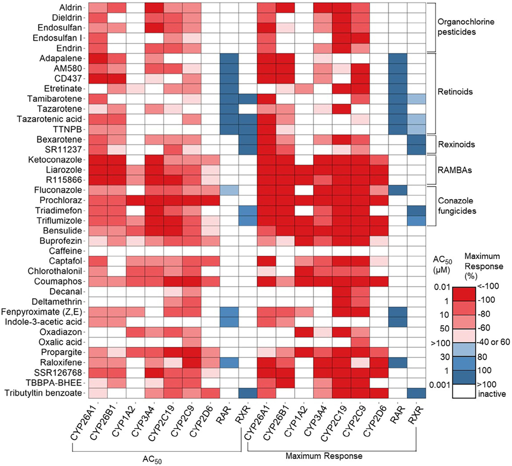
All-trans retinoic acid (atRA) is an endogenous ligand of the retinoic acid receptors, which heterodimerize with retinoid X receptors. AtRA is generated in tissues from vitamin A (retinol) metabolism to form a paracrine signal and is locally degraded by cytochrome P450 family 26 (CYP26) enzymes. The CYP26 family consists of three subtypes: A1, B1, and C1, which are differentially expressed during development. This study aims to develop and validate a high throughput screening assay to identify CYP26A1 inhibitors in a cell-free system using a luminescent P450-Glo assay technology. The assay performed well with a signal to background ratio of 25.7, a coefficient of variation of 8.9%, and a Z-factor of 0.7. To validate the assay, we tested a subset of 39 compounds that included known CYP26 inhibitors and retinoids, as well as positive and negative control compounds selected from the literature and/or the ToxCast/Tox21 portfolio. Known CYP26A1 inhibitors were confirmed, and predicted CYP26A1 inhibitors, such as chlorothalonil, prochloraz, and SSR126768, were identified, demonstrating the reliability and robustness of the assay. Given the general importance of atRA as a morphogenetic signal and the localized expression of Cyp26a1 in embryonic tissues, a validated CYP26A1 assay has important implications for evaluating the potential developmental toxicity of chemicals.

Keywords: CYP26; all‐trans retinoic acid (atRA); cytochrome P450 (CYP); retinoic acid receptor (RAR).


Conflict of interest statement

CONFLICT OF INTEREST STATEMENT

The authors declare no commercial or financial conflict of interest.

Figures

FIGURE 1
FIGURE 1
(A) General scheme of retinoic acid signaling pathway and metabolism by CYP26 enzymes. Retinol is converted to all-trans retinoic acid (atRA) via two oxidative steps, first to retinal mainly by retinol dehydrogenase 10 (RDH10) and then to atRA by retinal dehydrogenases 1 to 3 (RALDH1–3). atRA is further metabolized by the CYP26 and other CYP family of enzymes to primary metabolites, including 4-OH-RA, 18-OH-RA, and 5,6-epoxy-RA and are further metabolized to polar metabolites such as 4-oxo-RA. atRA is the activating ligand for the retinoic acid receptors (RARs), which heterodimerize with retinoid X receptors (RXRs) on the retinoic acid response element (RARE). In the inactive state, nuclear receptor co-repressor (CoR) complexes tighten and prevent the transcription of genes. Recruitment of co-activator (CoA) promotes transcription of genes downstream of the RARE; (B) Conversion of P450-Glo substrate by CYP26A1. CYP enzyme acts on a luminogenic P450-Glo substrate (first reaction) to produce a luciferin product that generates light with the luciferin detection reagent (second reaction). The amount of light produced is proportional to CYP activity. The red arrow indicates the site of modification by CYP enzyme.
FIGURE 2
FIGURE 2
Development of CYP26A1 assay. (A) Luminogenic substrate reactivities with or without CYP26A1. (B) Vmax and Km determined for luciferin-BE. (C) Time course of the CYP26A1 reaction with 2 μM luciferin-BE. (D) The rates of luciferin-BE reaction at varied CYP26A1 enzyme amounts. Data shown for A are single determinants, data shown for B, C, and D are the mean ± SD from three experiments.
FIGURE 3
FIGURE 3
Concentration-response curves. Testing the positive control compounds in the CYP26A1 assay under various conditions, such as (A) 0.1% BSA + 0.025% CHAPS, (B) 0.1% BSA + 0.01% Triton X-100, and (C) 0.1% BSA + 0.01% Tween-20. (D) Comparison of the inhbitory effects of all-trans retinoic acid (atRA) with its isomers, including 9-cis-RA and 13-cis-RA with 0.1% BSA + 0.025% CHAPS condition. Each value represents the mean ± SD from three experiments.
FIGURE 4
FIGURE 4
Concentration-response curves of the compounds tested in the CYP26A1 assay. (A) organochlorine pesticides: aldrin (IC50 = 12.8 μM), dieldrin (IC50 = 10 μM), endosulfan (IC50 = 12.1 μM), endosulfan I (IC50 = 9 μM), and endrin (IC50 = 2.1 μM); (B) retinoids: AM580 (IC50 = 4.1 μM), CD437 (IC50 = 0.1 μM), TTNPB (IC50 = 10.6 μM), adapalene (IC50 = 0.1 μM), tazarotene (IC50 = 54 μM), tamibarotene (IC50 = 4.5 μM), and tazarotenic acid (IC50 = 2.4 μM); (C) rexinoids: bexarotene (IC50 = 5.3 μM) and SR11237 (IC50 = 6.0 μM); (D) RAMBAs: ketoconazole (IC50 = 0.35 μM), liarozole (IC50 = 0.98 μM), and R115866 (IC50 = 0.05 μM); (E) conazole fungicides: fluconazole (IC50 = 20.2 μM), prochloraz (IC50 = 2.1 μM), triadimefon (IC50 = 1.1 μM), and triflumizole (IC50 = 9.5 μM); (F-H) diverse set of chemicals: bensulide (IC50 = 32.5 μM), buprofezin (IC50 = 80 μM), coumaphos (IC50 = 42.4 μM), captafol (IC50 = 4.6 μM), chlorothalonil (IC50 = 12.9 μM), fenpyroximate (IC50 = 34.8 μM), propargite (IC50 = 37.4 μM), indole-3-acetic acid (IC50 = 18.9 μM), raloxifene (IC50 = 7.2 μM), SSR126768 (IC50 = 8.6 μM), TBBPA-BHEE (IC50 = 119 μM), and tributyltin benzoate (IC50 = 55.5 μM); (I) negative controls. Each value represents the mean ± SD from three experiments.
FIGURE 5
FIGURE 5
Compound activities in various assays. The heatmap is colored based on the half maximum activity, (AC50, μM) in the left portion and maximum response (%) values displayed on the right. Inhibitory assays include all CYPs, and the activation assays are RAR and RXR. The compounds are classified into different groups based on their chemical structures and functions, such as organochlorine pesticides and conazole fungicides, or their mechanism of action, including retinoids, rexinoids, and RAMBAs.

Similar articles

Cited by

References

    1. Das BC., Thapa P., Kark R., Das S., Mahapatra S., Liu TC., Torregroza I., Wallac DP., Kambhampati S., Van Veldhuizen P., Verma A., Ray SK., & Evans T. (2014). Retinoic acid signaling pathways in development and diseases. Bioorganic & Medicinal Chemistry, 22(2), 673–683. - PMC - PubMed
    1. Bastien J, & Rochette-Egly C. (2004). Nuclear retinoid receptors and the transcription of retinoid-target genes. Gene, 328, 1–16. - PubMed
    1. Marill J, Idres N, Capron CC, Nguyen E, & Chabot GG (2003). Retinoic acid metabolism and mechanism of action: A review. Current Drug Metabolism, 4(1), 1–10. - PubMed
    1. Cunningham TJ, & Duester G. (2015). Mechanisms of retinoic acid signalling and its roles in organ and limb development. Nature Reviews Molecular Cell Biology, 16(2), 110–123. - PMC - PubMed
    1. McSorley LC, & Daly AK (2000). Identification of human cytochrome P450 isoforms that contribute to all-trans-retinoic acid 4-hydroxylation. Biochemical Pharmacology, 60(4), 517–526. - PubMed

MeSH terms

Substances

LinkOut - more resources