. 2024 Nov 14;45(6):1607-1616.
doi: 10.1093/jbcr/irae111.

FAS(APO), DAMP, and AKT Phosphoproteins Expression Predict the Development of Nosocomial Infection After Pediatric Burn Injury

Affiliations

FAS(APO), DAMP, and AKT Phosphoproteins Expression Predict the Development of Nosocomial Infection After Pediatric Burn Injury

Julia Penatzer et al. J Burn Care Res. .

Abstract

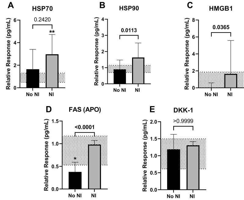
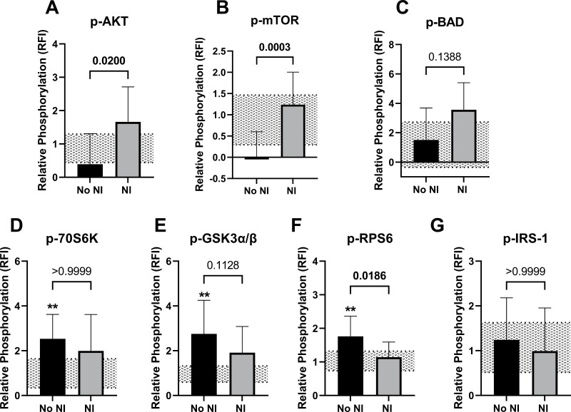
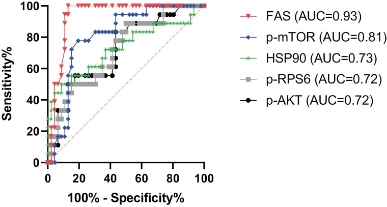
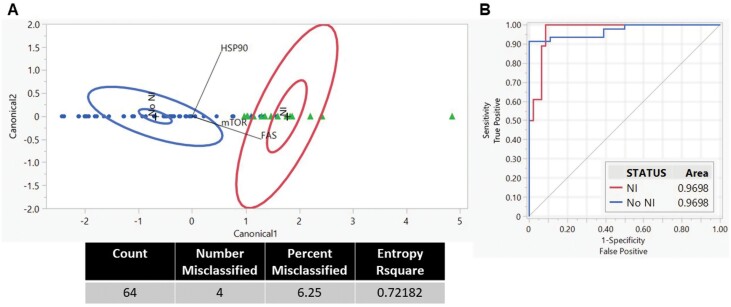
Pediatric burn injuries are a leading cause of morbidity with infections being the most common acute complication. Thermal injuries elicit a heightened cytokine response while suppressing immune function; however, the mechanisms leading to this dysfunction are still unknown. Our aim was to identify extracellular proteins and circulating phosphoprotein expression in the plasma after burn injury to predict the development of nosocomial infection (NI). Plasma was collected within 72 hours after injury from 64 pediatric burn subjects; of these, 18 went on to develop an NI. Extracellular damage-associated molecular proteins, FAS(APO), and protein kinase b (AKT) signaling phosphoproteins were analyzed. Subjects who went on to develop an NI had elevated high-mobility group box 1, heat shock protein 90 (HSP90), and FAS expression than those who did not develop an NI after injury (NoNI). Concurrently, phosphorylated (p-)AKT and mammalian target of rapamycin (p-mTOR) were elevated in those subjects who went on to develop an NI. Quadratic discriminant analysis revealed distinct differential profiles between NI and NoNI burn subjects using HSP90, FAS, and p-mTOR. The area under the receiver-operator characteristic curves displayed significant ability to distinguish between these 2 burn subject cohorts. These findings provide insight into predicting the signaling proteins involved in the development of NI in pediatric burn patients. Further, these proteins show promise as a diagnostic tool for pediatric burn patients at risk of developing infection while additional investigation may lead to potential therapeutics to prevent NI.

Keywords: FAS; danger-associated molecular protein; nosocomial infection; pediatric thermal injury; phosphoprotein.


Conflict of interest statement

The authors have no conflicts of interest to declare.

Figures

Figure 1.
Figure 1.
Relative cell death protein levels within the first 72 hours following pediatric thermal injury. Relative protein responses (normalized to healthy controls pg/mL) were observed for burn subjects who went on to develop a nosocomial infection (NI, gray bar) and those who recovered without developing an infection (NoNI, black bar). Healthy controls with standard error are noted as gray dotted line. HSP70 expression (A) was significantly increased in children who went on to develop NI compared to healthy controls. HSP90 (B) and HMGB1 (C) were increased in subjects who went on to develop an infection relative to those who did not. FAS(APO) was significantly decreased in subjects who did not develop an infection after burn injury relative to those who did and healthy controls (D). There were no significant differences for DKK-1 (E). Statistical analysis between groups was determined using Kruskal–Wallis test with Dunn’s multiple comparison. Differences between NI and NoNI are shown as line and brackets. Differences between burn groups and healthy controls are marked with an asterisk (*P < .05; **P < .01; ***P < .001; ****P < .0001). Responses are shown as median ± interquartile range. DKK-1: dickkopf-1; HMGB1: high-mobility group box 1 protein; HSP70: heat shock protein 70.
Figure 2.
Figure 2.
Relative phosphorylation within the first 72 hours following pediatric thermal injury. Relative fluorescence intensity (RFI) for phosphoprotein levels (normalized to healthy controls) was observed for burn subjects who went on to develop a nosocomial infection (NI, gray bar) and those who recovered without developing an infection (NoNI, black bar). Healthy controls with standard error are noted as gray dotted line. p-AKT (A), p-mTOR (B), and p-BAD (C) were increased in subjects who developed a NI relative to NoNI after pediatric burn injury. Burn subjects who did not develop an infection had higher p-70S6K (D), p-GSK3α/β (E), and p-RPS6 (F) from healthy controls. p-RPS6 was significantly elevated in burn subjects who did not develop an infection relative to those who did develop an infection (F). There were no significant differences for p-IRS-1 (G). Statistical analysis between groups was determined using Kruskal–Wallis test with Dunn’s multiple comparison. Differences between NI and NoNI are shown as line and brackets. Differences between burn groups and healthy controls are marked with an asterisk (*P < .05; **P < .01; ***P < .001; ****P < .0001). Responses are shown as median ± interquartile range. p-70S6K: phosphorylated p70 S6 kinase; p-AKT: phosphorylated protein kinase B; p-BAD: phosphorylated Bcl-2 agonist of cell death; p-GSK3α/β: phosphorylated glycogen synthase kinase 3; p-IRS-1: phosphorylated insulin receptor substrate 1; p-mTOR: phosphorylated mammalian target of rapamycin; p-RPS6: phosphorylated S6 ribosomal P phosphorylated glycogen synthase kinase 3.
Figure 3.
Figure 3.
Receiver-operating characteristics curve analysis within the first 72 hours following pediatric thermal injury. The area under the receiver-operating characteristic curves was highest for FAS (triangle) and p-mTOR (rhombus). This was followed by soluble HSP90 (star), p-RPS6 (square), and p-AKT (circle). The remaining markers had an AUC ≤0.70. AUC: area under the curve; HSP70: heat shock protein 70; p-AKT: phosphorylated protein kinase B; p-mTOR: phosphorylated mammalian target of rapamycin; p-RPS6: phosphorylated S6 ribosomal P phosphorylated glycogen synthase kinase 3.
Figure 4.
Figure 4.
Quadratic discriminant analysis (QDA) revealed distinct groupings for the development of infection in pediatric burn subjects (NI: triangle; NoNI: circle). The 12 proteins (5 cell death proteins and 7 phosphoproteins) were analyzed via QDA. The stepwise variable selection was chosen to optimize the model for the most crucial responses. Canonical scores for each protein (covariate) were calculated, and the 95% confidence interval is shown for NI (right) and NoNI (left) (A). The + symbol represents the mean of each group. Biplot rays describe the degree of association of a certain protein with the canonical variables. The performance of the stepwise-selected proteins was analyzed using receiver-operating characteristic (ROC) curves (B) and indicated an area under the curve of 0.97.

Similar articles

References

    1. American Burn Association. National Burn Repository 2016: Report of Data From 2006-2015. 2017. Accessed 2021. https://ameriburn.org/wp-content/uploads/2017/05/2016abanbr_final_42816.pdf
    1. Gonzalez R, Shanti CM.. Overview of current pediatric burn care. Semin Pediatr Surg. 2015;24(1):47–49. https://doi.org/ 10.1053/j.sempedsurg.2014.11.008 - DOI - PubMed
    1. World Health Organization. Burns. World Health Organization; 2018. Accessed August 5, 2021. https://www.who.int/news-room/fact-sheets/detail/burns
    1. Thakkar RK, Devine R, Popelka J, et al. Measures of systemic innate immune function predict the risk of nosocomial infection in pediatric burn patients. J Burn Care Res. 2020;42(3):488–494. https://doi.org/ 10.1093/jbcr/iraa193 - DOI - PMC - PubMed
    1. Devine RA, Diltz Z, Hall MW, Thakkar RK.. The systemic immune response to pediatric thermal injury. Int J Burns Trauma. 2018;8(1):6–16. - PMC - PubMed

MeSH terms